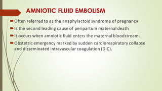
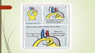
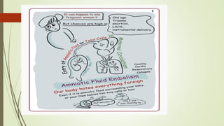
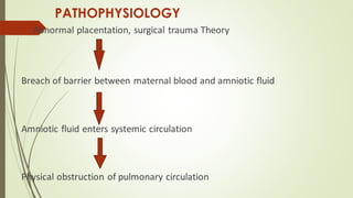
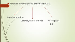
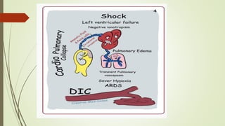
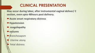
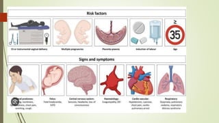
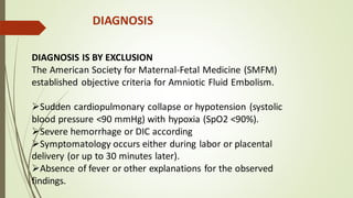
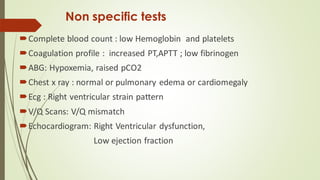
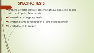
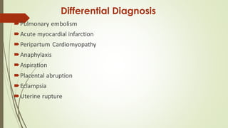
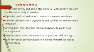
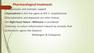
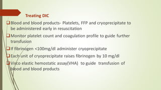
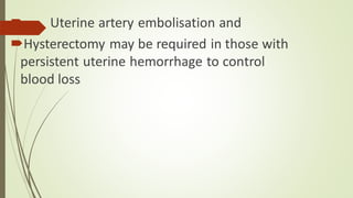
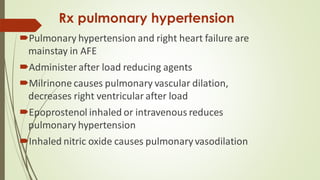
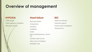
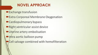
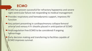
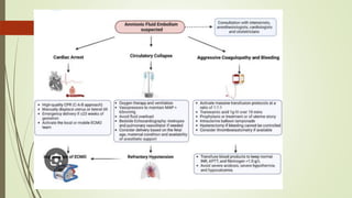
AMNIOTIC FLUID EMBOLISM
Dr SREENIVASULU YADAV SURINENI
Assistant Professor
SV Medical College, Tirupathi
AMNIOTIC FLUID EMBOLISM
Often referred to as the anaphylactoid syndrome of pregnancy
Is the second leading cause of peripartum maternal death
It occurs when amniotic fluid enters the maternal bloodstream.
Obstetric emergency marked by sudden cardiorespiratory collapse
and disseminated intravascular coagulation (DIC).
INCIDENCE
•The incidence ranges from 1.9 to 6.1 per 100,000 births,
•In the United States, affects 2.2 to 7.7 per 100,000 deliveries,
contributing to 7.5% of maternal deaths.
•In developing countries, the mortality rate ranges from 1.8 to
5.9 per 100,000 deliveries
RISK FACTORS
Maternal:
❑Older maternal age
❑Multiparity
❑After Abdominal trauma
❑During abortion
❑Amnio infusion
❑Placenta accreta
/Previa/Abruption
❑Tears in uterus, cervix
Fetal:
❑ Male baby
❑Fetal death
PATHOPHYSIOLOGY
 Abnormal placentation, surgical trauma Theory
Breach of barrier between maternal blood and amniotic fluid
Amniotic fluid enters systemic circulation
Physical obstruction of pulmonary circulation
Amniotic fluid in systemic circulation –second theory :
Activates inflammatory mediators
❑Platelet activating factor
❑Cytokines
❑Bradykinin
❑Leukotrienes
❑Thromboxane
Increased maternal plasma endothelin in AFE
Bronchoconstrictor
Coronary vasoconstrictor Procoagulant
DIC
CLINICAL PRESENTATION
May occur during labor, after instrumental vaginal deliver/ C
section, even upto 48hours post delivery.
Acute onset respiratory distress
Hypotension
 coagulopathy
seizures
Bronchosapsm
 Uterine atony
 Fetal distress
DIAGNOSIS
DIAGNOSIS IS BY EXCLUSION
The American Society for Maternal-Fetal Medicine (SMFM)
established objective criteria for Amniotic Fluid Embolism.
➢Sudden cardiopulmonary collapse or hypotension (systolic
blood pressure <90 mmHg) with hypoxia (SpO2 <90%).
➢Severe hemorrhage or DIC according
➢Symptomatology occurs either during labor or placental
delivery (or up to 30 minutes later).
➢Absence of fever or other explanations for the observed
findings.
The classic triad of AFE consists of hypoxia,
hypotension, and coagulopathy, with a normal body
temperature.
Fundus examination may detect minute bubbles in
retinal arteries.
 high-pitched murmur of tricuspid regurgitation.
Full-blown DIC is observed in approximately 83% of
patients.
Non specific tests
Complete blood count : low Hemoglobin and platelets
Coagulation profile : increased PT,APTT ; low fibrinogen
ABG: Hypoxemia, raised pCO2
Chest x ray : normal or pulmonary edema or cardiomegaly
Ecg : Right ventricular strain pattern
V/Q Scans: V/Q mismatch
Echocardiogram: Right Ventricular dysfunction,
Low ejection fraction
SPECIFIC TESTS
Broncho alveolar sample : presence of squamous cells coated
with neutrophils, Fetal debris
Elevated serum tryptase levels
Elevated plasma concentration of Zinc coproporphyrin
Elevated Sialyl Tn antigen
Differential Diagnosis
Pulmonary embolism
Acute myocardial infarction
Peripartum Cardiomyopathy
Anaphylaxis
Aspiration
Placental abruption
Eclampsia
Uterine rupture
MANAGEMENT
Goals of management:
Early recognition
Oxygenation
Resuscitation and hemodynamic stability
Maintain uterine tone
Correction of DIC
Delivery of Fetus
Taking care of ABCs
Secure the airway and administer 100% 02 with positive pressure
ventilation as early as possible
Optimise pre load and reduce pulmonary vascular resistance
Fluid resuscitation with crystalloids and colloids for hemodynamic
instability
Arterial line, Trans thoracic echocardiography to guide fluid
management
Vasoressors to maintain mean arterial pressure > 65 mm Hg
Plan for Packed cell transfusion in ongoing hemorrhage due to
uterine atony
Pharmacological treatment:
❑Vasopressors and Inotropic support
❑Epinephrine is first line agent as AFE is anaphylactoid
❑Noradrenaline and dopamine are other choices
❑In Right heart failure : Milrinone is considered
❑Steroids, to reduce inflammation induced by amniotic fluid
❑Uterotonic agents like Oxytocin
Methergine & Carboprost.
Treating DIC
❑Blood and blood products- Platelets, FFP and cryoprecipitate to
be administered early in resuscitation
❑Monitor platelet count and coagulation profile to guide further
transfusion
❑If fibrinogen <100mg/dl administer cryoprecipitate
❑Each unit of cryoprecipitate raises fibrinogen by 10 mg/dl
❑Visco elastic hemostatic assay(VHA) to guide transfusion of
blood and blood products
 Uterine artery embolisation and
Hysterectomy may be required in those with
persistent uterine hemorrhage to control
blood loss
Rx pulmonary hypertension
Pulmonary hypertension and right heart failure are
mainstay in AFE
Administer after load reducing agents
Milrinone causes pulmonary vascular dilation,
decreases right ventricular after load
Epoprostenol inhaled or intravenous reduces
pulmonary hypertension
Inhaled nitric oxide causes pulmonary vasodilation
Overview of management
HYPOXIA
100% oxygen
Positive pressure Ventilation
Refractory:ECMO
Heart failure
Fluid resuscitation
Vasopressors
Inotropes
Inodilators
Goals:
Mean arterial pressure > 65mm
Hg
Cardiac index>2 L/m2
Urine output 0.5ml/kg/hr
Pa02/fi02 > 250
DIC
Massive transfusion
Uterotonic agents
Uterine artery embolisation
Hysterectomy
NOVEL APPROACH
Exchange transfusion
Extra Corporeal Membrane Oxygenation
Cardiopulmonarybypass
Right ventricular assist device
Uterine artery embolisation
Intra aortic balloon pump
Cell salvage combined with hemofilteration
ECMO
ECMO has proven successful for refractory hypoxemia and severe
right ventricular failure not responding to medical management
Provides respiratory and hemodynamic support, improves RV
function
Any patient presenting in cardiopulmonary collapse femoral
arterial and venous 4 Fr sheaths placed in anticipation of ECMO
Anticoagulation free ECMO to be considered if ongoing
hemorrhage
Early decision making and transferring to facilities capable of
ECMO improves survival.
PROGNOSIS
Case fatality ranges between 11% to 26%
Death has been noted 1 to 12 hours after AFE
UK AFE registry reported 37% mortality rate
Early recognition and effective management of cardiac arrest
significantly improves survival rate.
REFERENCES
afe copy 2 (2).pdf anesthesiology ppt pd

afe copy 2 (2).pdf anesthesiology ppt pd

  • 1.
    AMNIOTIC FLUID EMBOLISM DrSREENIVASULU YADAV SURINENI Assistant Professor SV Medical College, Tirupathi
  • 2.
    AMNIOTIC FLUID EMBOLISM Oftenreferred to as the anaphylactoid syndrome of pregnancy Is the second leading cause of peripartum maternal death It occurs when amniotic fluid enters the maternal bloodstream. Obstetric emergency marked by sudden cardiorespiratory collapse and disseminated intravascular coagulation (DIC).
  • 4.
    INCIDENCE •The incidence rangesfrom 1.9 to 6.1 per 100,000 births, •In the United States, affects 2.2 to 7.7 per 100,000 deliveries, contributing to 7.5% of maternal deaths. •In developing countries, the mortality rate ranges from 1.8 to 5.9 per 100,000 deliveries
  • 5.
    RISK FACTORS Maternal: ❑Older maternalage ❑Multiparity ❑After Abdominal trauma ❑During abortion ❑Amnio infusion ❑Placenta accreta /Previa/Abruption ❑Tears in uterus, cervix Fetal: ❑ Male baby ❑Fetal death
  • 7.
    PATHOPHYSIOLOGY  Abnormal placentation,surgical trauma Theory Breach of barrier between maternal blood and amniotic fluid Amniotic fluid enters systemic circulation Physical obstruction of pulmonary circulation
  • 8.
    Amniotic fluid insystemic circulation –second theory : Activates inflammatory mediators ❑Platelet activating factor ❑Cytokines ❑Bradykinin ❑Leukotrienes ❑Thromboxane
  • 9.
    Increased maternal plasmaendothelin in AFE Bronchoconstrictor Coronary vasoconstrictor Procoagulant DIC
  • 12.
    CLINICAL PRESENTATION May occurduring labor, after instrumental vaginal deliver/ C section, even upto 48hours post delivery. Acute onset respiratory distress Hypotension  coagulopathy seizures Bronchosapsm  Uterine atony  Fetal distress
  • 15.
    DIAGNOSIS DIAGNOSIS IS BYEXCLUSION The American Society for Maternal-Fetal Medicine (SMFM) established objective criteria for Amniotic Fluid Embolism. ➢Sudden cardiopulmonary collapse or hypotension (systolic blood pressure <90 mmHg) with hypoxia (SpO2 <90%). ➢Severe hemorrhage or DIC according ➢Symptomatology occurs either during labor or placental delivery (or up to 30 minutes later). ➢Absence of fever or other explanations for the observed findings.
  • 16.
    The classic triadof AFE consists of hypoxia, hypotension, and coagulopathy, with a normal body temperature. Fundus examination may detect minute bubbles in retinal arteries.  high-pitched murmur of tricuspid regurgitation. Full-blown DIC is observed in approximately 83% of patients.
  • 17.
    Non specific tests Completeblood count : low Hemoglobin and platelets Coagulation profile : increased PT,APTT ; low fibrinogen ABG: Hypoxemia, raised pCO2 Chest x ray : normal or pulmonary edema or cardiomegaly Ecg : Right ventricular strain pattern V/Q Scans: V/Q mismatch Echocardiogram: Right Ventricular dysfunction, Low ejection fraction
  • 18.
    SPECIFIC TESTS Broncho alveolarsample : presence of squamous cells coated with neutrophils, Fetal debris Elevated serum tryptase levels Elevated plasma concentration of Zinc coproporphyrin Elevated Sialyl Tn antigen
  • 19.
    Differential Diagnosis Pulmonary embolism Acutemyocardial infarction Peripartum Cardiomyopathy Anaphylaxis Aspiration Placental abruption Eclampsia Uterine rupture
  • 20.
    MANAGEMENT Goals of management: Earlyrecognition Oxygenation Resuscitation and hemodynamic stability Maintain uterine tone Correction of DIC Delivery of Fetus
  • 21.
    Taking care ofABCs Secure the airway and administer 100% 02 with positive pressure ventilation as early as possible Optimise pre load and reduce pulmonary vascular resistance Fluid resuscitation with crystalloids and colloids for hemodynamic instability Arterial line, Trans thoracic echocardiography to guide fluid management Vasoressors to maintain mean arterial pressure > 65 mm Hg Plan for Packed cell transfusion in ongoing hemorrhage due to uterine atony
  • 22.
    Pharmacological treatment: ❑Vasopressors andInotropic support ❑Epinephrine is first line agent as AFE is anaphylactoid ❑Noradrenaline and dopamine are other choices ❑In Right heart failure : Milrinone is considered ❑Steroids, to reduce inflammation induced by amniotic fluid ❑Uterotonic agents like Oxytocin Methergine & Carboprost.
  • 23.
    Treating DIC ❑Blood andblood products- Platelets, FFP and cryoprecipitate to be administered early in resuscitation ❑Monitor platelet count and coagulation profile to guide further transfusion ❑If fibrinogen <100mg/dl administer cryoprecipitate ❑Each unit of cryoprecipitate raises fibrinogen by 10 mg/dl ❑Visco elastic hemostatic assay(VHA) to guide transfusion of blood and blood products
  • 24.
     Uterine arteryembolisation and Hysterectomy may be required in those with persistent uterine hemorrhage to control blood loss
  • 25.
    Rx pulmonary hypertension Pulmonaryhypertension and right heart failure are mainstay in AFE Administer after load reducing agents Milrinone causes pulmonary vascular dilation, decreases right ventricular after load Epoprostenol inhaled or intravenous reduces pulmonary hypertension Inhaled nitric oxide causes pulmonary vasodilation
  • 26.
    Overview of management HYPOXIA 100%oxygen Positive pressure Ventilation Refractory:ECMO Heart failure Fluid resuscitation Vasopressors Inotropes Inodilators Goals: Mean arterial pressure > 65mm Hg Cardiac index>2 L/m2 Urine output 0.5ml/kg/hr Pa02/fi02 > 250 DIC Massive transfusion Uterotonic agents Uterine artery embolisation Hysterectomy
  • 27.
    NOVEL APPROACH Exchange transfusion ExtraCorporeal Membrane Oxygenation Cardiopulmonarybypass Right ventricular assist device Uterine artery embolisation Intra aortic balloon pump Cell salvage combined with hemofilteration
  • 28.
    ECMO ECMO has provensuccessful for refractory hypoxemia and severe right ventricular failure not responding to medical management Provides respiratory and hemodynamic support, improves RV function Any patient presenting in cardiopulmonary collapse femoral arterial and venous 4 Fr sheaths placed in anticipation of ECMO Anticoagulation free ECMO to be considered if ongoing hemorrhage Early decision making and transferring to facilities capable of ECMO improves survival.
  • 30.
    PROGNOSIS Case fatality rangesbetween 11% to 26% Death has been noted 1 to 12 hours after AFE UK AFE registry reported 37% mortality rate Early recognition and effective management of cardiac arrest significantly improves survival rate.
  • 31.