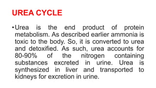
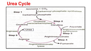
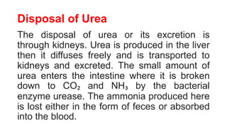
AMINO ACIDS
Miss Amanjot Kaur Sidhu
MSN (CCN)
DIGESTION OF PROTEINS
• The principle sources of protein are pulses, cereals,
peas, beans and nuts and principle animal sources are
milk and its products, meat, fish, liver, eggs etc.
• Primarily proteins are hydrolyzed from polypeptides
to dipeptides and then finally they are converted in
amino acids and absorbed in gut.
• Digestion of proteins begins in stomach and happens
at different levels in GI tract by help of different
digestive enzymes.
DIGESTION OF PROTEINS
•In mouth, no digestion of proteins takes place. Dietary
proteins are denatured on cooking, therefore digestion
becomes easy.
•Then, moves on to stomach and gastric juice in stomach
contains HCl & proteolytic enzymes such as pepsinogen
and mucin.
•In intestine, protein digestion takes place when food
enters duodenum in the form of acidic chyme, which
induce the release of cholecystokinin & secretin from
duodenum.
DIGESTION OF PROTEINS
• Secretin stimulates the secretion of bicarbonate which
increases pH of duodenum which makes it favorable for
catalytic activity of pancreatic enzymes.
• Trypsinogen is activated to trypsin by enterokinase
secreted by intestinal microvillus membrane.
• Active trypsin activates various proenzymozens. The
alkaline bile juice and pancreatic juice provide the optimum
pH i.e. 8 for pancreatic enzymes.
• In small intestine, trypsin, chymotrypsin act on proteins and
polypeptides.
DIGESTION OF PROTEINS
• Finally, protein digestion occurs in small intestine
producing mainly amino acids and some amount of
tripeptide and dipeptide.
• Intestinal juice contains enzymes such as leucine amino
peptidase, protein amino peptidase, dipeptidase and
tripeptidase.
• Amino peptidases break amino acid to produce free amino
acid, tripeptidase and dipeptidase to form amino acids.
• Finally, digestion of proteins produces mainly amino acids.
ABSORPTION OF PROTEINS
• Proteins are digested and finally converted into amino acids and
absorbed at the intestine through following two mechanisms:
• Simple passive diffusion: Absorption of D-isomers takes place
through simple passive diffusion.
• Active transportation: However, L-isomers are actively transported
across the intestine by the sodium dependent process; both L-amino
acid and sodium attach to carrier protein that is present on the
mucosal surface of microvilli and forms amino acids carrier, i.e.
sodium complex. The complex passes into the inner surface of
membrane and dissociate to liberate amino acids and sodium. Then,
sodium actively pumped out of the cell. The energy required for this
active transportation is acquired from hydrolysis of ATPs.
METABOLISM OF PROTEINS
•The amino acids undergo certain
common reactions like transamination
followed by deamination for the
liberation of ammonia.
•The amino group of the amino acids is
utilized for the formation of urea, an end
product of protein metabolism.
METABOLISM OF PROTEINS
•Transamination: The transamination, the name
indicates the transfer of an amino (-NH2) group
from an amino acid to a keto acid.
•This process involves the interconversion of an
amino acids and a pair of keto acids, catalyzed
by a group of enzymes called transaminases
also known as aminotransferases.
METABOLISM OF PROTEINS
•Deamination: Deamination means removal of
amino group from the amino acid as NH3.
•Deamination in its process removes amino group
along with the liberation of ammonia for urea
synthesis.
METABOLISM OF PROTEINS
•Transdeamination: The combined action of
aminotransferase and glutamate dehydrogenase
is referred to as transdeamination.
Transamination + deamination
•Transamination occurs in all tissues and
deamination only in liver.
METABOLISM OF PROTEINS
• Oxidative Deamination: Oxidative deamination is the liberation
of free ammonia from the amino group of amino acids coupled
with oxidation.
• Site: Liver and Kidney and Purpose of oxidative deamination is
to provide NH₃, for urea synthesis and α keto acid for a variety
of reactions, including energy generation.
• Glutamate serves as a collection center for amino groups in the
biological systems. Glutamate rapidly undergoes oxidation
deamination catalyzed by glutamate dehydrogenase (GDH) to
liberate ammonia.
METABOLISM OF PROTEINS
•Nonoxidative Deamination: Some of the
amino acids can be deaminated to liberate
NH₃, without undergoing oxidation.
METABOLISM OF AMMONIA
•Ammonia is constantly being liberated in the
metabolism of amino acids and other
nitrogenous compounds.
•Formation of Ammonia:
•The production of NH₃ occurs from amino acids,
i.e., transamination and deamination, biogenic
amines, amino group of purines and pyrimidine
and by the action of intestinal bacteria on urea.
METABOLISM OF AMMONIA
•Transport and storage of NH₃: Normal
plasma levels of NH₃ are 10-20 mg/dL. The
transport of ammonia between various
tissues and the liver mostly occurs in the
form of glutamine or alanine and not as free
ammonia.
•Disposal of Ammonia: Ammonia is
converted mostly to urea & uric acid.
UREA CYCLE
•Urea is the end product of protein
metabolism. As described earlier ammonia is
toxic to the body. So, it is converted to urea
and detoxified. As such, urea accounts for
80-90% of the nitrogen containing
substances excreted in urine. Urea is
synthesized in liver and transported to
kidneys for excretion in urine.
Urea Synthesis
•Urea synthesis is a five-step cyclic process.
The first two enzymes are present in
mitochondria and rest are localized in
cytosol.
Urea Cycle
Disposal of Urea
The disposal of urea or its excretion is
through kidneys. Urea is produced in the liver
then it diffuses freely and is transported to
kidneys and excreted. The small amount of
urea enters the intestine where it is broken
down to CO₂ and NH₃ by the bacterial
enzyme urease. The ammonia produced here
is lost either in the form of feces or absorbed
into the blood.
BIOLOGICALLY IMPORTANT COMPOUNDS
SYNTHESIZED FROM AMINO ACIDS
• GLUTATHIONE: Glutathione is a tripeptide of 3 amino acids. It is
synthesized in cytoplasm outside the ribosomes.
• SEROTONIN: Serotonin is synthesized in special "serotonin producing"
cells (also called Argentaffin cells or Kultchitsky's cells) of tissues like
gastric mucosa, intestine, brain, mast cells and probably platelets (or
stored in it).
• MELATONIN: It is a hormone of Pineal body and peripheral nerves of man.
It is synthesized from serotonin.
• NITRIC OXIDE: Nitric oxide (NO) is a wonder molecule having diverse
functions like PGs and is synthesized in the body from amino acid
arginine.
• CREATINE AND CREATININE: Creatine is found in the muscle, liver,
kidney and brain. It is synthesized in the liver and kidney from glycine,
arginine and methionine.
Disorders of Amino Acid Metabolism
• A number of disorders occur due to genetically determined
deficiency, absence or modification of a specific protein
(enzyme). A defective enzyme blocks a metabolic pathway
causing abnormalities in the normal metabolism. Some
most common errors of amino acid metabolism are
described below:
• Disorders of Aromatic Amino Acid Metabolism: Metabolic
blocks in the metabolic pathway of phenylalanine and
tyrosine cause a number of hereditary diseases like
phenylketonuria (PKU) alkaptonuria, tyrosinaemia, albinism,
etc. as shown in the following
Phenyl Ketonuria (PKU)
• This recessive disease is caused by complete or
partial deficiency of enzyme phenylalanine
hydroxylase. Classical type of PKU (1 in 10,000
live births) is due to absence of enzyme.
Phenylalanine and its alternative catabolites get
accumulated. Mousy odour of urine is due to
formation of phenyl acetyl glutamine.
• Treatment by giving diet having very low levels of
phenylalanine upto 6 years of age.
Alkaptonuria
• It prevents the body to break down the two amino acids
i.e. tyrosine & phenylalanine. This disorder is due to
deficiency of enzyme homogentisate oxidase resulting
in plasma accumulation and secretion of homogentisic
acid in urine. The urine darkens upon exposure to air
(oxidation to form benzaquinone acetate which is
polymerised to black alkapton bodies).
• This harmless condition does not need any treatment
except development of arthritis in the middle age or later
life.
Tyrosinaemia
•Disruption in the breakdown of amino acid,
tyrosine, a building block of most proteins
due to the deficiency of fumarylacetoacetate
hydrolase (FAH) that leads to abnormal liver
function and renal tubular dysfunction.
•Treatment is low protein diet.
Albinism
• This is due to inherited defects in the pigment cells
(melanocytes) of skin and eyes. Hypomelanosis is of
various types but can be divided into two major groups:
(i) Oculo-cutaneous albinism: There is decreased
pigmentation of skin and eyes. Biochemically, such
albinos are of two types: Tyrosinase negative
(unpigmented melanosomes) and tyrosinase positive
albinos (lightly pigmented melanosomes).
(ii) Ocular Albinism: It affects only eye and not the skin.
Causes of Proteinuria
• Dehydration
• Emotional stress
• Exposure to extreme cold
• Fever
• Strenuous exercise
Causes of Proteinuria
Diseases and conditions that can cause
persistently elevated levels of protein in urine,
which might indicate kidney disease, includes:
• Chronic kidney disease
• Diabetes
• Endocarditis
Causes of Proteinuria
• Immune disorders like lupus
• Acute inflammation of the kidney
(glomerulonephritis)
• Poisoning
• Kidney cancer
• Congestive heart failure
CAUSES OF HYPOPROTEINEMIA
• Non enough food protein in your diet: If you follow a vegetarian or
vegan diet, severe protein deficiency, called Kwashiorkor, may occur
as in developing countries.
• Improper absorption of protein from the foods you eat.
• Liver damage: Liver makes a protein called albumin, which makes
upto 60% of the total protein in your blood. Albumin carries vitamins,
hormones and other substances throughout your body. It also
prevents fluid from leaking out of your blood vessels. Damage to
your liver prevents it from making albumin.
• Kidney Damage
• Treatment: Taking Protein-Rich Foods.
CAUSES OF HYPER GAMMA
GLOBINEMIA
• Elevated levels of immunoglobulins in the blood:
• Any virus, bacteria, fungi, or condition that interferes with the
normal functioning of the immune system or the antibody
response can be a potential cause of hypergammaglobinemia.
• It might be the result of immune system dysfunction caused by
certain infections, such as – malaria, bacterial infections, viral
infections
• Other causes may include: rheumatoid arthritis, multiple
myeloma, liver diseases.
• Genetic disposition
PLASMA PROTEINS
• The plasma is the liquid part of blood (55-60%) after separation
of its cellular components.
• The serum is the liquid medium of blood, which separates out
after the blood clots or serum coagulates. It does not contain
fibrinogen and other clotting factors.
AMINO ACIDS.pptx

AMINO ACIDS.pptx

  • 1.
    AMINO ACIDS Miss AmanjotKaur Sidhu MSN (CCN)
  • 2.
    DIGESTION OF PROTEINS •The principle sources of protein are pulses, cereals, peas, beans and nuts and principle animal sources are milk and its products, meat, fish, liver, eggs etc. • Primarily proteins are hydrolyzed from polypeptides to dipeptides and then finally they are converted in amino acids and absorbed in gut. • Digestion of proteins begins in stomach and happens at different levels in GI tract by help of different digestive enzymes.
  • 3.
    DIGESTION OF PROTEINS •Inmouth, no digestion of proteins takes place. Dietary proteins are denatured on cooking, therefore digestion becomes easy. •Then, moves on to stomach and gastric juice in stomach contains HCl & proteolytic enzymes such as pepsinogen and mucin. •In intestine, protein digestion takes place when food enters duodenum in the form of acidic chyme, which induce the release of cholecystokinin & secretin from duodenum.
  • 4.
    DIGESTION OF PROTEINS •Secretin stimulates the secretion of bicarbonate which increases pH of duodenum which makes it favorable for catalytic activity of pancreatic enzymes. • Trypsinogen is activated to trypsin by enterokinase secreted by intestinal microvillus membrane. • Active trypsin activates various proenzymozens. The alkaline bile juice and pancreatic juice provide the optimum pH i.e. 8 for pancreatic enzymes. • In small intestine, trypsin, chymotrypsin act on proteins and polypeptides.
  • 5.
    DIGESTION OF PROTEINS •Finally, protein digestion occurs in small intestine producing mainly amino acids and some amount of tripeptide and dipeptide. • Intestinal juice contains enzymes such as leucine amino peptidase, protein amino peptidase, dipeptidase and tripeptidase. • Amino peptidases break amino acid to produce free amino acid, tripeptidase and dipeptidase to form amino acids. • Finally, digestion of proteins produces mainly amino acids.
  • 6.
    ABSORPTION OF PROTEINS •Proteins are digested and finally converted into amino acids and absorbed at the intestine through following two mechanisms: • Simple passive diffusion: Absorption of D-isomers takes place through simple passive diffusion. • Active transportation: However, L-isomers are actively transported across the intestine by the sodium dependent process; both L-amino acid and sodium attach to carrier protein that is present on the mucosal surface of microvilli and forms amino acids carrier, i.e. sodium complex. The complex passes into the inner surface of membrane and dissociate to liberate amino acids and sodium. Then, sodium actively pumped out of the cell. The energy required for this active transportation is acquired from hydrolysis of ATPs.
  • 7.
    METABOLISM OF PROTEINS •Theamino acids undergo certain common reactions like transamination followed by deamination for the liberation of ammonia. •The amino group of the amino acids is utilized for the formation of urea, an end product of protein metabolism.
  • 8.
    METABOLISM OF PROTEINS •Transamination:The transamination, the name indicates the transfer of an amino (-NH2) group from an amino acid to a keto acid. •This process involves the interconversion of an amino acids and a pair of keto acids, catalyzed by a group of enzymes called transaminases also known as aminotransferases.
  • 9.
    METABOLISM OF PROTEINS •Deamination:Deamination means removal of amino group from the amino acid as NH3. •Deamination in its process removes amino group along with the liberation of ammonia for urea synthesis.
  • 10.
    METABOLISM OF PROTEINS •Transdeamination:The combined action of aminotransferase and glutamate dehydrogenase is referred to as transdeamination. Transamination + deamination •Transamination occurs in all tissues and deamination only in liver.
  • 11.
    METABOLISM OF PROTEINS •Oxidative Deamination: Oxidative deamination is the liberation of free ammonia from the amino group of amino acids coupled with oxidation. • Site: Liver and Kidney and Purpose of oxidative deamination is to provide NH₃, for urea synthesis and α keto acid for a variety of reactions, including energy generation. • Glutamate serves as a collection center for amino groups in the biological systems. Glutamate rapidly undergoes oxidation deamination catalyzed by glutamate dehydrogenase (GDH) to liberate ammonia.
  • 12.
    METABOLISM OF PROTEINS •NonoxidativeDeamination: Some of the amino acids can be deaminated to liberate NH₃, without undergoing oxidation.
  • 13.
    METABOLISM OF AMMONIA •Ammoniais constantly being liberated in the metabolism of amino acids and other nitrogenous compounds. •Formation of Ammonia: •The production of NH₃ occurs from amino acids, i.e., transamination and deamination, biogenic amines, amino group of purines and pyrimidine and by the action of intestinal bacteria on urea.
  • 14.
    METABOLISM OF AMMONIA •Transportand storage of NH₃: Normal plasma levels of NH₃ are 10-20 mg/dL. The transport of ammonia between various tissues and the liver mostly occurs in the form of glutamine or alanine and not as free ammonia. •Disposal of Ammonia: Ammonia is converted mostly to urea & uric acid.
  • 15.
    UREA CYCLE •Urea isthe end product of protein metabolism. As described earlier ammonia is toxic to the body. So, it is converted to urea and detoxified. As such, urea accounts for 80-90% of the nitrogen containing substances excreted in urine. Urea is synthesized in liver and transported to kidneys for excretion in urine.
  • 16.
    Urea Synthesis •Urea synthesisis a five-step cyclic process. The first two enzymes are present in mitochondria and rest are localized in cytosol.
  • 17.
  • 18.
    Disposal of Urea Thedisposal of urea or its excretion is through kidneys. Urea is produced in the liver then it diffuses freely and is transported to kidneys and excreted. The small amount of urea enters the intestine where it is broken down to CO₂ and NH₃ by the bacterial enzyme urease. The ammonia produced here is lost either in the form of feces or absorbed into the blood.
  • 19.
    BIOLOGICALLY IMPORTANT COMPOUNDS SYNTHESIZEDFROM AMINO ACIDS • GLUTATHIONE: Glutathione is a tripeptide of 3 amino acids. It is synthesized in cytoplasm outside the ribosomes. • SEROTONIN: Serotonin is synthesized in special "serotonin producing" cells (also called Argentaffin cells or Kultchitsky's cells) of tissues like gastric mucosa, intestine, brain, mast cells and probably platelets (or stored in it). • MELATONIN: It is a hormone of Pineal body and peripheral nerves of man. It is synthesized from serotonin. • NITRIC OXIDE: Nitric oxide (NO) is a wonder molecule having diverse functions like PGs and is synthesized in the body from amino acid arginine. • CREATINE AND CREATININE: Creatine is found in the muscle, liver, kidney and brain. It is synthesized in the liver and kidney from glycine, arginine and methionine.
  • 20.
    Disorders of AminoAcid Metabolism • A number of disorders occur due to genetically determined deficiency, absence or modification of a specific protein (enzyme). A defective enzyme blocks a metabolic pathway causing abnormalities in the normal metabolism. Some most common errors of amino acid metabolism are described below: • Disorders of Aromatic Amino Acid Metabolism: Metabolic blocks in the metabolic pathway of phenylalanine and tyrosine cause a number of hereditary diseases like phenylketonuria (PKU) alkaptonuria, tyrosinaemia, albinism, etc. as shown in the following
  • 21.
    Phenyl Ketonuria (PKU) •This recessive disease is caused by complete or partial deficiency of enzyme phenylalanine hydroxylase. Classical type of PKU (1 in 10,000 live births) is due to absence of enzyme. Phenylalanine and its alternative catabolites get accumulated. Mousy odour of urine is due to formation of phenyl acetyl glutamine. • Treatment by giving diet having very low levels of phenylalanine upto 6 years of age.
  • 22.
    Alkaptonuria • It preventsthe body to break down the two amino acids i.e. tyrosine & phenylalanine. This disorder is due to deficiency of enzyme homogentisate oxidase resulting in plasma accumulation and secretion of homogentisic acid in urine. The urine darkens upon exposure to air (oxidation to form benzaquinone acetate which is polymerised to black alkapton bodies). • This harmless condition does not need any treatment except development of arthritis in the middle age or later life.
  • 23.
    Tyrosinaemia •Disruption in thebreakdown of amino acid, tyrosine, a building block of most proteins due to the deficiency of fumarylacetoacetate hydrolase (FAH) that leads to abnormal liver function and renal tubular dysfunction. •Treatment is low protein diet.
  • 24.
    Albinism • This isdue to inherited defects in the pigment cells (melanocytes) of skin and eyes. Hypomelanosis is of various types but can be divided into two major groups: (i) Oculo-cutaneous albinism: There is decreased pigmentation of skin and eyes. Biochemically, such albinos are of two types: Tyrosinase negative (unpigmented melanosomes) and tyrosinase positive albinos (lightly pigmented melanosomes). (ii) Ocular Albinism: It affects only eye and not the skin.
  • 25.
    Causes of Proteinuria •Dehydration • Emotional stress • Exposure to extreme cold • Fever • Strenuous exercise
  • 26.
    Causes of Proteinuria Diseasesand conditions that can cause persistently elevated levels of protein in urine, which might indicate kidney disease, includes: • Chronic kidney disease • Diabetes • Endocarditis
  • 27.
    Causes of Proteinuria •Immune disorders like lupus • Acute inflammation of the kidney (glomerulonephritis) • Poisoning • Kidney cancer • Congestive heart failure
  • 28.
    CAUSES OF HYPOPROTEINEMIA •Non enough food protein in your diet: If you follow a vegetarian or vegan diet, severe protein deficiency, called Kwashiorkor, may occur as in developing countries. • Improper absorption of protein from the foods you eat. • Liver damage: Liver makes a protein called albumin, which makes upto 60% of the total protein in your blood. Albumin carries vitamins, hormones and other substances throughout your body. It also prevents fluid from leaking out of your blood vessels. Damage to your liver prevents it from making albumin. • Kidney Damage • Treatment: Taking Protein-Rich Foods.
  • 29.
    CAUSES OF HYPERGAMMA GLOBINEMIA • Elevated levels of immunoglobulins in the blood: • Any virus, bacteria, fungi, or condition that interferes with the normal functioning of the immune system or the antibody response can be a potential cause of hypergammaglobinemia. • It might be the result of immune system dysfunction caused by certain infections, such as – malaria, bacterial infections, viral infections • Other causes may include: rheumatoid arthritis, multiple myeloma, liver diseases. • Genetic disposition
  • 30.
    PLASMA PROTEINS • Theplasma is the liquid part of blood (55-60%) after separation of its cellular components. • The serum is the liquid medium of blood, which separates out after the blood clots or serum coagulates. It does not contain fibrinogen and other clotting factors.