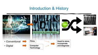
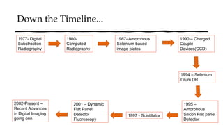
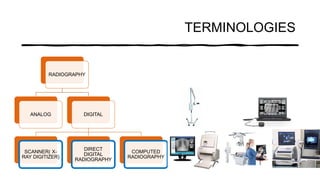
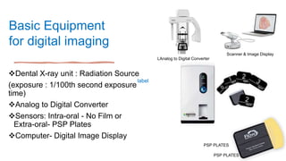
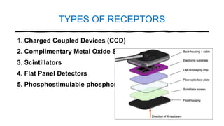
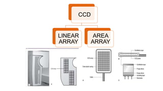
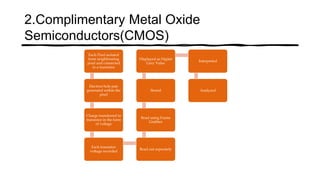
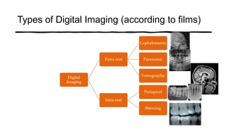
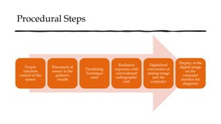
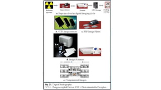
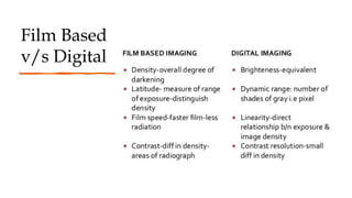
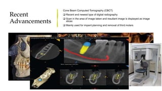
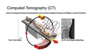
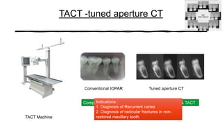
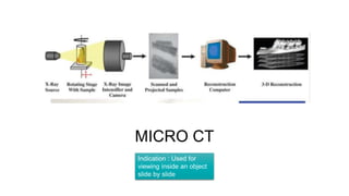
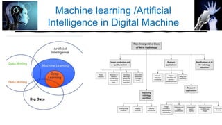
ADVANCES
IN
DIGITAL IMAGING
• Dr. Rupanjan Roy
• PG- 1st Year
• Department of Periodontology,
MCODS,MANIPAL
" A Picture is worth a Thousand
words"
Introduction & History
• Conventional
• Digital
Discovered by Dr. Francis Mouyens
Films
Computer
Technology
Used to store,
incorporate
and diagnose
Down the Timeline...
1977- Digital
Substraction
Radiography
1980-
Computed
Radiography
1987- Amorphous
Selenium based
image plates
1990 – Charged
Couple
Devices(CCD)
1994 – Selenium
Drum DR
1995 –
Amorphous
Silicon Flat panel
Detector
1997 - Scintillator
2001 – Dynamic
Flat Panel
Detector
Fluoroscopy
2002-Present –
Recent Advances
in Digital Imaging
going onn
TERMINOLOGIES
RADIOGRAPHY
DIGITAL
COMPUTED
RADIOGRAPHY
DIRECT
DIGITAL
RADIOGRAPHY
SCANNER( X-
RAY DIGITIZER)
ANALOG
label
Basic Equipment
for digital imaging
Dental X-ray unit : Radiation Source
(exposure : 1/100th second exposure
time)
Analog to Digital Converter
Sensors: Intra-oral - No Film or
Extra-oral- PSP Plates
Computer- Digital Image Display
PSP PLATES
LAnalog to Digital Converter
Scanner & Image Display
PSP PLATES
Analog to Digital Conversion
Sampling : Small range of voltage values grouped together into a
single value
Quantization – every sampled signal is assigned a value
Stored in computer and represent image- computer organizes
pixels-gives shades of grey-corresponds to no. assigned value
during quantization
TYPES OF RECEPTORS
1. Charged Coupled Devices (CCD)
2. Complimentary Metal Oxide Semiconductors(CMOS)
3. Scintillators
4. Flat Panel Detectors
5. Phosphostimulable phosphor plates(PSP)
CCD
LINEAR
ARRAY
AREA
ARRAY
2.Complimentary Metal Oxide
Semiconductors(CMOS)
Each Pixel isolated
from neighbouring
pixel and connected
to a transistor
Electron hole pair
generated within the
pixel
Charge transferred to
transistor in the form
of voltage
Each transistor
voltage recorded
Read out seperately
Read using Frame
Grabber
Stored
Displayed as Digital
Grey Value
Interpreted
Analyzed
3.Scintillator
Scintillator is a material that exhibits scintillation- property of a
material to exhibit luminescence when excited by ionizing radiation
They when stuck by an incoming material, absorbs it's energy and
scintillate I.e remit the absorbed energy in the form of light
They donot require charge transfer resulting in increased sensor
reliability and life span
Requires less system power
4. Flat
Panel
Detectors
Used for Medical Imaging, Extraoral imaging techniques
Provide large matrix area with less than 100 micrometer
Allows imaging of larger area including head
2 Types : Direct & Indirect
Indirect Flat Panel Detector – Sensitive to visible light
Direct Flat Panel Detector – Uses Selenium for efficient X-ray
absorption
It is a 'Sandwich' construction consisting of scintillator, an amorphous
silicon photodiode circuit layer and a Thin Film Transistor(TFT)
5.Photostimulable phosphor plates (PSP)
• Also known as Storage phosphor plates(SPP), Image plates or
computed radiography
• Flexible, wireless and indirect photography
• Available in the same size as intra-oral films
Digital Image Processing
Image Restoration
Image enhancement
Image analysis
Image Compression
Image Storage
Storage of image : magnetic or optical
File Size : 200KB (intraoral) and 6MB (extraoral)
Digital image can be modified or deleted
Backup media : External storage devices, pen drives, CD'S
, DVD'S
Types of Digital Imaging
Digital Imaging
Direct
Direct Digital
Imaging
Semi Direct Image
Plate System
Indirect
Image produced
using a CCD camera
instead of a flatbed
scanner
Types of Digital Imaging (according to films)
Digital
Imaging
Extra oral
Cephalometric
Panoramic
Tomographic
Intra oral
Periapical
Bitewing
Procedural Steps
Proper
infection
control of the
sensor
Placement of
sensor in the
patient's
mouth
Paralleling
Technique
used
Radiation
exposure with
conventional
radiographic
unit
Digitalized
conversion of
analog image
into the
computer
Display of the
digital image
on the
computer
monitor for
diagnosis
Film Based
v/s Digital
Indications
& Uses
Tumour
Detections
Detection of
Metastatic hard
tissue lesions
During
Instrumentation
Implant
Analysis
Caries detection
and periodontal
diseases
Detection of
Perforations
Advantages of Digital Imaging
3D
Reconstruction
Filtration
Time
Consuming
High Storage
capacity
Darkroom no
longer required
User Friendly
Tele
radiography
Dose
Reduction
Disadvantages of Digital Imaging
Cost
Medicolegal
Problems
Cross
infection
control
Sensor is
bulky
Radiovisuography
(RVG)
Recent
Advancements
Cone Beam Computed Tomography (CBCT)
 Recent and newest type of digital radiography
 Scan in the area of image taken and resultant image is displayed as image
slices
 Mainly used for impant planning and removal of third molars
Computed Tomography (CT)
TACT -tuned aperture CT
TACT Machine
Conventional IOPAR Tuned aperture CT
Comparison of the radiograph between conventional & TACT
Indications :
1. Diagnosis of Recurrent caries
2. Diagnosis of radicular fractures in non-
restored maxillary tooth
MICRO CT
Indication : Used for
viewing inside an object
slide by slide
DENTASCAN CT
Indications : To obtain true
cross sections of the
maxilla and the mandible
SIMPLANT
SIMPLANT is intended for
use as a software
interface and image
segmentation system for
the transfer of imaging
information from a
medical scanner such as
a CT scanner. It is also
intended as pre-planning
software for dental implant
placement and surgical
treatment.
Digital
Tomosynthesis
OPTICAL
COHERENCE
TOMOGRAPHY
• Layperson's explanation
• “a technique for
obtaining sub-surface
images of translucent or
opaque materials at a
resolution equivalent to
a low-
power microscope.”
Machine learning /Artificial
Intelligence in Digital Machine
Conclusion
• Innovative Technology to run a practice almost paper free
• Although, the conventional techniques like IOPAR&OPG are
Gold Standard to be used, still as research has progressed
incorporation of newer radiographic techniques have improved
Diagnosis, management of Diseases
"Be the Change You Want to see in the World – Mahatma Gandhi"
Advances in digital imaging

Advances in digital imaging

  • 1.
    ADVANCES IN DIGITAL IMAGING • Dr.Rupanjan Roy • PG- 1st Year • Department of Periodontology, MCODS,MANIPAL " A Picture is worth a Thousand words"
  • 2.
    Introduction & History •Conventional • Digital Discovered by Dr. Francis Mouyens Films Computer Technology Used to store, incorporate and diagnose
  • 3.
    Down the Timeline... 1977-Digital Substraction Radiography 1980- Computed Radiography 1987- Amorphous Selenium based image plates 1990 – Charged Couple Devices(CCD) 1994 – Selenium Drum DR 1995 – Amorphous Silicon Flat panel Detector 1997 - Scintillator 2001 – Dynamic Flat Panel Detector Fluoroscopy 2002-Present – Recent Advances in Digital Imaging going onn
  • 4.
  • 5.
    label Basic Equipment for digitalimaging Dental X-ray unit : Radiation Source (exposure : 1/100th second exposure time) Analog to Digital Converter Sensors: Intra-oral - No Film or Extra-oral- PSP Plates Computer- Digital Image Display PSP PLATES LAnalog to Digital Converter Scanner & Image Display PSP PLATES
  • 6.
    Analog to DigitalConversion Sampling : Small range of voltage values grouped together into a single value Quantization – every sampled signal is assigned a value Stored in computer and represent image- computer organizes pixels-gives shades of grey-corresponds to no. assigned value during quantization
  • 7.
    TYPES OF RECEPTORS 1.Charged Coupled Devices (CCD) 2. Complimentary Metal Oxide Semiconductors(CMOS) 3. Scintillators 4. Flat Panel Detectors 5. Phosphostimulable phosphor plates(PSP)
  • 8.
  • 9.
    2.Complimentary Metal Oxide Semiconductors(CMOS) EachPixel isolated from neighbouring pixel and connected to a transistor Electron hole pair generated within the pixel Charge transferred to transistor in the form of voltage Each transistor voltage recorded Read out seperately Read using Frame Grabber Stored Displayed as Digital Grey Value Interpreted Analyzed
  • 10.
    3.Scintillator Scintillator is amaterial that exhibits scintillation- property of a material to exhibit luminescence when excited by ionizing radiation They when stuck by an incoming material, absorbs it's energy and scintillate I.e remit the absorbed energy in the form of light They donot require charge transfer resulting in increased sensor reliability and life span Requires less system power
  • 11.
    4. Flat Panel Detectors Used forMedical Imaging, Extraoral imaging techniques Provide large matrix area with less than 100 micrometer Allows imaging of larger area including head 2 Types : Direct & Indirect Indirect Flat Panel Detector – Sensitive to visible light Direct Flat Panel Detector – Uses Selenium for efficient X-ray absorption It is a 'Sandwich' construction consisting of scintillator, an amorphous silicon photodiode circuit layer and a Thin Film Transistor(TFT)
  • 12.
    5.Photostimulable phosphor plates(PSP) • Also known as Storage phosphor plates(SPP), Image plates or computed radiography • Flexible, wireless and indirect photography • Available in the same size as intra-oral films
  • 13.
    Digital Image Processing ImageRestoration Image enhancement Image analysis Image Compression
  • 14.
    Image Storage Storage ofimage : magnetic or optical File Size : 200KB (intraoral) and 6MB (extraoral) Digital image can be modified or deleted Backup media : External storage devices, pen drives, CD'S , DVD'S
  • 15.
    Types of DigitalImaging Digital Imaging Direct Direct Digital Imaging Semi Direct Image Plate System Indirect Image produced using a CCD camera instead of a flatbed scanner
  • 16.
    Types of DigitalImaging (according to films) Digital Imaging Extra oral Cephalometric Panoramic Tomographic Intra oral Periapical Bitewing
  • 17.
    Procedural Steps Proper infection control ofthe sensor Placement of sensor in the patient's mouth Paralleling Technique used Radiation exposure with conventional radiographic unit Digitalized conversion of analog image into the computer Display of the digital image on the computer monitor for diagnosis
  • 19.
  • 20.
    Indications & Uses Tumour Detections Detection of Metastatichard tissue lesions During Instrumentation Implant Analysis Caries detection and periodontal diseases Detection of Perforations
  • 21.
    Advantages of DigitalImaging 3D Reconstruction Filtration Time Consuming High Storage capacity Darkroom no longer required User Friendly Tele radiography Dose Reduction
  • 22.
    Disadvantages of DigitalImaging Cost Medicolegal Problems Cross infection control Sensor is bulky
  • 23.
  • 24.
    Recent Advancements Cone Beam ComputedTomography (CBCT)  Recent and newest type of digital radiography  Scan in the area of image taken and resultant image is displayed as image slices  Mainly used for impant planning and removal of third molars
  • 25.
  • 26.
    TACT -tuned apertureCT TACT Machine Conventional IOPAR Tuned aperture CT Comparison of the radiograph between conventional & TACT Indications : 1. Diagnosis of Recurrent caries 2. Diagnosis of radicular fractures in non- restored maxillary tooth
  • 27.
    MICRO CT Indication :Used for viewing inside an object slide by slide
  • 28.
    DENTASCAN CT Indications :To obtain true cross sections of the maxilla and the mandible
  • 29.
    SIMPLANT SIMPLANT is intendedfor use as a software interface and image segmentation system for the transfer of imaging information from a medical scanner such as a CT scanner. It is also intended as pre-planning software for dental implant placement and surgical treatment.
  • 30.
  • 31.
    OPTICAL COHERENCE TOMOGRAPHY • Layperson's explanation •“a technique for obtaining sub-surface images of translucent or opaque materials at a resolution equivalent to a low- power microscope.”
  • 32.
  • 33.
    Conclusion • Innovative Technologyto run a practice almost paper free • Although, the conventional techniques like IOPAR&OPG are Gold Standard to be used, still as research has progressed incorporation of newer radiographic techniques have improved Diagnosis, management of Diseases "Be the Change You Want to see in the World – Mahatma Gandhi"