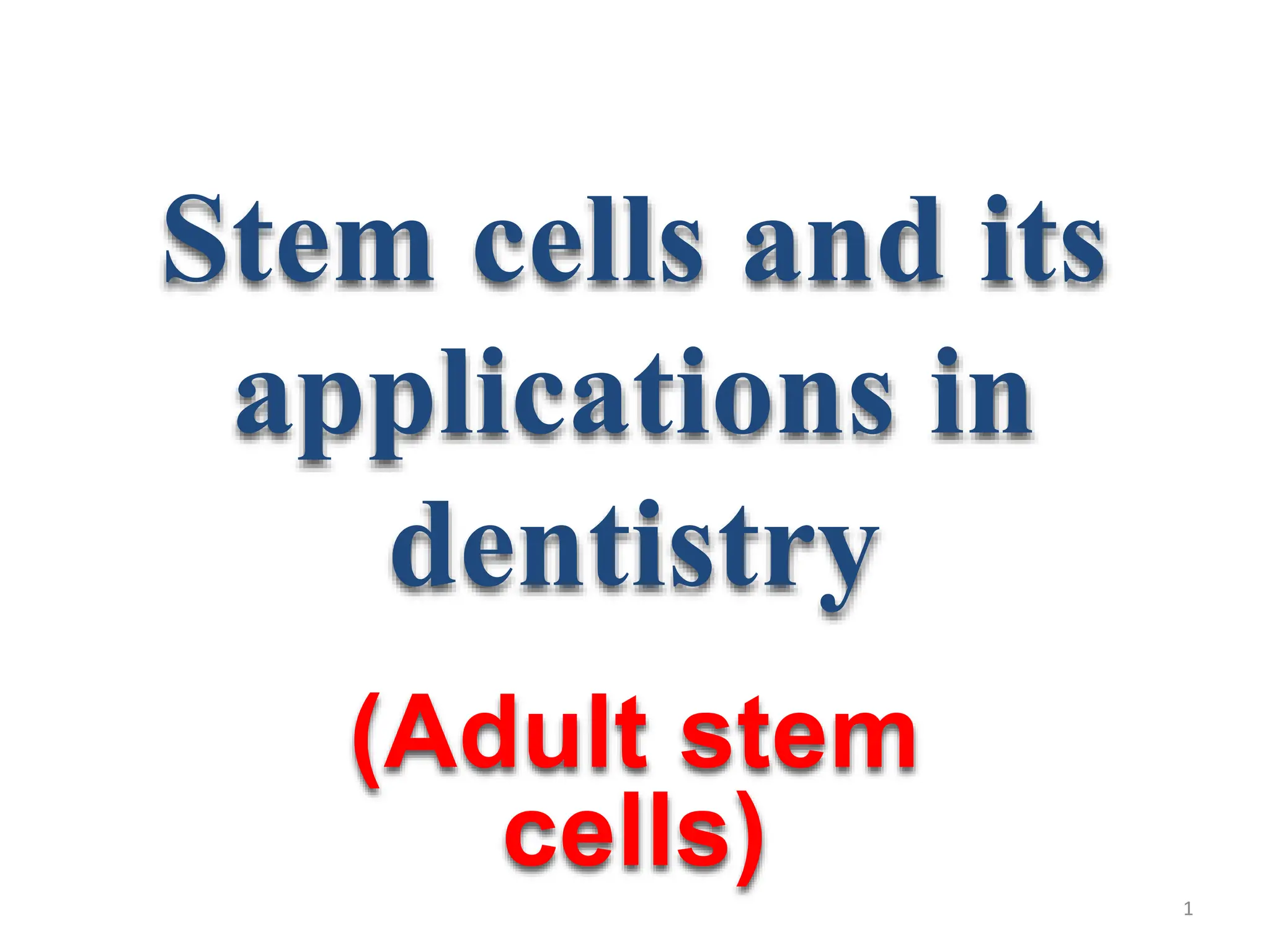
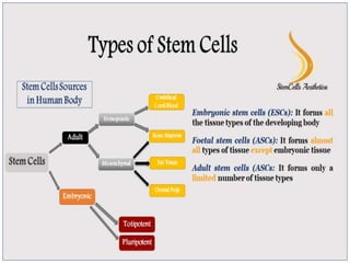
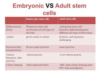
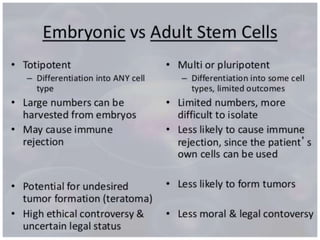
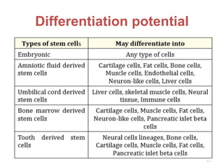
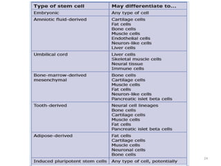
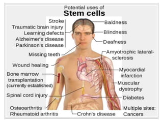
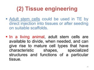
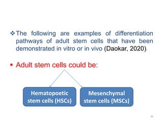
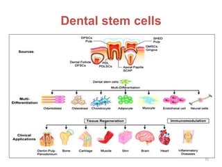
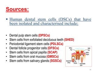
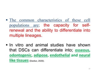
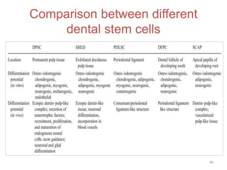
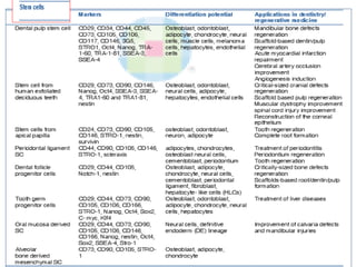
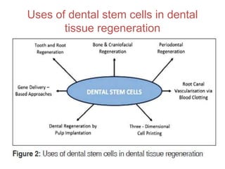
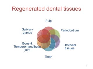
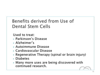
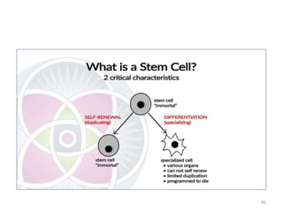
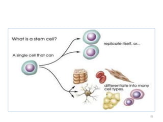
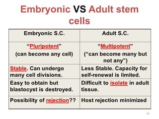
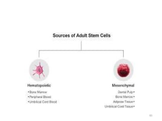
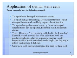
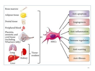
This document discusses stem cells and their applications in dentistry, focusing on adult stem cells. It defines stem cells as undifferentiated cells capable of becoming other cell types. Adult stem cells are found in adult tissues and can differentiate into multiple cell types. Sources of adult stem cells discussed include umbilical cord blood, amniotic fluid, bone marrow, adipose tissue, and dental tissues. Dental stem cells specifically can differentiate into bone, tooth, fat, nerve, and other tissues. Applications of adult stem cells in dentistry include cell-based therapy by direct injection or scaffold seeding for tissue engineering damaged tissues.
4. Introduction
A stem cell: is essentially a ‘blank’ cell,
capable of becoming another more
differential cell type in the body, such as a skin
cell, a muscle cell, or a nerve cell (Daokar, 2020).
In other words,
A stem cell: is a single cell that can replicate
itself or differentiate into many cell types (Google
scholar).
4
6. • Microscopic in size, stem cells are big news in
medical science circle because they can be
used to replace or even heal damaged tissues
and cells in the body.
• When called into action following an injury, a
stem cell self renews- undergoes cell division
and gives rise to one daughter stem cell and
one progenitor cell (Daokar, 2020).
6
7. • A progenitor cell is an intermediate cell type
formed before it achieves a fully
differentiating along a particular cellular
developmental pathway of stem cells (Daokar,
2020):
Stem cell > Stem cell + Progenitor cell >
Differentiated cell.
7
8. Types of stem cells
Based on their origin stem cells are
categorized either as;
(1) Embryonic stem cells (ESCs)
(2) Postnatal stem cells/somatic stem
cells/ adult stem cells (ASCs) (Daokar, 2020).
8
10. Characteristics
1. Totipotency: Generate all types of cells including germ cells
(ESCs).
2. Pluripotency: Generate all types of cells except cells of the
embryonic membrane.
3. Multipotency: Differentiate into more than one mature cell
(MSC).
4. Self renewal: Divide without differentiation and create
everlasting supply.
5. Plasticity: MSCs have plasticity and can undergo
differentiation. The trigger for plasticity is stress or tissue
injury which upregulates the stem cells and releases chemo-
attachments and growth factors (Daokar, 2020).
10
15. Definition
Adult stem cells are defined as: The
undifferentiated cells that are found in a
differentiated adult tissue, residing in a
specific area of each tissue (called a ‘stem
cell niche’) where they remain quiescent in
the body until they are activated by
epigenetic and/ or environmental factors,
such as mechanical forces, disease or
trauma (people’s journal of scientific research, 2009; Daokar, 2020).
15
16. The primary role of the adult stem cell:
is to maintain and repair the tissues in
which they are primarily found (Daokar, 2020).
16
17. Adult stem cells have been identified in many
organs and tissues, including: Brain, bone
marrow, peripheral blood, blood vessels,
skeletal muscle, skin, teeth, heart, gut, liver,
ovarian epithelium, and testis (Kolambkar et al., 2007).
Sources of adult stem cells include: The
umbilical cord, amniotic fluid, bone marrow,
adipose tissue, brain and teeth (Perin et al., 2007).
17
18. Adult stem cells come from
(extraoral & intraoral sources) as:
(1). Umbilical cord blood stem cells
(UCBSCs): Adult type stem cells can be
served from various pregnancy related
tissues.
• The greatest disadvantage of UCBSCs is that
there is only one opportunity to harvest them
from the umbilical cord at the time of birth.
• Similarly, amniotic stem cells can be sourced
only from amniotic fluid and are therefore
subject to time constraints (Laughlin, 2001).
18
19. (2). Amniotic fluid-derived stem cells
(AFSCs): These can be isolated from
aspirated of amniocentesis during genetic
screening.
• An increasing number of studies have
demonstrated that AFSCs have the capacity
for remarkable proliferation and
differentiation into multiple lineages (De Coppi et
al., 2007).
19
20. (3). Bone-marrow derived stem cells (BMSCs)
(Stem cell marker analysis fact sheet, 2019).
(4). Adipose derived stem cells (ASCs) (Estes et al.,
2006).
(5). Dental stem cells (DSCs): restored in 2003,
when Dr. Songtao Shi at the National Institues of
Health in Maryland discovered dental stem cells
in his daughter’s baby teeth (Karien, 2009).
20
21. (6). Body tissues: In adults and children, from
the moment we are born, stem cells are
present within virtually all tissues and organ
systems. (Daokar, 2020).
(7). Cadavers: Neural stem cells have been
removed from specific areas in postmortem
human brains as late as 20 hours following
death (Daokar, 2020).
21
22. According to source adult stem cells
could also be:
22
o Further details about each type is in our first ppt
(principles of TE)
Xenogenic
Allogenic
Autologous
26. (1) Cell based therapy
Adult stem cells (both non-dental and dental
stem cells) could be used as potential cell
based therapy for various diseases (liver,
bone, soft tissues of salivary glands & oral
mucosa, …….) (Mao, 2008; Daokar, 2020).
The following stem cell characteristics make
them good candidates for cell-based therapy:
• (1). Potential to be harvested from patients
• (2). High capacity of cell proliferation in culture.
26
27. • (3). Ease of manipulation to replace existing
nonfunctional genes via gene splicing method.
• (4). Ability to migrate to host target tissues (homing).
• (5). Ability to integrate into host tissues and interact
with the surrounding tissues (Daokar, 2020).
It is due to these unique characteristics that they
have, the potential to treat over 80 life threatening
diseases, and provide numerous benefits to the baby
is high.
27
29. (2) Tissue engineering
29
Adult stem cells could be used in TE by
direct injection into tissues or after seeding
on suitable scaffolds.
In a living animal, adult stem cells are
available to divide, when needed, and can
give rise to mature cell types that have
characteristic shapes, specialized
structures and functions of a particular
tissue.
30. The following are examples of differentiation
pathways of adult stem cells that have been
demonstrated in vitro or in vivo (Daokar, 2020).
Adult stem cells could be:
30
Hematopoetic
stem cells (HSCs)
Mesenchymal
stem cells (MSCs)
32. • Hematopoietic stem cells; give rise to all the
types of blood cells: red blood cells, B
lymphocytes, T lymphocytes, natural killer cells,
neutrophils, basophils, eosinophils, monocytes
and macrophages.
• Mesenchymal stem cells; give rise to a variety of
cell types: bone cells (osteocytes), cartilage cells
(chondrocytes), fat cells (adipocytes) and other
kinds of connective tissue cells such as those in
tendons (Daokar, 2020).
32
33. • Neural stem cells; in the brain give rise to three
major cell types: nerve cells (neuron), and two
categories of non-neuronal cells -astroctyes and
oligodendrocytes.
• Epithelial stem cells; in the lining of the digestive
tract occur in deep crypts and give rise to several cell
types: absorptive cells, goblet cells, panted cells and
enteroendocrine cells.
• Skin stem cells; occur in the basal layer of the
epidermis and at the base of hair follicles. The
epidermal stem cells give rise to keratinocytes, which
migrate to the surface of the skin and form a
protective layer. The follicular stem cells can give rise
to both the hair follicle and to the epidermis (Daokar,
2020).
34. Previous tables of differentiation potential
mentioned in slides no 23 and 24 are considered
an alternative or supportive to these
differentiation pathways.
34
36. Sources:
Human dental stem cells (DSCs) that have
been isolated and characterised include;
• Dental pulp stem cells (DPSCs)
• Stem cells from exfoliated deciduous teeth (SHED)
• Periodontal ligament stem cells (PDLSCs)
• Dental follicle progenitor cells (DFSCs)
• Stem cells from apical papilla (SCAP)
• Stem cells from oral mucosa (GMSCs)
• Stem cells from salivary glands (SGSCs)
36
37. The common characteristics of these cell
populations are; the capacity for self-
renewal and the ability to differentiate into
multiple lineages.
In vitro and animal studies have shown
that DSCs can differentiate into; osseous,
odontogenic, adipose, endothelial and neural
like tissues (Daokar, 2020).
37
38. The potential of dental MSC for tooth
regeneration and repair has been extensively
studied in the last years.
(1) Adult dental pulp stem cells (DPSC);
• Adult dental pulp stem cells (DPSCs) with ectomesenchymal
origin are the first type of DSCs isolated (in 2000) from adult
dental pulp of the extracted third molars or injured teeth (Lee
et al., 2014).
• They are the most common source of dental tissue derived
stem cells.
38
39. • DPSCs are multipotent and express STRO-1, CD 44 and CD 146
MSC markers.
• They are capable of differentiating into osteogenic,
odontogenic, myogenic, adipogenic and neurogenic
components both in-vitro and in-vivo and can produce pulp-
dentin complex in-vivo (Bluteau et al., 2008).
• Because of their ability to produce dental tissues in vivo
including dentin, pulp and crown like structures, theoretically,
a bio-tooth made from autogenous DPSCs should be the best
choice for clinical tooth reconstruction (Daokar, 2020).
39
40. (2) Stem cells from human exfoliated
deciduous teeth (SHED);
• Exfoliated deciduous teeth represent an
advantageous source of young stem cells for these
reasons (Lee et al., 2014):
• (1) Deciduous teeth are discarded after exfoliation, thus
collection of stem cells especially DPSCs avoids any type of
ethical considerations.
• (2) Have high proliferative capacity, faster proliferation rates
and increased population doublings.
• (3) High availability.
40
41. • Recent studies demonstrated that SHED∕immature DPSCs
(IDPSCs) expressed the embryonic stem cell markers Oct4,
Nanog, stage specific embryonic antigens (SSEA-3, -4), and
tumor recognition antigens (TRA-1-60 and TRA-1-81) (Lee et al.,
2014).
• So, although both SHED and DPSCs originate from the dental
pulp, they demonstrate differences regarding their
odontogenic differentiation and osteogenic induction.
• SHED cells can undergo osteogenic, adipogenic, neurogenic,
myogenic, chondrogenic, endothelial, and odontoblastic
differentiation. In vivo SHED cells can induce bone or dentin
formation (Miura et al., 2003; Paz et al., 2018).
41
42. • Also, the potential use of SHED as cell-based therapies has
expanded to a large number of tissues due to their high
degree of plasticity, stemness, and ability to cross lineage
boundaries (Lee et al., 2014).
42
43. (3) Periodontal ligament stem cells
(PDLSCs):
• The PDL is a specialized tissue located between the
cementum and the alveolar bone and has a role in the
maintenance and support of the teeth.
• Its continuous regeneration is thought to involve
mesenchymal progenitors arising from the dental follicle.
• PDL contains STRO-1 positive cells that maintain certain
plasticity since they can adopt adipogenic, osteogenic and
chondrogenic phenotypes in vitro.
• It is thus obvious that PDL itself contain progenitors, which
can be activated to self-renew and regenerate other tissues
such as cementum and alveolar bone (Gay et al., 2007). 43
44. (4) Stem cells from the dental follicle (DFSC):
• DFSCs have been isolated from follicle of human third molars
and express the stem cell markers Notch 1, STRO-1 and
nesting.
• These cells can be maintained in culture for at least 15
passages.
• STRO-1 positive DFSC can differentiate into cementoblasts in
vitro and are able to form cementum in vivo. Immortalised
dental follicle cells are able to re-create a new periodontal
ligament (PDL) after in vivo implantation (Yoko et al., 2007).
44
45. (5) Stem cells from the apical part of the
papilla (SCAP);
• Stem cells from the apical part of the human dental papilla
(SCAP) have been isolated and their potential to differentiate
into odontoblasts was compared to that of the periodontal
ligament stem cells (PDLSC).
• SCAP exhibit a higher proliferative rate and appears more
effective than PDLSC for tooth formation. Importantly, SCAP
are easily accessible since they can be isolated from human
third molars (Sonoyama et al., 2006; Paz et al., 2018).
45
46. (6) Oral mucosa derived stem cells
• It may either be oral epithelial stem cells or gingival stem
cells.
• The oral epithelial stem cells; are unipotent and develop only
into epithelial cells in-vivo. However, when used ex-vivo, they
develop a well stratified oral mucosal graft and are used for
grafting procedures involving the oral structures (Paz et al., 2018).
• Gingival stem cells; are multipotent, have reprogramming
capabilities, are more abundant, are easy to isolate and have a
rapid ex-vivo proliferation that makes their use clinically viable
(Paz et al., 2018).
46
47. (7) Salivary gland derived stem cells
• Cells derived from salivary gland have the potential to form
duct cells and acinar cells in-vitro and retain the capacity to
produce both mucin and amylase.
• They can therefore be used in rehabilitation of patients with
reduced salivary gland function following irradiation.
• However, an added difficulty in isolating the cells exists since
the harvested cells contain cells from the parenchymal,
stromal and blood vessel cells (Paz et al., 2018).
47
53. (A) Isolation of MSC From Different
Tissue Sources
• Traditionally, bone marrow (BM) has been the
prevailing source of MSC in humans.
• However, while BM is a rich source of
hematopoietic stem cells, it constitutes only a
rare MSC population.
• Additionally, due to the painful harvesting
procedure, which requires general anesthesia,
the supply of BM-derived MSC (bmMSC) and
their application in research and in the clinical
setting are limited (Li et al., 2016; Mushahary et al., 2018).
53
54. Over time, a number of other tissues have
been identified as alternative sources for
hMSC.
Today, MSC can be isolated from multiple
tissues including:
• Adipose tissue,
• Dental tissues,
• Skin and foreskin,
• Salivary gland,
• Limb buds,
• Perinatal tissues (Mushahary et al., 2018). 54
55. MSC Isolation procedures:
The procedures can generally be
divided into (Mushahary et al., 2018):
• 1. Primary explant technique.
• 2. Enzymatic disaggregation.
• 3. Mechanical disaggregation.
55
57. (1). Primary explant technique
• The explant culture (Fig. 4A) is one of the earliest
techniques of cell isolation and in vitro cell
cultivation.
• The source tissue is rinsed to remove blood cells and
then mechanically split (e.g., cutting or chopping)
into small pieces of no more than a few millimeter in
length. Reducing the size of tissue pieces improves
the diffusion of gases and nutrients toward the cells.
However, excessive mincing can lead to mechanical
destruction of the cells (Mushahary et al., 2018).
57
58. • The tissue pieces are then placed into plastic
culture vessels with growth medium. The MSC
grow out from tissue pieces onto the culture
dish surface. After several days the tissue
pieces can be removed.
58
60. (2). Enzymatic disaggregation
• Enzymatic protocol (Fig 4B) comprises
additional steps where the coarsely chopped
tissue is incubated with an enzyme solution
that degrades the extracellular matrix (ECM).
• Single cells or small cellular aggregates are
released from the tissue and transferred to
medium containing culture dishes (Mushahary et
al., 2018).
60
61. Explant procedures VERSUS Enzymatic
isolation methods
Although no comprehensive study to compare
the different isolation protocols is available to
date,
• several authors have compared specific
aspects of explant procedures versus
enzymatic isolation methods (Christodoulou et al.,
2013).
61
62. • The explant method has been reported to harvest
less heterogeneous cell populations exhibiting higher
proliferation rates and cell viability when compared
to the enzymatic method.
• This could be due to the presence of intact tissue
pieces and undissociated ECM during explant culture,
which keep the cells protected from proteolytic and
mechanical stress and hence provide favorable
environment for the migrating cells (Hendijani et al., 2017).
62
63. • Further advantages of explant culture lies in
the release of cytokines and growth factors
into the medium, higher yield of stromal cells,
shorter proliferation time, and simultaneous
expression of surface markers CD73, CD90,
and CD105, and absence of CD14, CD31,
CD34, and CD45 in all MSC populations (Hendijani
et al., 2017).
63
64. • The outcome of enzymatic digestion in terms of yield,
efficiency, and viability depends on the type and
concentration of the enzyme used for dissociation.
• Enzymatic digestion leads to dissociation of the ECM
resulting in low yield and double the time for cell
adherence.
Comparison between enzymatic digestion and
enzymatic with mechanical dissociation reported70%
increase in the yield using additional mechanical
method, suggesting the advantage of combining both
these methods (Mushahary et al., 2018).
64
65. (3). Mechanical
disaggregation
• For the disaggregation of soft tissues,
mechanical technique is usually employed.
• This technique basically involves careful
chopping or slicing of tissue into pieces and
collection of spill out cells.
65
66. The cells can be collected by two ways:
• i). Pressing the tissue pieces through a series of
sieves with a gradual reduction in the mesh size.
• ii). Forcing the tissue fragments through a syringe
and needle.
Although mechanical disaggregation involves the risk
of cell damage, the procedure is less expensive, quick
and simple.
• This technique is particularly useful when the
availability of the tissue is in plenty, and the
efficiency of the yield is not very crucial (google scholar).
66
67. Then detailed protocols that use suitable
materials and steps are followed for isolation
of stem cells from different tissues using
either method.
• The following protocol is an example of isolation of stem cells
using mechanical disaggregation method.
67
69. (B) Banking of stem cells
Stem cell banking or preservation is the
extraction, processing and storage of stem
cells, so that they may be used for treatment
in the future, when required.
Stem cell storage:
• Cryopreservation
• Magnetic freezing
69
71. a.) CRYOPRESERVATION (cryo – cold):
• It is considered to be a very effective method for maintaining
the viability of the transported stem cells by cooling to
subzero temperatures, typically –196°C (neelampari) where
the biological activity concludes leading to cell death.
• Procedures usually involves slow cooling at 1 to 2 °C/min in
the presence of a cryoprotectant, as Dimethyl SulphOxide
(DMSO) or Liquid nitrogen to avoid the damaging effects of
intracellular ice formation is considered standard.
• Liquid nitrogen is the most widely used material for
cryopreservation. It, not only preserves the cells but also
maintains their latency and potency.
71
72. b.) MAGNETIC FREEZING:
• Using a programmed freezer with a magnetic field. In this
method, a weak magnetic field is applied to the water and
tissues thereby lowering the freezing point of body up to 6-7°
C.
• It ensures distributed low temperature without the cell wall
damage resulted by ice expansion and nutrient drainage due
to capillary action, as caused by conventional freezing
methods.
• Magnetic freezing is considered to be more reliable as well as
relatively cheaper as compared with cryopreservation. It also
increases the survival rate of stored cells (Miura et al., 2017).
72
73. • Steps that involve centrifuging the cells, removing
supernatant, then suspending the cells with cell
banker are usually followed before banking them(Jindal
et al.,2019).
73
74. Commercial aspect of cell banking
• Utilization of these cells can be done best for the patients
from which they are harvested, and to some extent their
immediate family and blood relatives.
• As such, it is inevitable that the key to successful stem cell
therapy lies in being able to harvest the cells at the right point
of development and to safely store them until accident or
disease requires their usage.
• Needless to say, the cost and technical difficulty of storing for
decades properly make stem cell therapy using one’s own
cells a still uncertain bet.
• For dental stem cells, the trend of tooth banking is catching
up mainly in the developed countries but till date, it is not
very popular (Jindal et al.,2019).
74
75. References
• Bluteau. G, et al. European CE allies and Materials 16 (2008): 1-9.
• Christodoulou I, Kolisis FN, Papaevangeliou D, Zoumpourlis V. Comparative
evaluation of human mesenchymal stem cells of fetal (Wharton’s jelly) and adult
(adipose tissue) origin during prolonged in vitro expansion: Considerations for
cytotherapy. Stem Cells Int 2013;2013:246134.
• Daokar S. Adult Stem Cell - A Newer Dimension in Dentistry. ASDS,
(2020)10.31080-04.
• De Coppi P., et al. “Amniotic fluid and bone marrow derived mesenchymal stem
cells can be converted to smooth muscle cells in the cryoinjured rat bladder and
prevent compensatory hypertrophy of surviving smooth muscle cells”. Journal of
Urology 177.1 (2007): 369-376.
• Estes BT., et al. “Potent induction of chondrocytic differentiation of human
adipose-derived adult stem cells by bone morphogenetic protein 6”. Arthritis and
Rheumatology 54.4 (2006): 1222-1232.
• Gay I., et al. “Isolation and characterisation of multipotent human periodontal
ligament stem cells”. Orthodontics and Craniofacial Research 10 (2007): 149-160.
75
76. • Hendijani F. Explant culture: An advantageous method for isolation of
mesenchymal stem cells from human tissues. Cell Prolif 2017;50:e12334.
• Jindal L, Bhat N, Vyas D, Thakur K. “Stem Cells from Human Exfoliated Deciduous
Teeth (SHED) – Turning Useless into Miracle: A Review Article”. Acta Scientific
Dental Sciences 3.10 (2019): 49-54.
• Karien GA. “Dental Pulp Stem Cells, a New Era in Tissue Engineering”. Smile Dental
Journal 4 (2009): 6-9.
• Kolambkar YM., et al. “Chondrogenic differentiation of amniotic fluid-derived stem
cells”. Jouranl Molecular Pistology 38.5 (2007): 405-413.
• Laughlin MJ. “Umbilical cord blood for allogeneic transplantation in children and
adults”. Bone Marrow Transplant 27.1 (2001): 1-6.
• Lee SM, Zhang Q, Le AD. Dental Stem Cells: Sources and Potential Applications.
Curr Oral Health Rep (2014) 1:34–42
• Li H, Ghazanfari R, Zacharaki D, Lim HC, Scheding S. Isolation and characterization
of primary bone marrow mesenchymal stromal cells. Ann NY Acad Sci 2016;1370:
109–118.
76
77. • Mao JJ. “Stem cells and the future of dental care”. New York State Dental Journal
74.2 (2008): 20-24.
• Miura M, Gronthos S, Zhao M, Lu B, Fisher L. SHED: stem cells from human
exfoliated deciduous teeth. 71.3 (2017): 29-12.
• Mushahary D, Spittler A, Kasper C. Isolation, Cultivation, and Characterization of
Human Mesenchymal Stem Cells. Cytometry Part A 93A: 1931, 2018
• Paz A, Maghaireh H, Mangano F. Stem Cells in Dentistry: Types of Intra- and
Extraoral Tissue-Derived Stem Cells and Clinical Applications. Volume 2018, Article
ID 4313610, 14 pages.
• People’s journal of scientific research 2 (2009).
• Perin L., et al. “Renal differentiation of amniotic fluid stem cells”. Cell Prolix 40.6
(2007): 936-948.
• Sonoyama W., et al. “Mesenchymal Stem cell-mediated functional tooth
regeneration in swine”. PLos One 1 (2006): e79.
• Stem cell marker analysis fact sheet (2019).
• Yoko T., et al. “Establishment of immortalized dental follicle cells for generating
periodontal ligament in vivo”. Cell and Tissue Research 327 (2007): 301-311.
77