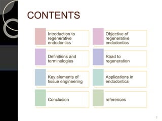
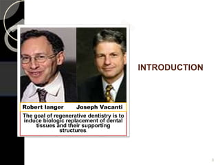
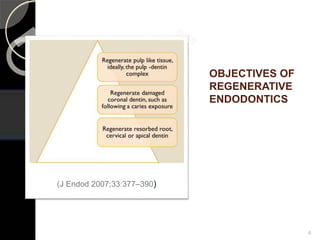
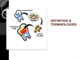
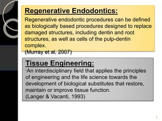
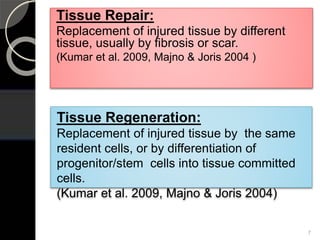
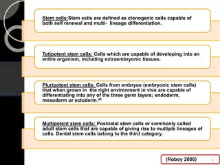
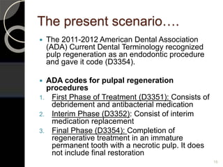
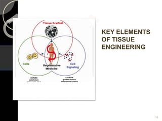
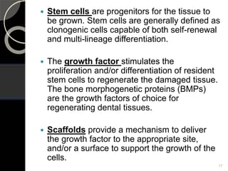
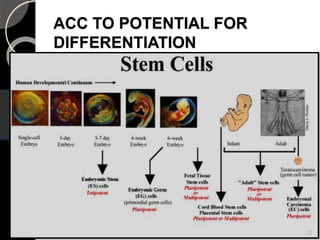
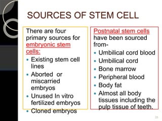
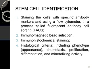
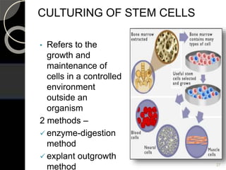
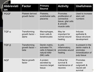
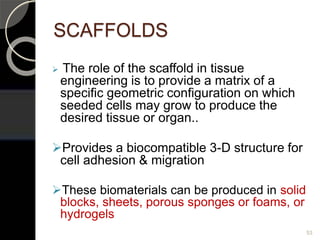
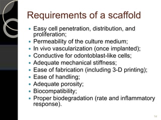
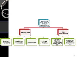
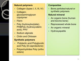
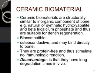
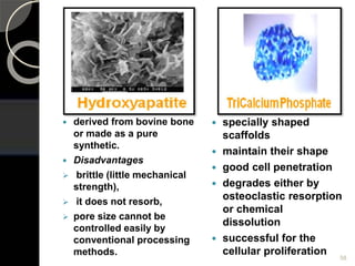
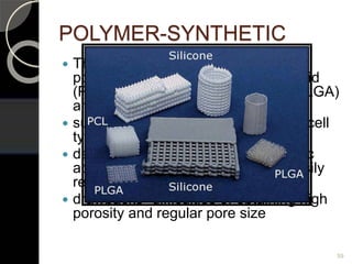
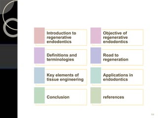
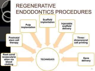
Regenerative endodontic procedures are designed to replace damaged structures, including dentin and root structures, as well as cells of the pulp-dentin complex
3. INTRODUCTION
The goal of regenerative dentistry is to
induce biologic replacement of dental
tissues and their supporting
structures.
Robert langer Joseph Vacanti
3
6. Regenerative Endodontics:
Regenerative endodontic procedures can be defined
as biologically based procedures designed to replace
damaged structures, including dentin and root
structures, as well as cells of the pulp-dentin
complex.
(Murray et al, 2007)
Tissue Engineering:
“An interdisciplinary field that applies the principles
of engineering and the life science towards the
development of biological substitutes that restore,
maintain or improve tissue function.
(Langer & Vacanti, 1993)
6
7. Tissue Repair:
Replacement of injured tissue by different
tissue, usually by fibrosis or scar.
(Kumar et al. 2009, Majno & Joris 2004 )
Tissue Regeneration:
Replacement of injured tissue by the same
resident cells, or by differentiation of
progenitor/stem cells into tissue committed
cells.
(Kumar et al. 2009, Majno & Joris 2004)
7
8. Stem cells:Stem cells are defined as clonogenic cells capable of
both self renewal and multi- lineage differentiation.
Totipotent stem cells: Cells which are capable of developing into an
entire organism, including extraembryonic tissues.
Pluripotent stem cells: Cells from embryos (embryonic stem cells)
that when grown in the right environment in vivo are capable of
differentiating into any of the three germ layers; endoderm,
mesoderm or ectoderm.45
Multipotent stem cells: Postnatal stem cells or commonly called
adult stem cells that are capable of giving rise to multiple lineages of
cells. Dental stem cells belong to the third category.
(Roboy 2000) 8
10. In 330 B.C, Aristotle
observed that a lizard
could grow back the lost
tip of its tail.
In late 1768, Spallanzani
reported that salamander
could regenerate a
complete limb after
surgical removal.
Trembley,in 1744,
demonstrated that a
bisected hydra gives rise
to two completely formed
individual
G. L. Feldman (1932) proposed
that through biological-aseptic
principle of tooth therapy,
regeneration of pulp might be
achieved and used dentine
fillings for stimulating pulp
regeneration
10
11. 1961 – Ostby studied the tissue
reorganization in the canal space filled
with blood clot.
1965 – Urist first demonstrated that new
bone could be formed at a non mineralizing
site after implantation of powder bone. This
led to isolation of active ingredient, Bone
morphogenetic protein (BMP).
1974 – Myers and Fountain observed
dental pulp regeneration in primate
using blood clot as scaffold.
1996 – The first dental pulp tissue
engineering was tested by Mooney et
al.
1996 – Sato et al first reported the
effectiveness of triple antibiotic
regimen to disinfect the root canal
space. 11
12. 1998 – Bohe et al reported
that pulp cells grown on
poly(glycolic) acid in vitro
resulted in high density
tissue similar to native
pulp.
1999 – Harada et al
localized stem cells in
cervical loop epithelium
of continuously growing
mouse incisor by Notch 1
expression.
2000 – Dental pulp stem
cells (DPSCs) were first
isolated and characterized
by Gronthoset al.
2001 – Iwaya et al
reported a case of
immature permanent
tooth with apical
periodontitis and sinus
tract undergoing
revascularisation.
2002 – Vacanti first
reported tooth
regeneration using
classical tissue
engineering technique.
12
13. 2006 – Yu et al published the first report of inducing dental pulp stem
cells differentiation into regular dentin-pulp complex by culturing in
tooth conditioned medium (contains tooth germ cells isolated from 2
day postnatal Sprague Dawley rat pulps).
2006 – Sonoyama et al isolated and characterized stem cells from
apical papilla (SCAP).
2004 – Ohazama et al studied the tooth forming ability by
recombination of non dental cell derived mesenchyme and embryonic
oral epithelium and Transfer of this embryonic tooth primordial into
the adult jaw resulted in development of tooth structures.
2004 – Seo et al suggested that periodontal ligament (PDL) contain
stem cells to generate cementum/PDL- like tissue in vivo.
2003 – Miura et al isolated and characterized stem cells from
exfoliating deciduous teeth (SHED).
13
14. 2008 – Huang et al
demonstrated the feasibility
of obtaining DPSCs from
supernumerary teeth.
2009 – karaoz et al isolated
dental pulp stem cells from
natal teeth.
2009 – Reynolds et al presented
a modified technique to avoid
discolouration of the crown
when using triple antibiotic paste
in revascularization approach by
sealing the dentinal tubules of
the chamber with flowable
composite
2009 – Honda et al developed
a protocol for the efficient
culture of enamel organ
epithelial progenitor cells
which facilitates the
engineering of enamel-tissue
in vivo.
2010 – Yagyuu et al
concluded in his study that
dental follicle and dental
papillae cells demonstrated
high proliferative and hard
tissue – forming ability even
after cryopreservation.
2014 – Hotwani proposed the
use of PRF as a therapeutic
material in regenerative
endodontics.
14
15. The present scenario….
The 2011-2012 American Dental Association
(ADA) Current Dental Terminology recognized
pulp regeneration as an endodontic procedure
and gave it code (D3354).
ADA codes for pulpal regeneration
procedures
1. First Phase of Treatment (D3351): Consists of
debridement and antibacterial medication
2. Interim Phase (D3352): Consist of interim
medication replacement
3. Final Phase (D3354): Completion of
regenerative treatment in an immature
permanent tooth with a necrotic pulp. It does
not include final restoration
15
17. Stem cells are progenitors for the tissue to
be grown. Stem cells are generally defined as
clonogenic cells capable of both self-renewal
and multi-lineage differentiation.
The growth factor stimulates the
proliferation and/or differentiation of resident
stem cells to regenerate the damaged tissue.
The bone morphogenetic proteins (BMPs)
are the growth factors of choice for
regenerating dental tissues.
Scaffolds provide a mechanism to deliver
the growth factor to the appropriate site,
and/or a surface to support the growth of the
cells.
17
18. STEM CELLS
Unspecialized cells
Give rise to more than 250 specialized
cells in the body
Serve as the body’s repair system
◦ Renew itself
◦ Replenish other cells
18
20. BASED UPON PLASTICITY
Stem cells Cell plasticity Source of stem
cells
Totipotent Each cell can
develop into a new
individual
Cells from early (1-
3 days) embryos
Pluripotent Cells can form any (over
200) cell types
Some cells of
blastocyst (5-14
days)
Multipotent Cells differentiated, but
can form a no. of other
tissues
Umbilical cord
blood, and
postnatal stem
cells including
dental pulp stem
cells
20
21. BASED ON THEIR SOURCE
STEM
CELLS
Autogenous – from patient’ts own
donor cells and have fewest problems
of immune rejection & pathogen
transmission.
Allogenic – From a donor of the same
species and Possibility of immune
rejection & pathogen transmission
and Costly, ethical & legal issues
Xenogenic – Isolated from another
species. Eg. Pigs, mice. High
possibility of rejection but removes
most of the legal & ethical issues.
21
23. SOURCES OF STEM CELL
There are four
primary sources for
embryonic stem
cells:
Existing stem cell
lines
Aborted or
miscarried
embryos
Unused In vitro
fertilized embryos
Cloned embryos
Postnatal stem cells
have been sourced
from-
Umbilical cord blood
Umbilical cord
Bone marrow
Peripheral blood
Body fat
Almost all body
tissues including the
pulp tissue of teeth.
25
24. STEM CELL IDENTIFICATION
1. Staining the cells with specific antibody
markers and using a flow cytometer, in a
process called fluorescent antibody cell
sorting (FACS)
2. Immunomagnetic bead selection
3. Immunohistochemical staining;
4. Histological criteria, including phenotype
(appearance), chemotaxis, proliferation,
differentiation, and mineralizing activity.
26
25. CULTURING OF STEM CELLS
• Refers to the
growth and
maintenance of
cells in a controlled
environment
outside an
organism
2 methods –
enzyme-digestion
method
explant outgrowth
method 27
26. enzyme-digestion method explant outgrowth
method
Sterile pulp
digested with
enzymes
cell
suspensions
cultured
using special
medium
cells
stimulated to
differentiate
Extracted pulp
tissue
Cut in 2-3 mm
cubes
Anchored with
microcarriers in
suitable
substrate
Incubated in
culture dishes
2 weeks is
needed for cells
to migrate
Haung et al. compared both methods and found that cells isolated by
enzyme-digestion had a higher proliferation rate than those isolated
by outgrowth 28
27. Eagle’s minimum
essential medium
(α- MEM) contains
10% fetal bovine
serum,
0.1 mg/ml
penicillin,
0.1 mg/ml
streptomycin
2.5 μg/ml
amphotericin B in a
humidified
atmosphere
containing 5% CO2
Dulbecco’s
modified Eagle’s
medium contains
15% fetal bovine
serum,
1 mM sodium
pyruvate,
0.1 mM non-essential
aminoacids,
4 Mm L- glutamine,
0.1 mM β
mercaptoethanol,
0.1 mg/ml penicillin,
0.1 mg/ml
streptomycin
2.5 μg/ml amphotericin
B at 37 degree
celscius in a
humidified 5% CO2
atmosphere 29
28. DIFFERENTIATION OF STEM
CELLS
Osteo/dentinogenic
medium –
dexamethasone,
glycerophosphate,
ascorbate phosphate
and
1,25 dihydroxy vitamin
D
Neurogenic induction –
B27 suplement
fibroblast GF,
epithelial GF
Adipogenic medium –
dexamethasone,
insulin
Generation of
specialized cells from
unspecialized stem
cells is a process
known as
differentiation, and is
triggered by signals
inside and outside the
cells
1. INTERNAL- Genes
interspersed across
long strands of DNA
2. EXTERNAL-
chemicals secreted
by other cells, 30
29. Types of stem cells
1) Stem cells of the apical papilla
(SCAP)
2) Dental pulp stem cells (DPSCs)
3) Stem cells from human exfoliated
deciduous teeth (SHED)
4) Periodontal ligament stem cells
(PDLSCs)
31
31. HUMAN DENTAL STEM CELLS
• 4 types of human dental stem cells have
been isolated and characterized
1. Dental pulp stem cells ( DPSC’s)
2. Stem cells from exfoliated deciduous teeth
(SHED’s)
3. Stem cells from apical papilla (SCAP’s)
4. Periodontal ligament stem cells (PDLSC’s)
DPSCs & SHEDs are from the pulp,
SCAPs from the pulp precursor tissue –
the apical papilla & PDLSCs from PDL. 33
33. STEM CELLS FROM HUMAN
EXFOLIATED DECIDUOUS TEETH
(SHED)
Isolated for the first time in 2003 by miura
et al.
Retrieved from a tissue that is disposable
and readily accessible
Differentiate into a variety of cell types
including neural cells, adipocytes,
osteoblast-like and odontoblast-like cells
Resulting tissue presented architecture and
cellularity that closely resemble those of a
physiologic dental pulp-Cordeiro et al
(2008)
35
34. 36
easily preserved
multiply rapidly and grow much faster
than the adult stem cells, suggesting
that they are less mature
With the rapid development of
advanced cryopreservation
technology, the first commercial tooth
bank was established as a venture
company at National Hiroshima
University in Japan in 2004
35. Collection, Isolation and
preservation of SHED
SHED banking is a proactive decision
made by the parents
put tooth fulfilling in sterile saline
solution
tooth exfoliated should have pulp red
in colour, indicating that the pulp
received blood flow up until the time of
removal, which is indicative of cell
viability
37
36. Transferred into the vial
containing a hypotonic
phosphate buffered
saline solution
The vial is then carefully
sealed and placed into
the thermette a
temperature phase
change carrier,
After which the carrier is
then placed into an
insulated metal
transport vessel.
The thermette along with
the insulated transport
vessel maintains the
sample in a hypothermic
state during
transportation-
SUSTENTATION.
Tooth surface is cleaned
by washing three times
with dulbecco’s
phosphate buffered
saline without ca++ and
mg++ (PBS).
Disinfection is done
with disinfection reagent
such as povidone iodine
and again washed with
PBS.
38
37. The pulp tissue is
isolated from the pulp
chamber with a sterile
small forceps or dental
excavator.
Contaminated Pulp
tissue is placed in a
sterile petridish which
was washed at least
thrice with PBS.
The tissue is then
digested with
collagenase Type I and
Dispase for 1 hour at
37ºC. Trypsin- EDTA
can also be used.
Isolated cells are
passed through a 70
um filter to obtain
single cell supensions.
Then the cells are
cultured in a
Mesenchymal Stem
Cell Medium( MSC)
medium which consists
of
Usually isolated
colonies are visible
after 24 hrs.
39
alpha modified minimal
essential medium with 2mM
glutamine
15% fetal bovine serum
(FBS),
0.1Mm L- ascorbic acid
phosphate,
100ug/ml penicillin.
100ug/ml streptomycin at
37ºC
5% CO2 in air
38. CRYOPRESERVATION MAGNETIC
FREEZING
preserving cells or
whole tissues by
cooling them to sub-
zero temperatures
typically -196 degree
Celsius
biological activity is
stopped
Cells harvested near
end of log phase
growth
preserved using liquid
nitrogen vapour at a
temperature of less
than -150ºC
programmed freezer
with a magnetic field,
the so called Cell Alive
System (CAS)
applying even a weak
magnetic field to water
or cell tissue will lower
the freezing point of
that body by up to 6-7
degrees Celsius.
object is uniformly
chilled, the magnetic
field is turned off and
the objects snap
freezes
40
39. DENTAL PULP STEM
CELLS(DPSC’s)
1st identified by Gronthos in 2000
isolated from the human adult third molars
,exfoliated deciduous teeth, supernumerary
teeth, crown fractured teeth that did not
require extraction and permanent tooth
germs
Huanget al (2006) conducted a study to
characterize human adult dental pulp cells
isolated and cultured in vitro and to
examine the cell differentiation potential
grown on dentin. It was concluded that
isolated human pulp stem cells may 41
40. Properties of human dental pulp stem cells:
◦ Self-renewal capability,
◦ Multilineage differentiation capacity,
◦ Clonogenic efficiency of human dental pulp stem
cells (DPSCs)
◦ DPSCs were capable of forming ectopic dentin
and associated pulp tissue in vivo.
42
• The stem cell population in the pulp is very
small; approximately 1% of the total cells
(Smith et al.2005) and the effect of aging
reduce the cell pool available to participate
in regeneration which reflects the better
healing outcomes seen in younger patients.
41. PERIODONTAL LIGAMENT
STEM CELLS (PDLSC’S)
Proliferative, Longer lifespan, and higher number of
population doublings in vitro
Develop into other cell lineages- differentiate into
cementoblast-like cells, adipocytes and collagen-
forming cells in vitro and the capacity to generate a
cementum/pdl-like structure in vivo.
Shi et al (2005) who demonstrated the generation
of cementum-like structures associated with PDL-
like connective tissue after transplanting PDLSCs
with hydroxyapatite/tricalcium phosphate particles
into immunocompromised mice.
43
42. Trubiani et al (2008) suggested that
PDLSCs had regenerative potential
when seeded onto a three dimensional
biocompatible scaffold, thus encouraging
their use in graft biomaterials for bone
tissue engineering in regenerative
dentistry
Li Y et al (2008) have reported
cementum and periodontal ligament-like
tissue formation when PDLSCs are
seeded on bioengineered dentin. 44
43. A new unique population of
mesenchymal stem cells (MSCs)
residing in the apical papilla of
permanent immature teeth, known as
stem cells from the apical papilla (SCAP)
discovered by Sonoyama et al (2008)
apical papilla is distinctive to the pulp in
terms of containing less cellular and
vascular components than those in the
pulp.
Cells in the apical papilla proliferated 2-
to 3-fold greater than those in the pulp in
organ cultures 45
STEM CELLS OF APICAL
PAPILLA (SCAP)
44. Stem cells in the apical papilla may also
explain a clinical phenomenon described
in a number of recent clinical case
reports showing that apexogenesis can
occur in infected immature permanent
teeth with periradicular periodontitis or
abscess.
It is likely that the SCAP residing in the
apical papilla survive such pulp necrosis
because of their proximity to the
vasculature of the periapical tissues.
Therefore, after endodontic disinfection,
and under the influence of the surviving
epithelial root sheath of hertwig, these
cells can generate primary odontoblasts
that complete root formation.
46
45. Role of SCAP
◦ Helps in continued root formation
◦ In pulp healing and regeneration
◦ In replantation and transplantation
47
46. GROWTH
FACTORS/MORPHOGENS
Growth factors/Morphogens are
extracellularly secreted signals governing
morphogenesis during epithelial-
mesenchymal interactions.
They are proteins that bind to receptors on
the cell and induce cellular proliferation
and/or differentiation.
Through their effects on gene expression in
the cell nucleus, mediated by transcription
and other factors, that the growth factors
influence cell behaviour and activity
48
47. Five major classes of evolutionary
conserved protein families- –
49
Bone
morphogenetic
proteins
(BMPs),
Fibroblast
growth factors
(FGFs),
Wingless- and
int-related
proteins
(Wnts),
Hedgehog
proteins
(Hhs), and
Tumor
necrotic factor
families
(TNF).
48. THE SOURCE, ACTIVITY &
USEFULNESS OF COMMON
GROWTH FACTORS
Abbreviatio
n
Factor Primary
source
Activity Usefulness
BMP Bone
morphogenetic
protein
Bone matrix Induces
diferentiation of
osteoblasts &
mineralization
of bone
To make stem
cells & secrete
mineral matrix
CSF Colony
stimulating
factor
A wide range
of cells
Are cytokines
that stimulate
the
proliferation of
specific
plueripotent
bone stem
cells.
Can be used to
increase stem
cell nos.
EGF Epidermal
growth factors
Submandibular
glands
Promote
proliferation of
mesenchymal,
glial &
Also used to
increase stem
cell nos.
50
49. Abbreviatio
n
Factor Primary
source
Activity Usefulness
FGF Fibroblast
growth factors
Wide range of
cells
Promotes
proliferation of
many cells
Used to
increase stem
cell numbers
IGF Insulin like
growth factors
I or II
I from liver
II from vareity
of cells
Promotes
proliferation of
many cells
Used to
increase stem
cell numbers
IL Interleukins
IL-1 to IL-13.
Leucocytes Are cytokines
which stimulate
the humoral &
cellular
immune
responses
Promotes
inflammatory
cell activity
51
50. Abbreviati
on
Factor Primary
Source
Activity Usefulness
PDGF Platelet derived
growth factor
Platelets,
endothelial cells,
placenta
Promotes
proliferation of
connective
tissue, glial cells
& smooth
muscle cells
Used to increase
stem cell
numbers
TGF-α Transforming
growth factor -
alpha
Macrophages,
brain cells &
keratinocytes
May be
important for
normal wound
healing
Induces
epithelial &
tissue structure
development
TGF-β Transforming
growth factor –
beta
Dentin matrix,
activated TH,
cells (t-Helper) &
natural killer
(NK) cells
Is anti-
inflammatory,
promotes wound
healing
Is present in the
dentin matrix &
promotes
mineralization of
pulp tissue
NGF Nerve growth
factor
A protein
secreted by
neurons target
tissue
Critical for
survival &
maintenance of
neurons
Promotes
neuron
outgrowth &
neural cell
survival 52
51. SCAFFOLDS
The role of the scaffold in tissue
engineering is to provide a matrix of a
specific geometric configuration on which
seeded cells may grow to produce the
desired tissue or organ..
Provides a biocompatible 3-D structure for
cell adhesion & migration
These biomaterials can be produced in solid
blocks, sheets, porous sponges or foams, or
hydrogels
53
52. Requirements of a scaffold
Easy cell penetration, distribution, and
proliferation;
Permeability of the culture medium;
In vivo vascularization (once implanted);
Conductive for odontoblast-like cells;
Adequate mechanical stiffness;
Ease of fabrication (including 3-D printing);
Ease of handling;
Adequate porosity;
Biocompatibility;
Proper biodegradation (rate and inflammatory
response).
54
54. Natural polymers
Collagen (types I, II, III, IV)
Collagen-
glycosaminoglycan
copolymer
Fibrin
Poly (hydroxybutylate),
PHBPoly (hydroxyvaleric
acid), PHV
Sodium alginate
Chitin and Chitosan
Synthetic polymers
Polylactic acid Polyglycolic
acid Poly (D-caprolactone)
Polyanhydrides Poly (ortho
esters)
Composites
Bone particles/natural or
synthetic polymers
Natural mineral
An organic bone (human
and bovine bone)
Reprocessed whole bone
An organic mineral
Hydroxyapatite
56
55. CERAMIC BIOMATERIAL
Ceramic biomaterials are structurally
similar to inorganic component of bone
e.g. natural or synthetic hydroxyapatite
and beta tricalcium phosphate and thus
are suitable for dentin regeneration.
Biocompatible
osteoconductive, and may bind directly
to bone.
They are protein-free and thus stimulate
no immunologic reaction.
Disadvantage- is that they have long
degradation times in vivo.
57
56. derived from bovine bone
or made as a pure
synthetic.
Disadvantages
brittle (little mechanical
strength),
it does not resorb,
pore size cannot be
controlled easily by
conventional processing
methods.
specially shaped
scaffolds
maintain their shape
good cell penetration
degrades either by
osteoclastic resorption
or chemical
dissolution
successful for the
cellular proliferation 58
57. POLYMER-SYNTHETIC
The synthetic materials include:
polylactic acid (PLA), polyglycolic acid
(PGA), polylactic-co-glycolic acid (PLGA)
and polycaprolactone (PCL),
support the growth of different stem cell
types
degrade to form lactic acid or glycolic
acid, a natural chemical which is easily
removed from body
drawbacks -difficulties of obtaining high
porosity and regular pore size
59
58. POLYMER-NATURAL
COLLAGEN- is a family of fibrous
insoluble proteins having a triple
helical conformation
good tensile strength
twisted or weaved into desired forms.
Collagen is a predominant component
of dentin and pulp tissues
60
59. PHYSICAL MODIFICATION OF COLLAGEN
STRUCTURES
Collagen structure is
temperature sensitive. Most
natural collagen is
insoluble.
Heated in dilute acid to
about 40°C, Collagen that
has been so denatured has
a random arrangement of
its macromolecules. Such
collagen is called
GELATIN.
The thrombogenic
properties of the collagen
can be changed by
eliminating the banded
structure by heat or
chemical treatment.
Porous collagen can be
obtained by freezing the
collagen fiber solution and
then evaporating the frozen
ice in a vacuum.
61
60. CHEMICAL MODIFICATION OF COLLAGEN
STRUCTURES
Dialdehyde and glutaraldehyde have been used to crosslink
collagen.
The average molecular weight between crosslinks could reach
about 70,000 g/mol after exposure for a few hours above
105°C.
The dehydration causes formation of interchain peptide
bonds.
Main method of changing the rate of degradation or resorption
of collagen is crosslinking via dehydration (to < 1% water
content) or by a chemical reagent.
62
61. CHITIN AND CHITOSAN
Chitin is a natural polymer that can be
obtained from crustacean exoskeletons
or via fungal fermentation processes. It is
a polysaccharide and can be digested by
lysozyme
Chitosan is an N-deacetylated derivative
of chitin that is prepared by treating the
chitin at 110— 120°C for 2-4 hours in a
40-50% NaOH solution.
Both chitin and chitosan can be made
into films, fibers and gels from dissolved
solutions.
63
63. PULP REGENERATION
Regenerating endodontium can be
based on two approaches
66
Creating a denovo
engineered tissue
construct in the
laboratory and
transplanting it into the
recipient tooth,
Inducing host stem cells
from the adjacent site to
mobilize and inhabit the
implanted/natural host marix
64. The cell line needs
to be grown and
expanded before
being implanted into
the root canal,
resulting in
protracted clinical
treatment times.
The implanted cells
then need to reliably
adhere to the
disinfected root
canal walls which
may dictate a
change in the way
clinicians currently
debride and disinfect
root canals.
The implanted tissue
lacks a crucial
vascular supply, and
it is technically
difficult to replant
the three-
dimensional
regenerated pulp
without damaging
the cells
IN VITRO PROCEDURES
67
68. Revascularization is the procedure
to reestablish the vitality in a
nonvital tooth to allow repair and
regeneration of tissues.
the goal from an endodontic
perspective is to regenerate a pulp-
dentin complex that
71
restores
functional
properties of
this tissue
prevents or
resolves
apical
periodontitis
fosters
continued
root
development
for immature
teeth
69. Term revascularization ???? Weisleder et
al (2003) maturogenesis
“ Maturogenesis has been defined as
physiologic root development, not
restricted to the apical segment. ”
72
70. Revascularization Protocol
73
1. Revascularization should be considered for
incompletely developed permanent tooth
that has an open apex. --Kling M et al
(1986) reported that the incidence of
revascularization was enhanced by 18%, if
the apex showed radiographic opening of
more than 1.1 mm.
2. Duration of the infection- the longer
standing of an infected pulp in immature
teeth there is the less survived pulp tissue
and stem cells may remain.
3. If no root development can be seen within
three months, the more traditional
apexification procedures can then be
started.
71. Disinfection protocol
an immature permanent tooth--blunderbuss shape
presence of thin, fragile dentinal walls that may be prone
to fracture during instrumentation or obturation
the risk of extruding material into the periradicular
tissues
Excessive instrumentation and dressing using cytotoxic
antiseptics may also remove pulp tissue that can survive in
the wide, well nourished apical area.
74
72. Irrigants-
• 2.5-5.25% NaOCl,
• 3% hydrogen
peroxide,
• povidine-iodine or
• 0.12%-2% CHX
Intracanal
medicaments-
• triple antibiotic paste
(a 1:1:1 mixture of
ciprofloxacin/metronid
azole/ minocycline or
• Ca(OH)2 alone or in
combination with
antibiotics or
formocresol.
75
73. Composition and mixing instructions for
the antibiotic paste (adapted from Hoshino et
al. 1996)
Antibiotics (3 Mix)
◦ Ciprofloxacin 200 mg
◦ Metronidazole 500 mg
◦ Minocvcline 100 mg
Carrier
◦ Macrogol ointment
◦ Propylene glycol
Protocol for preparation
Antibiotics (3 Mix) - be sure to not cross- contaminate
Remove sugar coating from tablets with surgical blade,
crush individually in separate mortars.
Open capsules, crush in individually in separate mortars
Grind each antibiotic to a fine powder
Combine equal amounts of antibiotics (1:1:1) on mixing
pad
76
74. Carrier
Equal amounts of macrogol ointment and
progylene glycol (1:1)
Using clean spatula, mix together on pad
Storage
Antibiotics must be kept separately in
moisture-tight porcelain containers
Macrogol ointment and propylene glycol
must be stored separately
Discard if mixture is transparent
(evidence of moisture contamination)
77
75. Clinical protocol of first appointment
Following
informed
consent,
the tooth is
anesthetize
d, isolated,
and
accessed.
Minimal
instrument
ation
should be
accomplish
ed, but the
use of a
small file to
"scout" the
root canal
system and
determine
working
length is
important.
The root
canal
system is
copiously
and slowly
irrigated
with 20 ml
of NaOCl
followed by
20 ml of
0.12% to
2%
chlorhexidi
ne (CHX).
The root
canal
system is
then dried
with sterile
paper
points, and
the
antimicrobi
al
medicamen
t is
delivered
into the
root canal
space.
After
antimicrobi
al
medicamen
t is placed,
the tooth is
then sealed
with a
sterile
sponge
and a
temporary
filling (e.g.,
Cavit), and
the patient
is
discharged
for 3 to 4
weeks
78
76. Second appointment
the patient is evaluated for
resolution of any signs or symptoms
of an acute infection (e.g., swelling,
sinus tract pain, etc.) that may have
been present at the first
appointment
Since revascularization-induced
bleeding will be evoked at this
appointment, the tooth should not be
anesthetized with a local anesthetic
containing vasoconstrictor.
Following isolation and
reestablishment of coronal access,
the tooth should be copiously and
slowly irrigated with 20 ml NaOCl,
possibly together with gentle
agitation with a small hand file to
remove the antimicrobial
medicament.
79
77. After drying the canal system with
sterile paper points, hemorrhage is
induced in the canal by penetrating
slightly into the remaining pulp
tissue or periapical tissue, about
2mm beyond the working length,
allowing the blood clot to form in
the canal at a level 3mm below CEJ.
Bleeding is induced with the help of
an endodontic explorer, endodontic
file or a 23 gauge needle.
About 3mm of MTA is then placed
over the blood clot. A small piece of
collacote may be placed at the pulp
chamber to serve as a resorbable
matrix to restrict the positioning of
MTA. The accessed cavity is then
sealed with glass ionomer or resin-
modified glass ionomer cement and
the tooth is followed up periodically
to observe the maturation of the
root.
If after several rounds of intra-canal
irrigation and medication the
clinical symptoms show no sign of
improvement, i.e., persistent
presence of sinus tract, swelling
and/or pain, apexification
procedure should then be carried
out.
80
78. The blood clot acts as a scaffold and source of
growth factors to facilitate the regeneration and
repair of tissues into the canal. Induction of
bleeding to facilitate healing is a common
surgical procedure
Currently, there is lack of histological evidence
showing that blood clot is required for the
formation of repaired tissues in the canal
space, nor are there systematic clinical studies
to show that this approach is significantly better
than without it.
However, these cases reports at least provide
some guidelines as to what extent the healing
potential these immature teeth are capable of.81
79. Mechanism of Revascularization
Another possible mechanism of continued
root development could be due to multipotent
dental pulp stem cells, which are present in
permanent teeth and might be present in
abundance in immature teeth. These cells
from the apical end might be seeded onto the
existing dentinal walls and might differentiate
into odontoblasts and deposit tertiary or 82
• It is possible that a few vital pulp cells remain at
the apical end of the root canal. These cells might
proliferate into the newly formed matrix and
differentiate into odontoblasts under the
organizing influence of cells of Hertwig's epithelial
root sheath, which are quite resistant to
destruction, even in the presence of inflammation.
80. Advantages
It requires a shorter treatment
time; after control of infection,
it can be completed in a single
visit.
It is also very cost-effective,
because the number of visits is
reduced, and no additional
material (such as TCP, MTA) is
required.
Obturation of the canal is not
required unlike in calcium
hydroxide-induced
apexification, with its inherent
danger of splitting the root
during lateral condensation.
achieving continued root
development (root lengthening)
and strengthening of the root
as a result of reinforcement of
lateral dentinal walls with
deposition of new dentin/hard
tissue.
83
81. Limitations
Long-term clinical results are
as yet not available.
It is possible that the entire
canal might be calcified,
compromising esthetics and
potentially increasing the
difficulty in future endodontic
procedures if required.
In case post and core are the
final restorative treatment
plan, revascularization is not
the right treatment option
because the vital tissue in
apical two thirds of the canal
cannot be violated for post
placement.
Concentration and
composition of cells trapped
in fibrin clot is unpredictable,
which may lead to variations
in treatment outcomes.
84
83. A major research obstacle for
regenerative endodontics is the
identification of a postnatal stem cell
source capable of differentiating into the
diverse cell population found in adult
pulp (e.g., fibroblasts, endothelial cells,
odontoblasts).
86
ODONTOBLAST
dental pulp
stem cells
(DPSC),
stem cells of
human
exfoliated
deciduous
teeth (SHED),
stem cells of
the apical
papilla
(SCAP),
dental follicle
progenitor
cells (DFPC),
bone
marrow-
derived
mesenchymal
stem cells
(BMMSC).
84. Technical obstacles include the
development of methods for harvesting
and any necessary ex vivo methods
required to purify and/or expand cell
numbers sufficiently for regenerative
endodontic applications
87
85. 88
One
possible
approach
would be
to use
dental
pulp
stem
cells
derived
from
autologous (patient's own) cells
that have been taken from a
buccal mucosal biopsy,
umbilical cord stem cells that
have been cryogenically stored
after birth
an allogenic purified pulp stem
cell line that is disease- and
pathogen-free; or
xenogenic (animal) pulp stem
cells that have been grown in the
laboratory
86. First, autogenous stem
cells are relatively easy to
harvest and to deliver by
syringe, and the cells have
the potential to induce new
pulp regeneration.
Second, this approach is
already used in
regenerative medical
applications, including
bone marrow replacement,
and a recent review has
described several potential
endodontic applications
First, the cells may
have low survival
rates.
Second, the cells
might migrate to
different locations
within the body,
possibly leading to
aberrant patterns of
mineralization.
89
88. In pulp implantation, replacement pulp tissue
is transplanted into cleaned and shaped root
canal systems.
The source of pulp tissue may be a purified
pulp stem cell line that is disease or
pathogen-free, or is created from cells taken
from a biopsy, that has been grown in the
laboratory.
The cultured pulp tissue is grown in sheets in
vitro on biodegradable membrane filters like
polymer nanofibers or on sheets of
extracellular matrix proteins such as collagen
I or fibronectin. Membrane filters will be
required to be rolled together to form a three-
dimensional pulp tissue, which can be
implanted into disinfected root canals.
91
89. The pulp stem cells are transduced with BMP gene and
attached to a defined scaffold to differentiate into
odontoblasts. The tubular dentin-pulp complex can be
transplanted on the exposed or amputated pulp in the cavity.
92
90. The cells are relatively
easy to grow on filters in
the laboratory.
Moreover, aggregated
sheets of cells are more
stable than dissociated
cells administered by
injection into empty root
canal systems.
The cells aggregated
together is that it localizes
the postnatal stem cells in
the root canal system
specialized procedures
may be required to ensure
that the cells properly
adhere to root canal walls
implantation of sheets of
cells may be technically
difficult. Since the sheets
are very thin and fragile,
research is needed to
develop reliable
implantation techniques.
The sheets of cells also
lack vascularity, so they
would be implanted into the
apical portion of the root
canal system with a
requirement for coronal
delivery of a scaffold
capable of supporting
cellular proliferation
93
92. A scaffold should contain growth factors to
aid stem cell proliferation and
differentiation, leading to improved and
faster tissue development.
The scaffold may also contain nutrients
promoting cell survival and growth, and
possibly antibiotics to prevent any bacterial
in-growth in the canal systems.
In addition, the scaffold may exert
essential mechanical and biological
functions needed by replacement tissue.
95
93. The assumption for the multistep
engineered tissue installments is
based on the concern that blood
vessel ingrowth can only occur from
the apical end.
A single installment, although it is
more ideal and will avoid chances of
introducing infection, may lead to the
cell death in the coronal third region
because of a lack of nutrients.
96
95. Hydrogels are injectable scaffolds that
can be delivered by syringe. Hydrogels
have the potential to be noninvasive and
easy to deliver into root canal systems.
In theory, the hydrogel may promote pulp
regeneration by providing a substrate for
cell proliferation and differentiation into
an organized tissue structure.
Controlled release of growth factors is
also possible by incorporating it into a
gelatin hydrogel which gradually
releases growth factor during in vivo
biodegradation
98
96. Past problems with hydrogels
included limited control over
tissue formation and
development, but advances in
formulation have dramatically
improved their ability to
support cell survival.
Modifying hydrogel polymers
with peptides like arginine,
glycine or aspartic acid
have helped in improving cell
adhesion and matrix synthesis
rendering them suitable for
use.
Despite these advances,
hydrogels at are at an early
stage of research, and this
type of delivery system,
although promising, has yet to
be proven to be functional in
vivo
99
98. In theory, an ink-jet-like device is used to
dispense layers of cells suspended in a
hydrogel to recreate the structure of the
tooth pulp tissue.
The three- dimensional cell printing
technique can be used to precisely
position cells, and this method has the
potential to create tissue constructs that
mimic the natural tooth pulp tissue
structure.
The ideal positioning of cells in a tissue
engineering construct would include placing
odontoblastoid cells around the periphery to
maintain and repair dentin, with fibroblasts
in the pulp core supporting a network of 101
99. Disadvantages
• careful orientation of the pulp
tissue construct according to its
apical and coronal asymmetry
would be required during
placement into cleaned and
shaped root canal systems.
• Highly complex and variable
internal anatomy amongst 32
teeth with variations from tooth
to tooth and individual to
individual makes the task quite
ardent.
102
101. Gene therapy is recently used as a means of
delivering genes for growth factors,
morphogens, transcription factors,
extracellular matrix molecules locally to
somatic cells of individuals with a resulting
therapeutic effect.
The gene can stimulate or induce a natural
biological process by expressing a molecule
involved in regenerative response for the
tissue of interest.
Precise delivery and efficient transfer of
genes into target tissue cells, prompt
assessment of gene expression at required
times and appropriate levels and the
minimization of undesirable systemic toxicity
are essential for successful gene therapy.
104
102. Nonviral delivery systems of plasmids,
peptides, cationic liposomes, DNA-
ligand complex, gene gun,
electroporation, and sonoporation have
been developed to address safety
concerns such as immunogenicity and
insertional mutagenesis. Most of the
risks of gene therapy may arise from
the vector system rather than the gene
expressed.
105
104. CHALLENGES IN
REGENERATIVE
ENDODONTICS
Disinfection and shaping of the root canals in a fashion to permit regenerative
endodontics.
Creation of replacement pulp-dentin tissue.
Delivery of replacement pulp-dentin tissues.
Nerve and vascular regeneration.
Measuring appropriate
clinical outcomes
107
105. CONCLUSION
108
The hope of
research to date
rests on the
ability that the
use of naturally
occurring cells
at the site of
injury may
lessen side-
effect risks.
There are no
clinical studies
that can be
routinely
performed in an
effort that will
lead to dentin-
pulp repair and
regeneration.
The aspect of
dentin-pulp
tissue
engineering is
of great interest,
with a large
number of
studies
performed over
the past several
years. However,
Tissue
regeneration
and engineering
is the most
challenging part
of a tissue
repair/
regeneration
program
Better understanding of the dentin-pulp complex biology will
lead to an exciting era of the development of cell-based
approaches.
106. REFERENCES
The Hidden Treasure in Apical Papilla: The Potential Role
in Pulp/Dentin Regeneration and BioRoot Engineering
JOE — Volume 34, Number 6
,2008
Regeneration Potential of the Young Permanent Tooth:
What Does the Future Hold? JOE — Volume 34, Number 7S
Regenerative Endodontics: A Review of Current Status
and a Call for Action JOE — Volume 33, Number 4,
2007
Apexogenesis in an Incompletely Developed Permanent Tooth
with Pulpal Exposure February 2003 ORAL
HEALTH
A paradigm shift in endodontic management of immature
teeth: Conservation of stem cells for regeneration
J Dentistry 2008
Revascularization of Immature Permanent Teeth
107. •Dental pulp tissue engineering with stem cells from
exfoliated deciduous teeth
J Endod Aug 2008
•Differentiation potential of dental papilla, dental pulp,
and apical papilla progenitor cells.
JOE VOL 36 , NO 5,
2010
•Regenerative Endodontics: A Review of Current Status
and a Call for Action
JOE — Volume 33, Number 4, April
2007
•Mesenchymal stem cells derived from dental tissues vs.
those from other sources: their biology and role in
regenerative medicine.
J DENT RES 2009, VOL 88 ,
NO, 9
• Library dissertation on regenerative endodontics by Dr.
Niti Shah
• Regenerative endodontics – DCNA July 2012 vol 56
Current endodontic therapy aims to maintain the health of the pulp in cases of inflammation, but a much desired objective is the regeneration of a healthy pulp-dentin complex.
Late in 1980 proposed that it might be possible to generate a tissue or an organ by seeding the cells that make this tissue into a biodegradable scaffold
In Greek mythology, the Titan Prometheus stole fire from Zeus and gave it to mankind. As punishment, Zeus ordered Prometheus chained to a rock and sent an eagle to eat his liver every day. However, the liver of Prometheus was able to regenerate itself, enabling him to survive, only to endure the torture again the next day.1The legend mentions for the first time the potential of the liver to regenerate an thus leading to an age of tissue engineering.
In 2004, Banchs and Trope proposed a clinical protocol for revascularization of infected immature teeth
Platelet rich fibrin
TRIAD OF RE
The plasticity of the stem cell defines its ability to produce cells of different tissues.
Ethical and legal issues
Syngeneic or isogenic cells:-
Isolated from genetically identical organisms such as twins, clones or highly inbred research animal models
EARLY EMBRYONIC SC- Early stem cell
BLASTOCYST EMBRYONIC STEM CELLS- ethical and legal issues-not used
FETAL STEM CELL -8 week
UMBLICAL CORD STEM CELL-
POST NATAL/ADULT SC- Proginetor cells- DPSC, SHED ,SCAP
as the blood, skin, lining of the gut, the brain and eyes.
BASED UPON POTENTIAL FOR DIFFERENTIATION
PLUIRI N MULTIPOTENT
FACS together with the protein marker CD34 is widely used to separate human stem cells expressing CD34 from peripheral blood, umbilical cord, blood, and cell cultures.
ISOLATED
Eagle’s minimum essential medium or Dulbecco’s modified Eagle’s medium can be used maintain cells in tissue culture.
All are mesenchymal stem cells (MSC’s) and possess different levels of capacities to become specific tissue forming cells
AS THE NAME SUGGESTS THEY R OBTAINED FROM DECIDUOUS TEETH AFTER THEIR EXFOLIATION.
appear at the 6th week during the embryonic stage of human development.
. If the pulp is gray in color, it is likely that blood flow to the pulp has been compromised, and thus, the stem cells are likely necrotic and are no longer viable for recovery.
Teeth that become very mobile, either through trauma or disease (e.g. Class III or IV mobility), often have a severed blood supply, and are not candidates for stem cell recovery
Pulpal stem cells should not be harvested from teeth with apical abscesses, tumors or cysts
COLLECTION
Placing a tooth into this vial at room temperature induces hypothermia.
PHENYLBENZ IMIDAZOLE SULFONIC ACID
The viability of the stem cells is both time and temperature sensitive, and careful attention is required to ensure that the sample will remain viable. The time from harvesting to arrival at the processing storage facility should not exceed 40 hours
ISOLATION
Stem cell rich pulp can also be flushed out with salt water from the center of the tooth.
PRESERVATION
sample is divided into four cryo-tubes and each part is stored in a separate location in cryo-genic system so that even in the unlikely event of a problem with one of storage units, there will be another sample available for use
The idea of CAS is to completely chill an object below freezing point without freezing occuring, thus ensuring, distributed low temperature without the cell wall damage caused by ice expansion and nutrient drainage due to capillary action, as normally caused by conventional freezing methods
Pulp cells, after being seeded onto mechanically and chemically treated dentin surface, appeared to establish an odontoblast-like morphology with a cytoplasmic process extending into a dentinal tubule revealed by scanning electron microscopy analysis.
specific functions
increase stem cell numbers-platelet- derived growth factor (PDGF), fibroblast growth factor (FGF), insulin-like growth factor (IGF), colony-stimulating factor (CSF) and epidermal growth factor (EGF).
modulate the humoral and cellular immune responses -(interleukins 1- 13)
regulators of angiogenesis, such as vascular endothelial growth factor (VEGF),
wound healing and tissue regeneration engineering, -transforming growth factor alpha and beta.
Humoral immunity, also called the antibody-mediated beta cellularisimmune system, is the aspect of immunity that is mediated by macromolecules (as opposed to cell-mediated immunity) found in extracellular fluids such as secreted antibodies, complement proteins and certain antimicrobial peptides.
Biodegradability is essential, since scaffolds need to be absorbed by the surrounding tissues without the necessity of surgical removal
The rate at which degradation occurs has to coincide as much as possible with the rate of tissue formation
CALCIUM MONOPHOSPHATE , DI , TRI
CALCIUM SULFATE
CERAMIC BIOMATERIALS
CERAMIC BIOMATERIAL
POLYMER-SYN OR NATURAL
POLY GLYCOLIC ACID –INSOLUBLE IN WATER—GLYCOLIC ACID CAUSE TISSUE ACIDOSIS AND DAMAGE
POLYLACTIC ACID- HYDROPHOBIC THAN ABOVE –LOCALLY TOXIC TO TISSUE
Another material of interest is Platelet rich plasma (PRP) and Platelet rich fibrin (PRF) which could serve as potentially viable scaffold materials as they are rich in preexisting growth factors like PDGF, TGF f, are biodegradable and easy to prepare in a dental setting. these may be considered as a scaffold matrix
Remaining part –to be continued
Engineering dental pulp will require application of the basic principles of tissue engineering already described i.e.
Molecular signals---- induce the differentiation
Cells---- respond to the signals
Scaffolds---- either carry or attract these cells and provide an environment where they can proliferate, differentiate and develop a tissue with the characteristics and function of normal pulp
Capitalizing on the aforementioned principles of tissue engineering, technologies for
The various in vitro procedures for the regeneration of the pulpal tissue pose certain problems
Dental pulp tissue engineering was first tested by Mooney's group (1996).75 They described a technique to engineer new pulp-like tissues utilizing cultured cells and synthetic extracellular matrices. Fibroblasts were obtained from human adult dental pulps and multiplied in culture. These cells were subsequently seeded onto synthetic matrices fabricated from fibers (approximately 15 microns in diameter) of polyglycolic acid (PGA). The results indicated that the pulp-derived fibroblasts adhered to the fibers, proliferated, and formed a new tissue over 60 days in culture with a cellularity similar to that of native pulp.
Bohl KS (1998)71 described a tissue engineering approach to dental pulp tissue replacement utilizing cultured cells seeded upon synthetic extracellular matrices. Human pulp fibroblasts were obtained and multiplied in culture. These cells were then seeded onto three different synthetic matrices: scaffolds fabricated from polyglycolic acid (PGA) fibers, a type I collagen hydrogel, and alginate in an effort to examine which matrix is most suitable for dental pulp tissue formation. It was seen that culturing cells on PGA resulted in a very high cell density tissue with significant collagen deposition. No cell proliferation was observed on alginate, and the growth of cells in collagen gels after 45 days was only moderate. It was concluded that dental pulp-like tissues can be engineered, and this may provide the first step to engineering a complete tooth.
Huang et al (2006)52 conducted a study to characterize human adult dental pulp cells isolated and cultured in vitro and to examine the cell differentiation potential grown on dentin. Pulp cells, after being seeded onto mechanically and chemically treated dentin surface, appeared to establish an odontoblastlike morphology with a cytoplasmic process extending into a dentinal tubule revealed by scanning electron microscopy analysis. It was concluded that isolated human pulp stem cells may differentiate into odontoblasts on dentin in vitro.
Cordeiro et al. (2008)62 seeded SHED and endothelial cells onto biodegradable scaffolds within human tooth slices then implanted them into immunocompromised mice. It was observed that cells differentiated into odontoblast-like and endothelial-like cells in vivo with the resulting tissue closely resembling dental pulp with a viable blood supply.
Several major areas of research have been identified that might have application in the development of regenerative endodontic techniques.
It is emphasized here that most of these technologies are hypothetical and do not currently have FDA approval for use in patients
Although the trauma literature has used the term revascularization to describe this treatment's outcome,
(accepted term)
----to use when dealing with the treatment of immature teeth with vital pulps.
The continued deposition of the dentin occurs throughout the length of the root, providing greater strength and resistance to fracture
no guideline that can be established to help clinicians determine which condition of cases that can be treated with this conservative approach
evidence-based
The large diameter of the immature (open) apex may foster the ingrowth of tissue into the root canal space and may be indicative of a rich source of mesenchymal stem cells of the apical papilla (SCAP)
These tissues are likely lacerated during the evoked bleeding step and constitute one likely source of mesenchymal stem cells delivered into the root canal space.
the lack of instrumentation would also be effective to have the benefit of avoiding a smear layer that could occlude the dentinal walls or tubules. Therefore, the disinfection solely relies on irrigants and intracanal medicaments.
especially in the case of apexification
. However, the use of calcium hydroxide is controversial as intra-canal medicament for revascularization as it damages the remaining pulp tissue, apical papilla and HERS.
The triple antibiotic paste could be a valuable adjunct for revascularization procedures, since it could be used to create an environment favorable for the ingrowth of vasculature and regenerative cells by reducing or eradicating bacteria in the canal space of teeth with necrotic pulps and incompletely formed apices.
Polyethylene glycol
Tetracyclines are effective against most spirochaetes, and many anaerobic and facultative bacteria
Metronidazole- against anaerobic cocci, as well as gram-negative and gram-positive bacill
Ciprofloxacin has very potent activity against gram-negative pathogens but very limited activity against gram-positive bacteria.
tetracycline enhances the growth of host cells on dentin, not by an antimicrobial action, but via exposure of embedded collagen fibers or growth factors
Minocycline has a potential to stain teeth, thus making restorative and esthetic management challenging. This can be minimized by using a delivery system that restricts the drug below the mentoenamel junction (CEJ). Discolouration can also be avoided by sealing the dentinal tubules within the chamber.
minocycline can be either left out thus using a bi-antibiotic paste or Cefaclor can be used as a substitute for minocycline.
When it does occur, it can be reduced or eliminated by a walking bleach method with sodium perborate.
Tetracyclines are effective against most spirochaetes, and many anaerobic and facultative bacteria
Metronidazole- against anaerobic cocci, as well as gram-negative and gram-positive bacill
Ciprofloxacin has very potent activity against gram-negative pathogens but very limited activity against gram-positive bacteria.
tetracycline enhances the growth of host cells on dentin, not by an antimicrobial action, but via exposure of embedded collagen fibers or growth factors. However, it is not yet known if minocycline shares this effect and whether these additional properties might contribute to successful revascularization. Minocycline has a potential to stain teeth, thus making restorative and esthetic management challenging. This can be minimized by using a delivery system that restricts the drug below the mentoenamel junction (CEJ). Discolouration can also be avoided by sealing the dentinal tubules within the chamber. minocycline can be either left out thus using a bi-antibiotic paste or Cefaclor can be used as a substitute for minocycline. When it does occur, it can be reduced or eliminated by a walking bleach method with sodium perborate.
(place the needle into the apical third and irrigate using needles with a closed end and side-port vents (e.g., Max-I- Probe needles).
together with a slow rate of infusion, to help to reduce any irrigants passing through the open apex
Instead, 3% mepivacaine can be used, which will facilitate the ability to trigger bleeding into the root canal system.
Absorbable wound dressing ( zimmer dental, ca
The newly formed odontoblasts can lay down atubular dentin at the apical end, causing apexogenesis (elongation of root), as well as on lateral aspects of dentinal walls of the root canal, reinforcing and strengthening the root.
The third possible mechanism could be attributed to the presence of stem cells in the periodontal ligament
to stem cells from the apical papilla or the bone marrow.
At least five different types of postnatal mesenchymal stem cells have been reported to differentiate into odontoblast-like cells, including
. In pulp-exposed teeth, dentin chips have been found to stimulate reparative dentin bridge formation. Dentin chips may provide a matrix for pulp stem cell attachment and also be a reservoir of growth factors
Collagen has been considered as a convenient pulp cell carrier and could conveniently be injected into canal space to regenerate pulp clinically, yet collagen as the matrix has been found to contract significantly when carrying pulp cells as described earlier, which may considerably affect pulp tissue regeneration.
In vivo experiments using mouse as a study model showed that when pulp cells were cast into collagen gel and placed into a canal space, the contraction interfered in the pulp regeneration. This observation was also noted in vitro, where cells/collagen gel filled the entire canal space right after casting but underwent contraction over time.
Contraction-resistant scaffolds such as PLG (D,Llactide and glycolide) appear to be a more suitable carrier for pulp cells. Seven weeks after seeding the DPSCs onto PLG scaffolds, no observable contraction was noted in vitro, and the cells attached well onto the scaffold surface
The final approach for creating replacement pulp tissue may be to create it using a three-dimensional cell printing technique. Merely combining all three elements generates a tissue with haphazardly distributed cells and a questionable functional capacity.
To overcome these deficiencies and in the hope of creating a more structured and functional tissue, an engineered three dimensional model is advocated
safety concerns –addresed---overcome
pulp stem cells, periodontal stem cells, and fibroblasts do not adhere and grow in infected root canal systems
Presence of a smear layer - inhibit the adherence of implanted pulp stem cells, potentially causing the regenerative endodontic treatment to fail
Chemical chelating agents :-
17% solution of ethylenedia-minetetraacetic acid (EDTA)
doxycycline
Tetracyclinecingener
Citric acid
MTAD is an aqueous solution of
3% doxycycline,
4.25% citric acid,
0.5% polysorbate
80 detergent
In this product, doxycycline hyclate is used instead of its free base, doxycycline monohydrate, to increase the water solubility of this broad spectrum antibiotic.
Tissue engineered pulp has been implanted- not ethical to remove functioning tissues to conduct a histological analysis.
Rely on the noninvasive tests in use today,
laser Doppler blood flowmetry in teeth
pulp testing involving heat, cold, and electricity
lack of signs or symptoms.
Magnetic resonance imaging : potential to distinguish between vital and nonvital tooth pulps - expensive