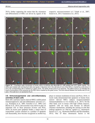
This document discusses adipose-derived regenerative cells and their potential for use in regenerative medicine. It summarizes key findings from studies conducted by the authors: (1) mesenchymal stem cells can be isolated from various tissues including adipose tissue and differentiated into cells from all three germ layers, supporting the hypothesis that a universal stem cell exists; (2) the microenvironment determines the orientation and differentiation of mesenchymal stem cells; (3) these stem cells can be obtained from small amounts of adipose tissue using appropriate isolation techniques and applied to patients without processing or manipulation. The document argues that use of a patient's own adipose-derived regenerative cells has potential as a new generation of regenerative
![Alt et al. Adipose-derived regenerative cells
3
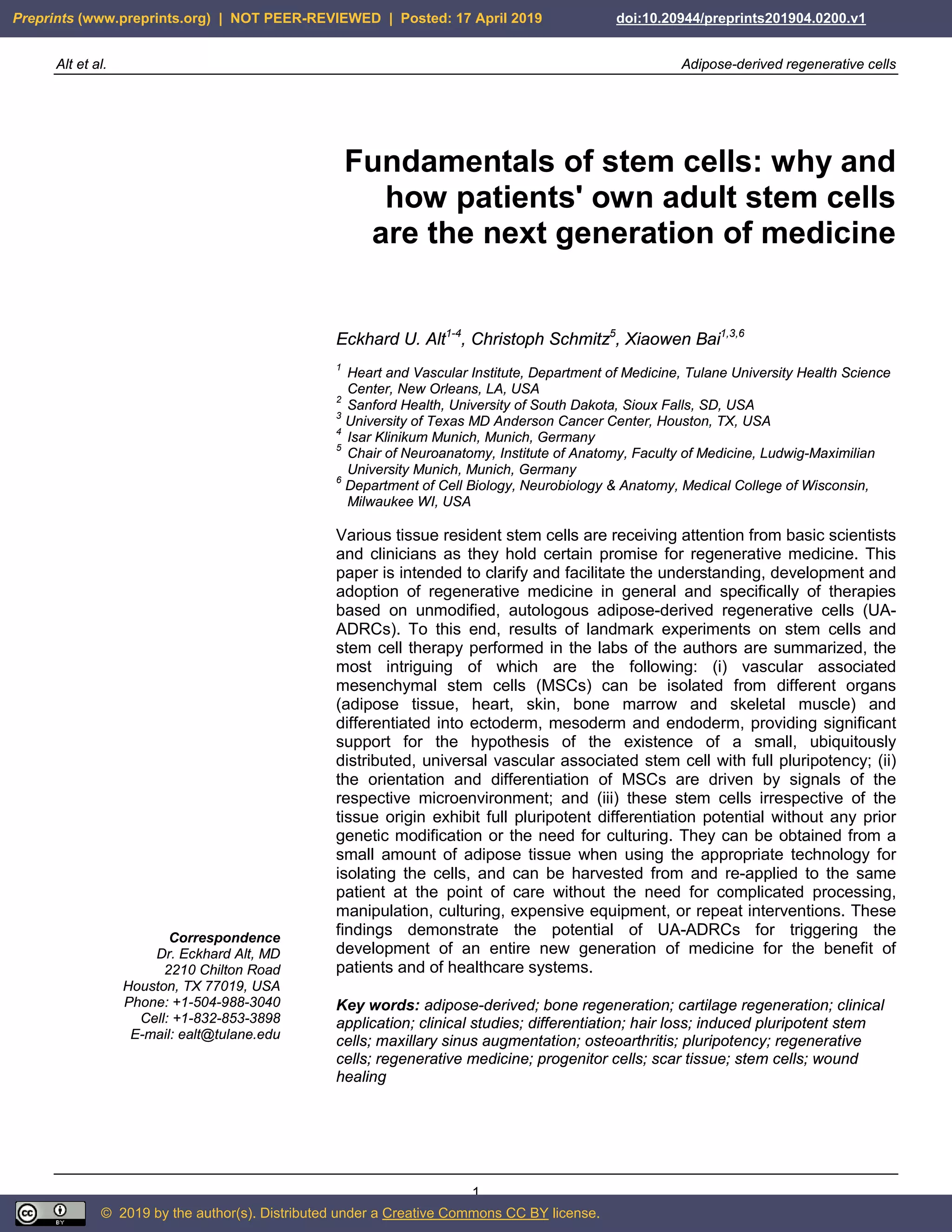
2. The definition of the term stem cells
2.1. The current state of confusion about the term
stem cells
Approximately 50 years ago Friedenstein et al. (1968;
1974) described multipotent cells derived from bone
marrow stroma that were adherent to plastic and had the
ability to differentiate into cells other than hematopoietic
lineages. Since then, our knowledge about stem cells has
been steadily growing. However, the current definition of
the term stem cells as given by Dominici et al. (2006), that
describes MSCs as being adherent to plastic, expressing
the surface markers CD73, CD90 and CD105, and having
the ability to differentiate into osteoblasts, adipocytes and
chondrocytes, is not quite complete. This is partly due to
the fact that, for example, fibroblasts express these surface
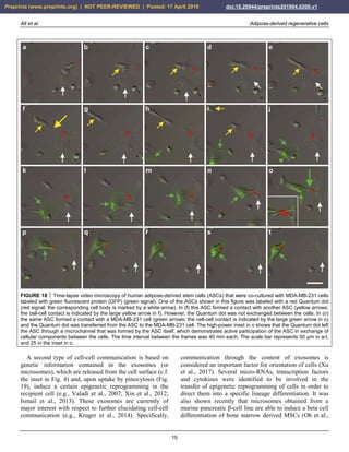
markers as well, without having the ability to
transdifferentiate into other lineages or being MSCs (Alt
et al., 2011). The definition of the term stem cells
primarily by surface markers (c.f. Dominici et al., 2006) is
not sufficient, especially as we have learned in the
meantime that the true pluripotent stem cells do not yet
express CD73, CD90 and CD105 (outlined in detail in
Section 3 of this article). Rather, expression of cell surface
markers is a dynamic process. When cultured in fetal
bovine serum (FBS) or platelet lysate culture media,
MSCs can turn on new surface markers. Alternatively,
MSCs in culture can lose their surface marker expression,
such as for example the loss of the previously expressed
progenitor marker CD34 or the endothelial progenitor
marker CD31. In addition, the specific type of culture
media (FBS or defined serum free media [SFM]) and the
surface characteristics of a culture dish strongly impact on
visual appearance, growth rate and surface marker
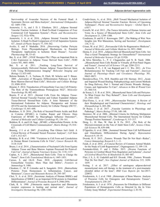
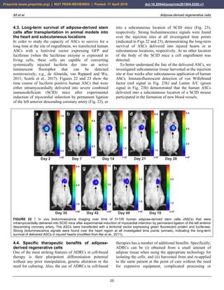
expression of MSCs. For example, MSCs are adherent to
plastic and display a typical, spindle shaped appearance
when cultured in FBS (Fig. 2a,c). In contrast, they form
spheroids when cultured in SFM (Fig. 2b,d). Besides this,
the cellular growth rate determined by the G1, S and M
phases of the cell cycle considerably differ between MSCs
cultured in FBS (high growth rate) compared with MSCs
cultured in SFM (very low growth rate) (data not shown).
FIGURE 2 Change in the morphology of rat adipose-derived stem cells cultured first in fetal bovine serum (FBS) for 14 days (a), then
in serum free medium (SFM) for 24 hours (b), and then again in FBS for one week (c). Afterwards, then transfer to SFM again induced
a spheroid like appearance (d). The scale bar represents 100 µm.
One of the major current problems in understanding
stem cells is based on different and varying definitions of
the meaning of the term stem cells (e.g., Pittenger et al.,
1999; Phinney and Prockop, 2007; Bourin et al., 2013).
The way the term stem cells is often used by researchers
and the public confuses true stem cells with progenitor
cells. True stem cells are defined as being able to
differentiate into all three germ layers (e.g., Bai et al.,
2007; Ratajczak, 2015). In contrast, progenitor cells are
already on a pre-determined pathway to become a
differentiated cell and have lost their ability to decide
what they want to be ‘in life’. In other words, in contrast
to true stem cells, progenitor cells are typically determined
to differentiate and develop into a defined cell type and
primarily have lost their multipotency. For example, in
bone marrow, more than 99% of the cells are not true stem
cells, but primarily hematopoietic progenitor cells (e.g.,
Song, Prantl and Alt, 2010; Bruno et al., 2014).
Accordingly, progenitor cells have already started a
pathway of lineage-committed differentiation. In case of
bone marrow derived cells, these hematopoietic progenitor
cells (often incorrectly labeled as stem cells) started to
differentiate into future hematopoietic cells of the white,
red or platelet lineage. Pending on their progress in
maturation in this differentiation process, these cells are
no longer able to revert their pathway of differentiation.
At best, they are able to vary somewhat within the same
germ layer of differentiation, but typically stay within the
same lineage (Ricci-Vitiani et al., 2010; Wang et al.,
2010). Much of the confusion about the term stem cells
Preprints (www.preprints.org) | NOT PEER-REVIEWED | Posted: 17 April 2019 doi:10.20944/preprints201904.0200.v1](https://image.slidesharecdn.com/preprints201904-210911022822/85/Fundamentals-of-Stem-Cells-3-320.jpg)
![Alt et al. Adipose-derived regenerative cells
4
originates from this mix-up of the definition of true stem
cells with progenitor cells in the literature (c.f., e.g.,
Bourin et al., 2013; Caplan, 2017; Velten et al., 2017; see
also Alt, 2015).
2.2. The limited significance of induced
pluripotent stem cells for the practice of
medicine
Another problem is the incorrect public belief that
naturally no stem cells would exist that are able to
differentiate into all three lineages without being first
modified or genetically altered. The euphoria around the
so-called induced pluripotent stem cells (iPS cells) (that
resulted in the granting of the Nobel Prize in Physiology
or Medicine 2012) has led to a direction in the last couple
of years in research that will likely not significantly
change the practice of medicine. For more than ten years it
has been demonstrated many times that naturally there are
pluripotent stem cells which are able to differentiate into
all three germ layers without being first genetically
modified (c.f., e.g., Pittenger et al., 1999; Zuk et al., 2001;
Song, Prantl and Alt, 2011). Hence, it becomes clear that
the need for genetically manipulated iPS cells (in which
first an artificial [induced] overexpression of embryonic
genes like Oct4, Klf4, Sox2 and/or cMyc is necessary; c.f.,
e.g., Jullien et al., 2011; Heffernan, Sumer and Verma,
2011; Shi et al., 2017) is not required for the practice of
medicine. Rather, the universally distributed true stem
cells we refer to in this article are naturally able to
differentiate into all three germ layers and into any tissue
of the body under guidance of the local
microenvironment, without requiring prior overexpression
of embryonic genes or any prior artificial genetic
manipulation.
Another important aspect regarding iPS cells is that the
genetic manipulation of overexpression of embryonic
genes in these cells makes them less likely to enter the
practice of medicine due to the complexity of the
procedure. Besides this, there are concerns that iPS cells
can demonstrate features similar to cancer cells (c.f., e.g.,
Takahashi et al., 2007; Yu et al., 2007; Lee et al., 2013).
While the iPS cell technology will most likely not advance
to a stage where therapeutic transplants are deemed safe
(Lee et al., 2017), iPS cells are certainly supportive in
helping to better understand differentiation pathways of
stem cells and patient-specific bases of diseases, as well as
to develop personalized drug discovery efforts (c.f., e.g.,
Marsoner, Koch and Ladewig, 2018).
Dr. Paul Knoepfler and his team at UC Davis School of
Medicine (Davis, CA, USA) were the first to demonstrate
that induced pluripotency and oncogenic transformation
are related processes when comparing the transcriptomes
of iPS cells with the transcriptomes of cancer cells (Riggs
et al., 2013; see also Steinemann, Göhring and
Schlegelberger, 2013; Tung and Knoepfler, 2015).
Collectively, these studies have established a need for
caution when conducting clinical trials using iPS cell
based therapies.
2.3. Important differences regarding pluripotency
and multipotency between embryonic stem cells,
adult stem cells and cancer cells
There are important differences regarding totipotency,
pluripotency and multipotency between embryonic, fetal
and adult stem cells. A totipotent cell (found in the morula
stage of embryonic development) can differentiate into
any type of cell in the body. A pluripotent cell (found in
the blastocyst stage of embryonic development) possesses
the intrinsic pattern and existing guidelines for building a
new organ which is not yet existing. Pluripotency here
means that the embryonic stem cells can develop into any
tissue and organ of the three germ layers (i.e., ectoderm,
mesoderm and endoderm), without that these tissues or
organs yet exist.
In a similar way as in embryonic stem cells,
pluripotency is naturally present in adult stem cells as
evidenced by their ability to differentiate into ectodermal,
mesodermal and endodermal lineages. The difference
compared to embryonic stem cells, however, is that adult
stem cells cannot form new tissue on their own. They miss
the intrinsic program of embryonic stem cells but instead
depend on the signaling from the respective
microenvironment (outlined in detail in the following
sections). This means that adult stem cells may obtain any
of the three lineages but depend on constant induction of
differentiation and re-confirmation by signals released and
communicated from the local microenvironment (c.f., e.g.,
Rezza, Sennett and Rendl, 2014; Dong et al., 2015; Wabik
and Jones, 2015). If this information and confirmation is
missing or ceases, adult stem cells stop differentiating
(Trosko, 2014; Zhang et al., 2015).
In contrast, cancer cells with upregulated embryonic
genes like Oct4 can overcome this tissue
microenvironment dependency and form a tissue of their
initial tissue type, resulting in a metastasis (Tai et al.,
2004; Ilmer et al., 2014). For example, pancreatic tumor
cells can grow in the liver, forming a metastasis. In
contrast, normal pancreatic progenitor cells stop
differentiation and undergo apoptosis when implanted into
the liver. A continuous lineage specific differentiation
only occurs if it is supported by continuous signaling from
the respective microenvironment. For example, the
injection of cells derived from the bone marrow (which
for their majority consist of hematopoietic progenitor
cells) into a knee would result in a significant
Preprints (www.preprints.org) | NOT PEER-REVIEWED | Posted: 17 April 2019 doi:10.20944/preprints201904.0200.v1](https://image.slidesharecdn.com/preprints201904-210911022822/85/Fundamentals-of-Stem-Cells-4-320.jpg)
![Alt et al. Adipose-derived regenerative cells
9
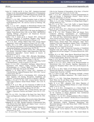
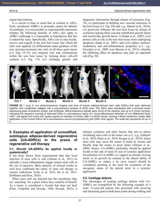
FIGURE 8 Expression of surface markers of mesenchymal stem cells in their primary, secondary and tertiary niche.
FIGURE 9 Aggregation of human adipose-derived stem cells
(a), leading to the formation of a spheroid (b) that resembles the
appearance of an embryoid body formed by embryonic stem
cells. The scale bar represents 20 µm in (a) and 75 µm in (b).
The understanding of the mode of differentiation of
MSCs and their surface markers has been confusing.
Specifically, markers such as CD44, CD73, CD90 and
CD105 – typically believed to be indicative of true stem
cells (Bourin et al., 2013) – are only present in cells that
have already left their silenced location and started to
enter the next developmental phase in order to attain
progenitor status. Besides this, CD44, CD90 and CD105
are also expressed in fibroblasts that exhibit no plasticity
at all (Alt et al., 2011).
We performed surface analysis of the antigen profiles of
human ASCs that were cultured for four days in serum
free media. Immunofluorescent analysis showed that these
cells were immunonegative for CD11b, CD14, CD31,
CD34, CD45 and HLA-DR (Fig. 10). Corresponding flow
cytometric analysis revealed that the relative numbers of
cells that were immunopositive for these surface markers
were smaller than 1.5% (relative number of cells that were
immunopositive for CD11b [CD11b+]: 1.1%; CD14+:
0.6%; CD31+: <0.1%; CD34+: 0.4%; CD45+: 1.3% and
HLA-DR: 0.2%). These results confirmed that cells that
express the following markers were not present in this cell
culture: macrophages (which typically are CD11b+),
hematopoietic progenitor cells (CD14+), endothelial
progenitor cells (CD31+), progenitor cells in general
(CD34+), cells expressing the pan-leukocyte marker
(CD45+) and cells expressing HLA-DR.
Preprints (www.preprints.org) | NOT PEER-REVIEWED | Posted: 17 April 2019 doi:10.20944/preprints201904.0200.v1](https://image.slidesharecdn.com/preprints201904-210911022822/85/Fundamentals-of-Stem-Cells-9-320.jpg)
![Alt et al. Adipose-derived regenerative cells
19
location where they are most needed. From these
considerations it also becomes clear that stem cell therapy
is not only directed to a specific organ, tissue or disease,
but will take the function of replacing and repairing tissue
and organs that suffer from a lack of repair, renewal and
rejuvenation.
4. Key advantages of adipose-derived
regenerative cells over bone marrow
derived stem cells for cell based
therapies
4.1. Comparison of bone marrow derived stem
cells with adipose-derived stem cells
For almost a decade bone marrow was the primary source
of MSCs for research into and development of therapies
based on adult stem cells. Bone marrow derived MSCs
exhibit significant potential for promoting tissue
regeneration, protection of ischemic tissue at risk, and
modulation of inflammation and autoimmunity (Murphy,
Moncivais and Caplan, 2013). However, utilizing bone
marrow derived MSCs for therapeutic purposes typically
requires to first isolate these cells and expand them in
culture. Because of the overwhelming presence of
hematopoietic progenitor cells in bone marrow that aim to
form new blood cells (Al-Drees et al., 2015; Ratajczak,
2015), only a small fraction of the cells in fresh bone
marrow aspirate are true pluripotent stem cells. In
contrast, other tissues such as adipose tissue yield orders
of magnitude more MSCs per unit volume than bone
marrow (e.g., Izadpanah et al., 2006; Yoshimura et al.,
2006; Bruno et al., 2014). In fact, as highlighted in the
previous sections of this article, adipose tissue is an organ
that is highly vascularized and contains a significant
number of MSCs. Thus, adipose tissue may be utilized as
a fresh cell preparation, rich in MSCs, without the need
for expansion in cell culture. There is also less damage to
the tissue where the stem cells are taken from, and
typically, the stem cells in adipose tissue have not been
challenged by ischemia, trauma or infections.
Compared to other sources of adult stem cells, adipose
tissue has the following specific advantages: (i) adipose
tissue is readily available in most individuals; (ii) small
amounts of adipose tissue (25 to 100 ml) can be harvested
using a simple liposuction procedure with low
invasiveness, with tolerable discomfort and low donor-site
damage; (iii) considerably larger amounts of MSCs can be
obtained from adipose tissue than from the same amount
of bone marrow; and (iv) the latter allows the usage of
ASCs without further need of culturing (named adipose-
derived regenerative cells [ADRCs] in order to
differentiate from cultured ASCs; c.f. Fig. 1). Given these
advantages, unmodified, autologous ADRCs (UA-
ADRCs) appear to be the most promising candidate for
repair and regeneration of many tissues, including chronic
wounds, soft tissue defects, bone and cartilage defects,
non-healing fractures, tendinopathies, diseased or injured
myocardium, urological conditions such as incontinence,
and neurological conditions (c.f., e.g., Schäffler and
Büchler, 2007; Gimble, Guilak and Bunnell, 2010; Altman
et al., 2010; Alt et al., 2011; Gir et al., 2012; Klein et al.,
2016).
4.2. Differences in the effectiveness of various
systems and methods that are available for
isolating adipose-derived regenerative cells
Different techniques and protocols were described for
releasing ADRCs for therapeutic use (c.f., e.g., Oberbauer
et al., 2015; Condé-Green et al., 2016; Van Dongen et al.,
2018). Collagenase I and II containing enzyme
preparations that degrade collagen are commonly used.
However, in order to release the vascular associated MSCs
from their binding site in the extracellular matrix inside
the blood vessels (and hereby to release the cells from
their 'hibernating' or silenced state) collagenases are only
partially effective. The addition of a neutral protease to a
collagenase enzyme preparation can significantly increase
the number of ADRCs recovered from a given volume of
adipose tissue. This was achieved by developing the
proprietary Matrase enzymatic reagent (InGeneron Inc.,
Houston, TX, USA). Isolating ADRCs with the Matrase
enzymatic reagent and the Transpose RT system
(InGeneron) appears advantagous to other commercial cell
separation systems (c.f. Haenel et al., 2018). Specifically,
ADRCs that were isolated with the Matrase enzymatic
reagent and the Transpose RT system may contain
approximately 40% of cells that are immunopositive for
CD29 and CD44, which are markers of ASCs (Haenel et
al., 2018). These authors also reported a colony forming
units (CFU) frequency (considered to be an indicator of
stemness) of approximately 11% of ADRCs isolated with
the Matrase enzymatic reagent and the Transpose RT
system. Metcalf et al. (2016) reported relative CFU values
of approximately 8% when isolating ADRCs from equine
adipose tissue. In contrast, relative CFU values between
0.2% and 1.7% were reported for ADRCs that were
isolated in head-to-head comparisons with four other
commercial cell separation systems (Aronowitz &
Ellenhorn, 2013; Aronowitz et al., 2016). However, a
direct comparison of the CFU values is hardly possible
due to significant methodological differences how the
respective numbers of CFUs were determined.
Preprints (www.preprints.org) | NOT PEER-REVIEWED | Posted: 17 April 2019 doi:10.20944/preprints201904.0200.v1](https://image.slidesharecdn.com/preprints201904-210911022822/85/Fundamentals-of-Stem-Cells-19-320.jpg)
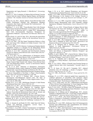
![Alt et al. Adipose-derived regenerative cells
23
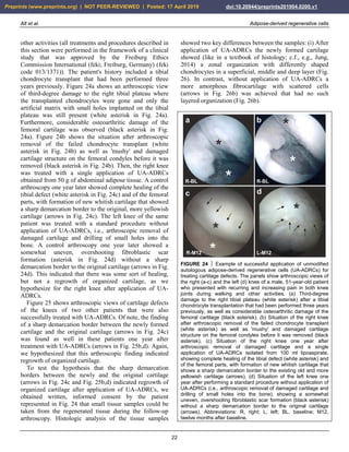
FIGURE 25 Other examples of successful application of
unmodified autologous adipose-derived regenerative cells (UA-
ADRCs) for treating cartilage defects. The panels show
arthroscopic views of the knees of two patients (Patient 1 [male,
45 years old]: a,b; Patient 2 [male, 55 years old]: c,d) before (a,
c) and one year after (b,d) arthroscopic removal of damaged
cartilage (asterisks in a,c) and application of UA-ADRCs isolated
from 50 ml lipoaspirate. The arrows in (b,d) indicate the sharp
demarcation border between the newly formed and the original
cartilage. The white arrow in (a) points to the arthroscopic
instrument that was used to remove damaged cartilage.
(ii) Furthermore, after application of UA-ADRCs the
contact zone between the newly formed cartilage and bone
showed (also like in a textbook of histology) typical
chondrocytes with a small nucleus and a hollow space
around (arrows in Fig. 26c). In contrast, without
application of UA-ADRCs the contact zone between the
newly formed cartilage and bone showed an infiltration
with inflammatory cells, fibroblasts (arrows in Fig. 26d)
and small blood vessels (arrowheads in Fig. 26d).
Analysis of the tissue sample taken during arthroscopic
inspection of the right knee of the patient represented in
Fig. 24 at one year after arthroscopic removal of damaged
cartilage and a single application of UA-ADRCs from 50
g of his adipose tissue with polarized light microscopy
demonstrated that the collagen fiber bundles in the deep
and middle layers of the newly formed cartilage show a
more vertical orientation (i.e., perpendicular to the border
between bone and cartilage), whereas the collagen fiber
bundles in the superficial layer showed a more horizontal
orientation (i.e., parallel to the surface) (Fig. 27). This
finding is in line with the description of the physiologic
orientation of collagen fiber bundles in articular cartilage
in the literature when analyzed with polarized light
microscopy (e.g., Rieppo et al., 2008).
To our knowledge, the results presented in this section
go beyond the state-of-the-art in the field of regenerating
damaged cartilage with ADRCs and ASCs. In a recent
review (Damia et al., 2018) a number of clinical studies
were listed in which cartilage defects in the human knee
were treated with ASCs (Jo et al., 2014; Pers et al., 2016;
Freitag et al., 2017). Of note, in all of these studies ASCs
were applied, whereas we have been using fresh
uncultured ADRCs (for the advantages of ADRCs over
ASCs see Sections 4.1. and 4.4.). The maximum follow-
up period in the studies by Jo et al. (2014) and Pers et al.
(2016) was only six months after application of ASCs
(MRI, arthroscopy and histologic analysis in both studies;
n=18 patients in both studies). In the single case report by
Freitag et al. (2017) MRI was performed at twelve months
after application of ASCs, but no arthroscopy and, thus, no
histologic analysis. Furthermore, in none of these studies
tissue samples were ever investigated with polarized light
microscopy.
5.3. Chronic, recalcitrant low back pain caused
by lumbosacral facet syndrome
Lumbosacral facet syndrome is a term used to describe a
painful condition caused by inflammation and irritation of
the zygapophyseal (facet) joints of the spine (Fig. 28a-c).
It is most commonly caused by chronic degenerative
changes in the lumbosacral spine. Symptoms include low
back pain with or without referral to the lower extremities
(Alexander and Dulebohn, 2018). To our knowledge,
reports on cell-based therapies for chronic low back pain
caused by lumbosacral facet syndrome have not yet been
published.
Figure 28d-f shows typical wear and tear that occurred
in the spine of three former professional ski racers (all
treatments and procedures described in this section were
performed in the framework of clinical assessment). The
green arrows indicate normal intervertebral disc structure
between the lumbar vertebrae L3 and L4, demonstrating
that there was a certain level of water content, reflected by
the white signal in the MRI. In contrast, the yellow arrows
show that between vertebrae L4 and L5 in all three
athletes there was a reduction in height of the
intervertebral disc due to the diminished overall volume of
the intervertebral disc. The increased body weight now
resting on the facet joints results in an inflammatory
reaction of the facet joints. All three athletes were treated
with UA-ADRCs. The cells were injected into the right
and left facet joints between L4 and L5 as well as between
L5 and S1 within a single procedure of harvesting and
injection that lasted about two hours. This treatment
resulted in a significant and long lasting (now more than
three years) pain reduction that enabled these athletes to
return to successful competitive sports.
Preprints (www.preprints.org) | NOT PEER-REVIEWED | Posted: 17 April 2019 doi:10.20944/preprints201904.0200.v1](https://image.slidesharecdn.com/preprints201904-210911022822/85/Fundamentals-of-Stem-Cells-23-320.jpg)
![Alt et al. Adipose-derived regenerative cells
24
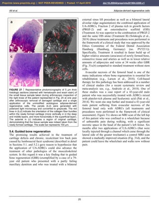
FIGURE 26 Representative photomicrographs of 5 µm thick histologic sections (stained with toluidin blue [a,b] or hematoxylin and
eosin stain [c,d]) of the small tissue samples taken during arthroscopic inspection of the knees of the patient represented in Fig. 24 at
one year after arthroscopic removal of damaged cartilage and a single application of unmodified autologous adipose-derived
regenerative cells (UA-ADRCs) (right knee) (a,c) or after performing a standard procedure without application of UA-ADRCs (i.e.,
arthroscopic removal of damaged cartilage and drilling of small holes into the bone) (left knee) (b,d), respectively. The dotted lines in (a)
indicate a zonal organization of the newly formed cartilage with differently shaped chondrocytes in a superficial (SL), middle (ML) and
deep layer (DL). The arrows in (b) point to scattered cells within newly formed amorphous fibrocartilage. The arrows in (c) indicate
typical chondrocytes with a small nucleus and a hollow space around in the contact zone between the newly formed cartilage and bone,
whereas the arrows in (d) point to an infiltration with inflammatory cells in the contact zone between the newly formed cartilage and
bone. The arrowheads in (d) indicate small blood vessels. The scale bar represents 100 µm.
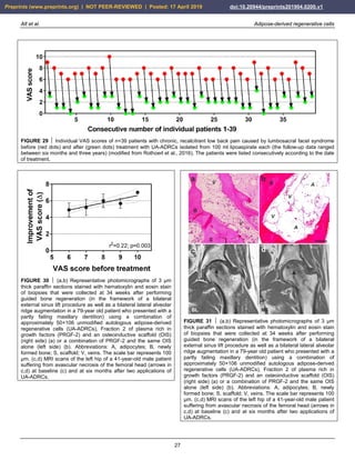
Figure 29 shows individual pain scores on a VAS scale
(with 0 representing no pain and 10 representing
maximum, unbearable pain) of n=39 patients with chronic,
recalcitrant low back pain caused by lumbosacral facet
syndrome before (red dots in Fig. 29) and one year after
(green dots in Fig. 29) treatment with UA-ADRCs isolated
from 100 ml lipoaspirate each (the follow-up interval
ranged between twelve months and more than three years)
(modified from Rothoerl et al., 2016) (all treatments and
procedures described in this section were performed in the
framework of a clinical study that was approved by the
Freiburg Ethics Commission International (feki; Freiburg,
Germany) (feki code 016/1252)). The mean and standard
error of the mean of the VAS scores before and after
treatment was 7.21 ± 0.17 and 1.80 ± 0.17; this difference
was highly statistically significant (Wilcoxon matched-
pairs signed rank test; p < 0.001). As a consequence, the
quality of life of these patients was significantly
improved.
Figure 30 shows mean and standard deviation of the
individual improvement of the VAS score after treatment
as a function of the VAS score before treatment of these
39 patients. Linear regression analysis showed a
statistically significant relationship between the VAS
score before treatment and the individual improvement of
the VAS score after treatment, with the best results
obtained with the highest VAS scores before treatment (r2
= -0.22; p = 0.003). This impressively demonstrates the
potential of UA-ADRCs for the treatment of chronic,
recalcitrant low back pain caused by lumbosacral facet
syndrome, and perhaps opens the possibility for obtaining
comparable results for other chronic pain conditions of the
musculoskeletal system.
Preprints (www.preprints.org) | NOT PEER-REVIEWED | Posted: 17 April 2019 doi:10.20944/preprints201904.0200.v1](https://image.slidesharecdn.com/preprints201904-210911022822/85/Fundamentals-of-Stem-Cells-24-320.jpg)

![Alt et al. Adipose-derived regenerative cells
30
development of an entire new generation of medicine for
the benefit of patients and of healthcare systems.
FIGURE 33 Examples of successful application of unmodified
autologous adipose-derived regenerative cells (UA-ADRCs) for
reducing scar tissue. (a,b) Male, 48-year-old patient; scar tissue
formation on the upper arm after an injury caused by a car
accident; single application of UA-ADRCs isolated from 100 ml
lipoaspirate. (c,d) Male, 50-years-old patient; scar tissue
formation (arrows) in the face after an acid attack; three
treatments with UA-ADRCs isolated from 100 ml lipoaspirate on
days 1, 90 and 180. Abbreviations: BL, baseline; M1/M12,
one/twelve months after application of UA-ADRCs.
Ethics
The different studies during which cells were collected for
performing the experiments shown in Figs 2-6, 9-20 and
22-23 were approved by the Institutional Review Boards
(IRB) for Baylor College of Medicine and Affiliated
Hospitals (Houston, TX, USA) (Protocol # H-18357) and
University of Texas M. D. Anderson Cancer Center
(Houston, TX, USA) (Protocol # 03-08-03031 and 03-08-
03031).
Killing mice by cervical dislocation for isolating cells
from subcutaneous fat tissue (Muehlberg et al., 2009),
killing neonatal rats by decapitation for isolating
cardiomyocytes (Sadat et al., 2007) and killing adult rats
by decapitation for isolating cells (Alt and Bai,
unpublished data) did not require IRB approval but was
performed following the guidelines of Veterinary
Medicine & Surgery at the University of Texas M. D.
Anderson Cancer Center and the U.S. National Institutes
of Health.
Hallo
FIGURE 34 Examples of successful application of unmodified
autologous adipose-derived regenerative cells (UA-ADRCs) for
treating hair loss. (a,b) 46-year-old female patient; sparse hair;
single application of UA-ADRCs isolated from 100 ml
lipoaspirate. (c-f) 23-years-old female patient suffering from
atopic dermatitis and alopecia totalis for three years without
spontaneous improvement; three treatments with UA-ADRCs
isolated from 100 ml lipoaspirate each at baseline (c) and at
three and six month after the first treatment. Results are shown
at one (M1; d), three (M3; e) and seven (M7; f) month after the
first treatment.
References
Ader, M. and E.M. Tanaka. 2014. "Modeling human development in
3D culture." Current Opinion in Cell Biology 31, 23–28.
Al-Drees, M. A. et al. 2015. „Making Blood: The Haematopoietic
Niche Throughout Ontogeny.” Stem Cells International 2015,
571893.
Alexander, C. E. and S. C. Dulebohn. 2018. „ Lumbosacral Facet
Syndrome”, StatPearls [Internet], Treasure Island (FL): StatPearls
Publishing; 2018-. 2017 Nov 5. (available online at
https://www.ncbi.nlm.nih.gov/books/NBK441906/).
Alt, E. et al. 2011. „Fibroblasts Share Mesenchymal Phenotypes with
Stem Cells, but Lack their Differentiation and Colony-Forming
Potential”, Biology of the Cell 103, 197–208.
Alt, E. 2015. „Mesenchymal Stem Cell - What Is in the Name?”,
Modern Medicine 22, 193–196.
Altman, A. M. et al. 2009. " IFATS Collection: Human Adipose-
Derived Stem Cells Seeded on a Silk Fibroin-Chitosan Scaffold
Enhance Wound Repair in a Murine Soft Tissue Injury Model", Stem
Cells 27:250–258.
Altman, A. M. et al. 2010. "Human Tissue-Resident Stem Cells
Combined with Hyaluronic Acid Gel Provide Fibrovascular-
Integrated Soft-Tissue Augmentation in a Murine Photoaged Skin
Model", Plastic and Reconstructive Surgery 125, 1–11.
Altman, A. M. et al. "Adipose Tissue–Derived Stem Cells Enhance
Bioprosthetic Mesh Repair of Ventral Hernias", Plastic and
Reconstructive Surgery 126, 845–854.
Altman, A. M. et al. 2011. „Wound Microenvironment Sequesters
Adipose-Derived Stem Cells in a Murine Model of Reconstructive
Surgery in the Setting of Concurrent Distant Malignancy”, Plastic
and Reconstructive Surgery 127, 1467–1477.
Andriolo, L. et al. 2018. „Regenerative Therapies Increase
Preprints (www.preprints.org) | NOT PEER-REVIEWED | Posted: 17 April 2019 doi:10.20944/preprints201904.0200.v1](https://image.slidesharecdn.com/preprints201904-210911022822/85/Fundamentals-of-Stem-Cells-30-320.jpg)