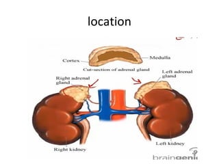
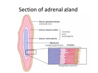
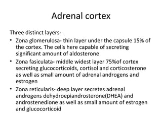
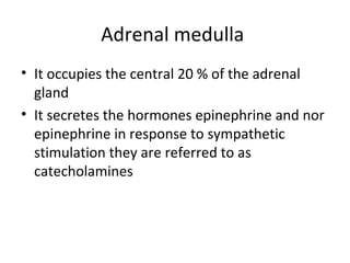
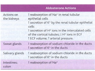
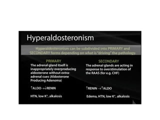
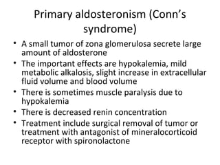
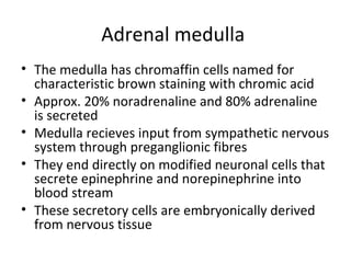
Adrenal gland
Anatomy
• There are 2 adrenal glands each weighing
about 4 gms
• They lie at the superor poles of the two
kidneys
• Each gland is composed of two distinct parts
adrenal cortex and adrenal medulla
• Blood supplied by superior ,inferior and
middle adrenal arteries
location
Section of adrenal gland
Adrenal cortex
Three distinct layers-
• Zona glomerulosa- thin layer under the capsule 15% of
the cortex. The cells here capable of secreting
significant amount of aldosterone
• Zona fasiculata- middle widest layer 75%of cortex
secreting glucocorticoids, cortisol and corticosterone
as well as small amount of adrenal androgens and
estrogen
• Zona reticularis- deep layer secretes adrenal
androgens dehydroepiandrosterone(DHEA) and
androstenedione as well as small amount of estrogen
and glucocorticoid
Adrenal medulla
• It occupies the central 20 % of the adrenal
gland
• It secretes the hormones epinephrine and nor
epinephrine in response to sympathetic
stimulation they are referred to as
catecholamines
Biosynthesis of adrenal steroids
• Important steroid products of adrenal cortex are
aldosterone , cortisol and androgens
• All the steps of syntheis occurs in mitochondria and
endoplasmic reticulum
• Approx 90-95% of cortisol in plasma binds to plasma
protein globulin called transcortin and to albumin
• Binding serves as a reservoir to lessen rapid
fluctuations in free hormone concentration
• Adrenal steroids are degraded mainly by liver and
conjugated to glucoronic acid or sulphates
• 25% of these conjugates are excreted in the bile and
then in feces and remaining in urine
Mineralocorticoid- aldosterone
• Aldosterone is the principal mineralocorticoid
secreted by the adrenal glands
• Aldosterone exerts 90% of the mineralocorticoid
activity of adrenal cortex rest 10 % is by cortisol
• Aldosterone increases the reabsorption of
sodium ions in the ECF
• It acts on principal cells of collecting ducts in the
kidney increasing sodium in exchange of
potassium and hydrogen ions
Mechanism of action
Renin angiotensin aldosterone system
(RAAS)
• Aldosteone secretion is mainly controlled by
angiotensin and potassium acting directly on
adrenocortical cells
Factors that play a role in regulation of
aldosterone
• Increased potassium ion concentration in the
extracellular fluid
• Increased angiotensin II concentration in ECF
• Increased sodium ion concentation in ECF
• ACTH from anterior pituitary
Primary aldosteronism (Conn’s
syndrome)
• A small tumor of zona glomerulosa secrete large
amount of aldosterone
• The important effects are hypokalemia, mild
metabolic alkalosis, slight increase in extracellular
fluid volume and blood volume
• There is sometimes muscle paralysis due to
hypokalemia
• There is decreased renin concentration
• Treatment include surgical removal of tumor or
treatment with antagonist of mineralocorticoid
receptor with spironolactone
Aldosterone cell signalling pathway
• activation of MR receptor can be antagonized
with Spironolactone
• Amiloride is a drug that can block sodium
reabsorption
• This is used in treatment of primary
aldosteronism
Glucocorticoids
• 95% of glucocorticoid activity occurs from
secretion of cortisol known as hydrocortisone
• Cortisol has effects on
– Carbohydrate metabolism
– Protein metabolism
– Fat metabolism
– Antiinfammatory effects
Regulation of cortisol secretion
• ACTH stimulates cortisol secretion
– Secretion of cortisol is controlled entirely by ACTH
• ACTH activates by increase in cAMP
– ACTH acts on adrenaocortical cells to activate
adeny cyclase
– Long term activation can cause hypertrophy
• Inhibitory effect of cortisol
– Cortisol sends negative feedback to hypothalamus
and pituitary to regulate cortisol levels
Regulation of cortisol secretion
Different types of stress that increase
cortisol release
• Trauma of any type
• Infection
• Intense heat or cold
• Injection of norepinephrine
• Surgery
• Any debilitating disease
Effect of cortisol in preventing
inflammation
• Cortisol stabilizes the lysosomal membrane
• Cortisol decreases the permeability of the
capillaries preventing loss of plasma into tissues
• Cortisol decreases migration of WBCs into
inflamed area and phagocytosis of damaged cells
• Cortisol suppresses the immune system causing
lymphocyte reproduction to decrease
• Cortisol attenuates fever as it reduces the release
of interleukin-1
Adrenal androgens
• Adrenal androgens mainly dehydroepisterone
is continully secreted by adrenal cortex during
fetal life
• Small amount of estrogen and progesterone
are also secreted
• It is believed early devlopment of male sex
organs result from adrenal androgens
• Alrenal androgens are also responsible for
axillary and pubic hair in females
Hypoadrenalism (Addison’s disease)
• This is due to insufficient adrenocortical
hormones
• Most common cause is primary atrophy or
injury, tuberculous destruction of the gland or
cancer
• Disturbances cause mineralocorticoid
deficiency, Glucocorticoid deficiency and
melanin pigmentation
Features of Addison’s disease
Addison’s disease
• Mineralocorticoid deficiency
– Results in loss of sodium ions, chloride ions and water to be lost
into urine
– This results in hyponatremia,hypercalemia and mild acidosis
– Plasma voliume falls, cardiac output and blood pressure
decreases and patient dies in shock
• Glucocorticoid deficiency
– No proper synthesis of glucose, reduced mobilization of
proteins and fats
• Melanin pigmentation
– Melanin pigmentation of mucous membrane and skin
– Melanin deposited in blothes in thin areas of the skin due to
increased sectretion of MSH
Addison’s disease
Hyperadrenalism- Cushing’s syndrome
Hypercorticolism can occur due to-
• Adenomas of the anterior pitutary secreting increased
ACTH
• Abnormal function of hypothalamus causing increased
CRH and thereby increased ACTH
• Ectopic secretion of ACTH by a tumor somewhere else
in the body
• Adenomas of the adrenal cortex
When cushing’s syndrome is secondary to excess
secretion of ACTH by the anterior pituitary this is called
Cushing’s disease
Adrenal medulla
• The medulla has chromaffin cells named for
characteristic brown staining with chromic acid
• Approx. 20% noradrenaline and 80% adrenaline
is secreted
• Medulla recieves input from sympathetic nervous
system through preganglionic fibres
• They end directly on modified neuronal cells that
secrete epinephrine and norepinephrine into
blood stream
• These secretory cells are embryonically derived
from nervous tissue
Syntheis of catecholamines
Degradation of catecholamines
Factors controlling secretion of
catecholamines
• Position of individual- standing increases
norepinephrine
• Exercise
• Hypoglycemia stronly stimulates epinephrine secretion
• Cigarette smoking
• Surgery – both epinephrine and norepinephrine
secretion
• Ketoacidosis
• Myocardial infarction
• Anesthesia
Adrenergic receptors
• There are 2 types of adrenergic receptors alpha
and beta receptors
• Beta receptors are type 1,2 and 3 while alpha are
type 1 and 2
• Norepinephrine and epinephrine both of which
are secreted into the blood have slightly different
effects in exciting alpha and beta receptors
• Norepinephrine exites mainly alpha receptors but
beta also to lesser extent epinephrine exites both
• Effect is determined by type of receptors in the
organs
Adrenergic receptors
Alarm or stress response of
symphathetic nervous system
• Increased arterial pressure
• Increased blood flow to active muscles for motor
activity
• Increased rate of cellular metabolism
• Increased glucose concentation
• Increased glycolysis in liver and muscles
• Increased muscle strength
• Increased mental activity
• increased rate of blood coagulation
Pheochromocytoma
• Tumors arise from chromaffin cells that
secrete catecholamines
• 90% tumors arise in adrenal medulla
• They are rare and equal occurance in males
and females
• If present in children it may be heriditary
• Most common feature is hypertension
Tests for adrenal dysfunction

Adrenal gland

  • 1.
  • 2.
    Anatomy • There are2 adrenal glands each weighing about 4 gms • They lie at the superor poles of the two kidneys • Each gland is composed of two distinct parts adrenal cortex and adrenal medulla • Blood supplied by superior ,inferior and middle adrenal arteries
  • 3.
  • 4.
  • 5.
    Adrenal cortex Three distinctlayers- • Zona glomerulosa- thin layer under the capsule 15% of the cortex. The cells here capable of secreting significant amount of aldosterone • Zona fasiculata- middle widest layer 75%of cortex secreting glucocorticoids, cortisol and corticosterone as well as small amount of adrenal androgens and estrogen • Zona reticularis- deep layer secretes adrenal androgens dehydroepiandrosterone(DHEA) and androstenedione as well as small amount of estrogen and glucocorticoid
  • 6.
    Adrenal medulla • Itoccupies the central 20 % of the adrenal gland • It secretes the hormones epinephrine and nor epinephrine in response to sympathetic stimulation they are referred to as catecholamines
  • 7.
    Biosynthesis of adrenalsteroids • Important steroid products of adrenal cortex are aldosterone , cortisol and androgens • All the steps of syntheis occurs in mitochondria and endoplasmic reticulum • Approx 90-95% of cortisol in plasma binds to plasma protein globulin called transcortin and to albumin • Binding serves as a reservoir to lessen rapid fluctuations in free hormone concentration • Adrenal steroids are degraded mainly by liver and conjugated to glucoronic acid or sulphates • 25% of these conjugates are excreted in the bile and then in feces and remaining in urine
  • 9.
    Mineralocorticoid- aldosterone • Aldosteroneis the principal mineralocorticoid secreted by the adrenal glands • Aldosterone exerts 90% of the mineralocorticoid activity of adrenal cortex rest 10 % is by cortisol • Aldosterone increases the reabsorption of sodium ions in the ECF • It acts on principal cells of collecting ducts in the kidney increasing sodium in exchange of potassium and hydrogen ions
  • 10.
  • 11.
    Renin angiotensin aldosteronesystem (RAAS) • Aldosteone secretion is mainly controlled by angiotensin and potassium acting directly on adrenocortical cells
  • 13.
    Factors that playa role in regulation of aldosterone • Increased potassium ion concentration in the extracellular fluid • Increased angiotensin II concentration in ECF • Increased sodium ion concentation in ECF • ACTH from anterior pituitary
  • 16.
    Primary aldosteronism (Conn’s syndrome) •A small tumor of zona glomerulosa secrete large amount of aldosterone • The important effects are hypokalemia, mild metabolic alkalosis, slight increase in extracellular fluid volume and blood volume • There is sometimes muscle paralysis due to hypokalemia • There is decreased renin concentration • Treatment include surgical removal of tumor or treatment with antagonist of mineralocorticoid receptor with spironolactone
  • 17.
  • 18.
    • activation ofMR receptor can be antagonized with Spironolactone • Amiloride is a drug that can block sodium reabsorption • This is used in treatment of primary aldosteronism
  • 20.
    Glucocorticoids • 95% ofglucocorticoid activity occurs from secretion of cortisol known as hydrocortisone • Cortisol has effects on – Carbohydrate metabolism – Protein metabolism – Fat metabolism – Antiinfammatory effects
  • 22.
    Regulation of cortisolsecretion • ACTH stimulates cortisol secretion – Secretion of cortisol is controlled entirely by ACTH • ACTH activates by increase in cAMP – ACTH acts on adrenaocortical cells to activate adeny cyclase – Long term activation can cause hypertrophy • Inhibitory effect of cortisol – Cortisol sends negative feedback to hypothalamus and pituitary to regulate cortisol levels
  • 23.
  • 24.
    Different types ofstress that increase cortisol release • Trauma of any type • Infection • Intense heat or cold • Injection of norepinephrine • Surgery • Any debilitating disease
  • 25.
    Effect of cortisolin preventing inflammation • Cortisol stabilizes the lysosomal membrane • Cortisol decreases the permeability of the capillaries preventing loss of plasma into tissues • Cortisol decreases migration of WBCs into inflamed area and phagocytosis of damaged cells • Cortisol suppresses the immune system causing lymphocyte reproduction to decrease • Cortisol attenuates fever as it reduces the release of interleukin-1
  • 26.
    Adrenal androgens • Adrenalandrogens mainly dehydroepisterone is continully secreted by adrenal cortex during fetal life • Small amount of estrogen and progesterone are also secreted • It is believed early devlopment of male sex organs result from adrenal androgens • Alrenal androgens are also responsible for axillary and pubic hair in females
  • 27.
    Hypoadrenalism (Addison’s disease) •This is due to insufficient adrenocortical hormones • Most common cause is primary atrophy or injury, tuberculous destruction of the gland or cancer • Disturbances cause mineralocorticoid deficiency, Glucocorticoid deficiency and melanin pigmentation
  • 28.
  • 29.
    Addison’s disease • Mineralocorticoiddeficiency – Results in loss of sodium ions, chloride ions and water to be lost into urine – This results in hyponatremia,hypercalemia and mild acidosis – Plasma voliume falls, cardiac output and blood pressure decreases and patient dies in shock • Glucocorticoid deficiency – No proper synthesis of glucose, reduced mobilization of proteins and fats • Melanin pigmentation – Melanin pigmentation of mucous membrane and skin – Melanin deposited in blothes in thin areas of the skin due to increased sectretion of MSH
  • 30.
  • 31.
    Hyperadrenalism- Cushing’s syndrome Hypercorticolismcan occur due to- • Adenomas of the anterior pitutary secreting increased ACTH • Abnormal function of hypothalamus causing increased CRH and thereby increased ACTH • Ectopic secretion of ACTH by a tumor somewhere else in the body • Adenomas of the adrenal cortex When cushing’s syndrome is secondary to excess secretion of ACTH by the anterior pituitary this is called Cushing’s disease
  • 35.
    Adrenal medulla • Themedulla has chromaffin cells named for characteristic brown staining with chromic acid • Approx. 20% noradrenaline and 80% adrenaline is secreted • Medulla recieves input from sympathetic nervous system through preganglionic fibres • They end directly on modified neuronal cells that secrete epinephrine and norepinephrine into blood stream • These secretory cells are embryonically derived from nervous tissue
  • 36.
  • 37.
  • 38.
    Factors controlling secretionof catecholamines • Position of individual- standing increases norepinephrine • Exercise • Hypoglycemia stronly stimulates epinephrine secretion • Cigarette smoking • Surgery – both epinephrine and norepinephrine secretion • Ketoacidosis • Myocardial infarction • Anesthesia
  • 39.
    Adrenergic receptors • Thereare 2 types of adrenergic receptors alpha and beta receptors • Beta receptors are type 1,2 and 3 while alpha are type 1 and 2 • Norepinephrine and epinephrine both of which are secreted into the blood have slightly different effects in exciting alpha and beta receptors • Norepinephrine exites mainly alpha receptors but beta also to lesser extent epinephrine exites both • Effect is determined by type of receptors in the organs
  • 40.
  • 41.
    Alarm or stressresponse of symphathetic nervous system • Increased arterial pressure • Increased blood flow to active muscles for motor activity • Increased rate of cellular metabolism • Increased glucose concentation • Increased glycolysis in liver and muscles • Increased muscle strength • Increased mental activity • increased rate of blood coagulation
  • 42.
    Pheochromocytoma • Tumors arisefrom chromaffin cells that secrete catecholamines • 90% tumors arise in adrenal medulla • They are rare and equal occurance in males and females • If present in children it may be heriditary • Most common feature is hypertension
  • 44.
    Tests for adrenaldysfunction