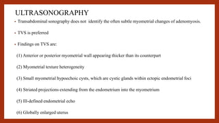
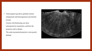
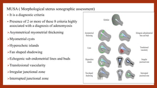
Transvaginal ultrasound and MRI are used to diagnose adenomyosis. On transvaginal ultrasound, the uterus may appear globally enlarged with heterogeneous myometrial texture and poorly defined endometrial-myometrial junction. MRI can identify adenomyosis as increased junctional zone thickness over 12mm with T2 hyperintense foci. Hysteroscopy with biopsy can confirm the diagnosis but is rarely used. Treatment options include NSAIDs, hormonal therapy like combined oral contraceptives or progestogens to reduce bleeding and pain, and long-acting reversible contraceptives like the levonorgestrel IUS.

![MRI
• MR imaging may be equal or slightly superior to TVS (Dueholm, 2001; Reinhold, 1996). Thus, MR imaging may be most
appropriate when the diagnosis is inconclusive, when further delineation would affect patient management, or when
coexisting uterine myomas distort anatomy (American College of Obstetricians and Gynecologists, 2014b).
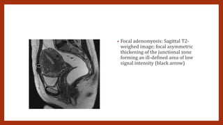
• T2-weighted sequences are key for diagnosing adenomyosis
• Adenomyosis appears as increased thickness of the junctional zone, forming an ill-defined area of low
signal intensity on T2, representing the smooth muscle hyperplasia accompanying the heterotopic
endometrial tissue. This aspect is frequently associated with bright foci on T2-weighted images, which
represent foci of heterotopic endometrial tissue, cystic dilatation of endometrial glands or haemorrhagic
foci.
• Adenomyosis is mainly located in the fundus [20] and commonly observed in the posterior wall. The
typical appearance is a large, rand asymmetric uterus, with a maximum junctional zone thickness of at
least 12 mm and punctate high-intensity myometrial foci [17].
• There are two forms of adenomyosis: diffuse, in which foci of adenomyosis are distributed throughout the
uterus (Fig. 1), and focal form, also named adenomyoma, when it affects a limited area (Fig. 2). The most
frequent finding for the diagnosis of adenomyosis is thickening of the junctional zone, with a thickness
exceeding 12 mm being highly predictive of the diagnosis](https://image.slidesharecdn.com/adenomyosisppt1-231011142309-5e730477/85/ADENOMYOSIS-PPT-1-pptx-6-320.jpg)