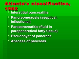
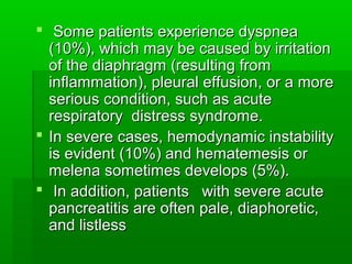
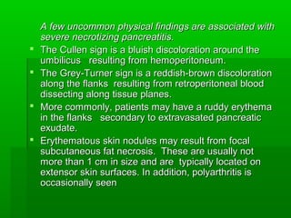
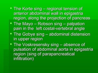
The document discusses acute pancreatitis, which occurs when the pancreas becomes inflamed. It describes the pancreas's normal functions and the pathophysiology of acute pancreatitis, which involves premature activation of digestive enzymes that digest the pancreas itself. Symptoms include severe abdominal pain. Complications can include multi-organ failure if the inflammation spreads beyond the pancreas. Risk factors, signs, symptoms, classification, complications, and treatment approach are outlined.
The document provides an overview of acute pancreatitis including:
- It begins with anatomy and blood supply of the pancreas.
- Pathophysiology involves autodigestion from inappropriately activated pancreatic enzymes.
- Etiology is commonly gallstones, alcohol, or idiopathic.
- Clinical presentation includes severe abdominal pain and systemic complications can involve multiple organ systems.
This document discusses the etiology and diagnosis of acute pancreatitis. It lists various etiological factors including mechanical obstruction, alcohol, hypertriglyceridemia, genetic mutations, drugs, infections, and trauma. It describes the diagnosis of acute pancreatitis based on abdominal symptoms, lipase or amylase levels, and imaging findings. It also discusses local complications like acute peripancreatic fluid collection, pancreatic pseudocyst, acute necrotic collection, and walled-off necrosis. Organ failure is defined using the Modified Marshall Scoring System.
This document provides an overview of acute pancreatitis, including its definitions, etiology, pathogenesis, and diagnostic assessment. It discusses the major causes of acute pancreatitis such as alcohol use, gallstones, medications, and genetic factors. The pathogenesis involves the abnormal activation of pancreatic enzymes leading to immune response and microcirculatory disturbances. Diagnosis is based on clinical features, elevated serum amylase and lipase levels, and imaging findings on ultrasound or CT scan. Several scoring systems are described to assess the severity of acute pancreatitis, including ATLANTA criteria, Ranson score, APACHE-II score, and Marshall score. Biochemical markers like CRP, PCT, and hematocrit can also help predict
This document discusses pancreatitis, including its anatomy, physiology, etiology, clinical presentation, diagnosis, prognosis, management, and complications. Pancreatitis is defined as inflammation of the pancreas and can be acute or chronic. Acute pancreatitis is commonly caused by gallstones or alcohol and may range from mild to severe, with severe cases involving pancreatic necrosis and multi-organ failure. Diagnosis involves blood tests measuring amylase and lipase along with imaging like CT. Management depends on severity but generally involves hospitalization, IV fluids, pain control, and monitoring for complications.
This document discusses acute pancreatitis, including its anatomy and physiology, causes, pathogenesis, clinical presentation, predictors of severity, management, and treatment algorithms. It provides details on:
- The exocrine functions of the pancreas and mechanisms that normally protect it from premature enzyme activation.
- Etiologies of acute pancreatitis including gallstones, alcohol use, and other associated conditions.
- Scoring systems like Ranson criteria, CT severity index, and APACHE II that are used to predict severity and guide management.
- Diagnostic tests including serum amylase and lipase levels, CT scans, and C-reactive protein to evaluate for necrosis or infection.
- Initial supportive management focusing on fluid
The document discusses the embryology, anatomy, clinical features, investigations and imaging findings of acute pancreatitis. Regarding embryology, it describes how the pancreas develops from dorsal and ventral buds that fuse. For anatomy, it outlines the relationships of different parts of the pancreas. It also summarizes the etiology, pathophysiology and scoring systems used to classify severity of acute pancreatitis. Imaging findings on ultrasound, CT and MRI are summarized to diagnose and characterize acute pancreatitis and its complications.
15 cm in length, 60-140 gm, consists of head, body & tail; pancreatic duct empty into duodenum or common bile duct
Histologically, consists of 2 components:
1) Exocrine: 80-85%, consists of numerous glands (acini) lined by columnar basophilic cells containing zymogen granules, which form lobules; ductal system
Trypsin, chemotrypsin, aminopeptidase, amylase, lipase
2) Endocrine: islets of Langerhans, which are invaded by capillaries. Islets consist of:
4 main cell types: B (insulin), A (glucagon), D (somatostatin), PP cells (pancreatic polypeptide)
2 minor cell types: D1 (VIP) & enterochromaffin cells (serotonin
A 42-year-old woman presents with acute onset of epigastric pain radiating to the back along with nausea and vomiting. Physical exam reveals mid-epigastric tenderness and mild ileus on abdominal radiography. Ultrasonography of the abdomen is the most appropriate next step, as it can detect gallstones, the most common cause of acute pancreatitis in the United States. Serum lipase and amylase are also elevated in most cases of pancreatitis and help make the diagnosis.
- The patient has a history of chronic pancreatitis dating back to 2010 when she was diagnosed with a large pancreatic duct stone. She has undergone multiple procedures to treat this including ERCP and pancreaticojejunostomy.
- On current imaging the pancreatic duct remains dilated with a large stone present. The patient presents with ongoing upper abdominal pain.
- Surgical management options discussed include further endoscopic procedures, drainage procedures such as longitudinal pancreaticojejunostomy, or resectional procedures depending on the extent of disease. Pain control and enzyme supplementation are also important aspects of ongoing medical management.
George, a 40-year-old male with a history of chronic alcoholism and gallstones, presented with severe abdominal pain after starting sulfasalazine for ulcerative colitis. Lab results showed elevated amylase, lipase, and white blood cell count. The physician's diagnosis was acute pancreatitis, likely caused by sulfasalazine triggering the condition. Due to the severity of symptoms and lab abnormalities, the patient should be admitted to the ICU and given IV fluids, analgesics, and monitored closely for complications of acute pancreatitis.
This document summarizes acute and chronic pancreatitis. It covers the anatomy and physiology of the pancreas, pathophysiology, causes, symptoms, signs, diagnostic testing including labs and imaging, medical and surgical treatment options, and prognosis. The main causes of acute pancreatitis are biliary disease and alcohol use, while chronic pancreatitis is primarily caused by alcohol use long-term. Complications can include pseudocysts, abscesses, pancreatic necrosis, and pancreatic duct disruption. Treatment depends on the severity and includes pain control, antibiotics, nutritional support, enzyme replacement, and possible surgery.
This document provides an overview of acute pancreatitis, including:
- The epidemiology, with higher rates in certain countries and among males and older populations. Alcohol and gallstones are leading causes.
- The pathophysiology, involving premature activation of digestive enzymes in the pancreas causing self-digestion and inflammation.
- Clinical presentation typically includes severe abdominal pain that is relieved by forward leaning or sitting, as well as nausea, vomiting, and fever.
- Diagnosis is based on abdominal pain, elevated pancreatic enzymes, and imaging findings. Severity is classified by organ dysfunction.
- Treatment focuses on supportive care, pain management, and treating any underlying causes or complications like infections. Prognosis depends on
1. Acute pancreatitis is inflammation of the pancreas that can range from mild to severe. It is most often caused by gallstones or excessive alcohol use.
2. Diagnosis is supported by laboratory tests showing elevated pancreatic enzymes in blood and urine, along with abdominal imaging showing swelling or inflammation of the pancreas.
3. The clinical course and severity can be predicted using scoring systems like Ranson's criteria that evaluate markers of organ failure over the first 48 hours. Early identification of severe cases allows for more aggressive management to reduce mortality risk.
This document provides information about acute pancreatitis including its anatomy, pathogenesis, clinical presentation, diagnosis, severity assessment, complications and management. Some key points:
- Acute pancreatitis can range from mild to severe and is commonly caused by gallstones or alcohol use.
- Diagnosis involves elevated pancreatic enzymes and imaging such as CT scan which can also assess severity. Several scoring systems exist to evaluate prognosis.
- Management of mild cases is usually conservative while severe cases require ICU monitoring, IV fluids, nutritional support and antibiotics if infected necrosis is present.
- Complications include fluid collections, pancreatic necrosis, pseudocysts and vascular issues which may require drainage or surgical debridement.
This document provides an overview of acute pancreatitis. It begins by describing the pancreas and its functions. Acute pancreatitis is defined as reversible pancreatic injury associated with inflammation. It then discusses the epidemiology, pathogenesis, causes, symptoms, diagnosis, management, complications, and monitoring of acute pancreatitis. The presentation notes that acute pancreatitis is the most common gastrointestinal diagnosis for inpatients in the US. Causes include gallstones, alcohol use, hypertriglyceridemia, among others. Diagnosis involves abdominal pain, elevated pancreatic enzymes, and imaging findings. Management focuses on fluid resuscitation, pain control, preventing organ failure, and monitoring for complications like necrosis, fluid collections, and systemic effects.
severe acute pancreatitis has high mortality rate and there is always confusions in between physicians. This topic is about management of acute pancreatitis its complications and ongoing controvercies. hope this will help and clear the doubts among physicians, residents and medical students
This document provides an overview of pancreatitis and acute pancreatitis. It discusses the anatomy and functions of the pancreas, etiologies of acute pancreatitis including alcohol use and genetic disorders, clinical features, methods for assessing severity, management approaches for mild versus severe cases, and complications. The key points covered are:
- The pancreas has both exocrine and endocrine functions. Acute pancreatitis can be caused by factors like heavy alcohol use, genetic mutations, medications, and endoscopic procedures.
- Symptoms include severe abdominal pain but physical exam may be variable. Imaging like CT scans are used to diagnose and assess for complications like necrosis.
- Severity is classified using criteria like the revised Atlanta
The pancreas is located behind the stomach and has two main functions - an exocrine function that aids digestion by producing enzymes, and an endocrine function that regulates blood sugar through hormones like insulin and glucagon. Diseases of the pancreas include pancreatitis, which is inflammation that can be acute or chronic, and pancreatic cancer. Pancreatitis has causes like gallstones, alcohol use, and genetic factors. Pancreatic cancer develops from cells of the exocrine pancreas and is difficult to detect early since symptoms often only appear at late stages. Precancerous conditions like cysts and tumors can be indicators of developing cancer.
The document discusses acute pancreatitis, including its increasing incidence, mortality rates, definition, etiology, pathophysiology, clinical features, diagnosis, and assessments like Ranson's criteria. It notes that acute pancreatitis is defined as acute inflammation of the pancreatic parenchyma leading to injury or destruction of acinar cells. Common causes include gallstones, alcohol abuse, metabolic factors like hypertriglyceridemia, drugs, trauma, and infections. Clinical features may include epigastric pain, nausea, vomiting, and jaundice. Diagnosis involves blood tests like serum amylase and lipase levels, as well as imaging like ultrasound and CT scan. Prognosis can be assessed using tools like Ranson
Revised Atlanta classification of Acute PancreatitisDr M Venkatesh
The most important change in Atlanta classification is the categorization of the various pancreatic collections.
In acute IEP, collections that do not have an enhancing capsule are called APFCs; after development of a capsule, they are referred to as
pseudocysts
In necrotizing pancreatitis,a collection without an enhancing capsule is called an ANC (usually in the first 4 weeks) and thereafter a WON, which has an enhancing capsule.
The most important distinction between collections in necrotizing pancreatitis and those associated with acute IEP is the presence of nonliquefied material in collections due to necrotizing pancreatitis.
This document provides an overview of pancreatitis, including its epidemiology, pathophysiology, etiology, clinical presentation, workup, severity scoring systems, treatment, prognosis, and complications. It defines acute and chronic pancreatitis and describes the reversible inflammation of the pancreas that occurs in acute pancreatitis. Key points include that the annual incidence is 13-45 per 100,000 people, the pathophysiology involves premature activation of digestive enzymes within the pancreas rather than the intestines, and treatment depends on the severity but generally involves IV rehydration and pain management for mild cases and more aggressive monitoring and support in an ICU for severe cases.
The document provides information on acute pancreatitis including its definition, causes, pathogenesis, clinical manifestations, complications, diagnostic criteria, imaging, and severity scoring systems. Key points include:
- Acute pancreatitis is defined as pancreatic inflammation initiated by pancreatic injury and activation of enzymes.
- Common causes are gallstones, alcohol use, and other structural/metabolic issues.
- Diagnosis requires abdominal pain plus elevated pancreatic enzymes or imaging findings. Severity is evaluated using Ranson or Glasgow criteria.
- Complications can include pancreatic necrosis, fluid collections, vascular issues, and multi-organ failure. Management involves treatment of the underlying cause, pain control, and monitoring for complications.
This document provides an overview of acute pancreatitis, including its epidemiology, pathophysiology, etiology, clinical presentation, workup, severity scoring systems, treatment, prognosis, and complications. Acute pancreatitis is defined as an acute condition presenting with abdominal pain associated with raised blood or urine pancreatic enzymes due to pancreatic inflammation. It can be classified as mild or severe based on the presence of organ failure and local or systemic complications. Common causes include gallstones, alcohol use, metabolic factors like hyperlipidemia, genetic disorders, mechanical injury, vascular issues, and certain drugs. The pathophysiology involves premature activation of pancreatic enzymes within the pancreas leading to autodigestion.
1) Acute pancreatitis is an inflammation of the pancreas that can range from mild to severe. It involves autodigestion of the pancreas by its own enzymes.
2) There are two main types - edematous pancreatitis which is mild and necrotizing/hemorrhagic pancreatitis which is more severe and can lead to loss of pancreatic function.
3) Causes include gallstones, alcohol abuse, medications, trauma, hyperlipidemia and sometimes the cause is unknown. Clinical features include severe abdominal pain, nausea and tenderness on examination. Investigations include blood tests and imaging. Management involves IV fluids, nil by mouth, antibiotics if infected, and sometimes
1) Acute pancreatitis is defined as an acute inflammatory process of the pancreas with variable involvement of other tissues. It is diagnosed when a patient presents with abdominal pain consistent with the disease as well as serum amylase or lipase levels over three times the upper limit of normal.
2) Common causes of acute pancreatitis include gallstones, alcohol use, hypertriglyceridemia, endoscopic retrograde cholangiopancreatography (ERCP), trauma, postoperative complications, and certain drugs.
3) Management involves adequate hydration, analgesia, monitoring for organ failure, cautious administration of fluids and insulin, and consideration of endoscopic procedures or surgery in severe cases with complications like necrosis
This document summarizes acute pancreatitis (AP), including its causes, presentation, diagnosis, severity assessment, treatment, and complications. AP ranges from mild to severe and is commonly caused by gallstones or alcohol abuse. Clinically it presents with abdominal pain and elevated pancreatic enzymes. Imaging like CT can help determine severity and guide management, which involves supportive care, pain control, and treating any underlying conditions or complications like pancreatic necrosis. More severe cases may require antibiotics, minimally invasive drainage procedures, or surgery.
This document summarizes acute pancreatitis, including its definition, causes, symptoms, pathogenesis, complications, diagnostic tests, severity scoring systems, and management. Acute pancreatitis is characterized by inflammation of the pancreas and is most commonly caused by gallstones or alcoholism. It presents with severe epigastric pain and other gastrointestinal symptoms. The pathogenesis involves premature activation of digestive enzymes within the pancreas that can lead to autodigestion. Complications include pseudocysts, abscesses, necrosis, and systemic complications like shock. Diagnosis involves blood tests showing elevated pancreatic enzymes and imaging tests. Severity is assessed using scoring systems like Ranson criteria, APACHE II, and CT severity index. Treatment focuses on fluid
A 22-year-old male presented with sudden onset of epigastric pain radiating to the back with no significant past medical history. On examination, he was in pain with normal vital signs and abdominal tenderness. This raises concern for acute pancreatitis. The document discusses definitions, diagnosis, assessment of severity, management of fluid replacement, antibiotics, nutrition, and other issues related to acute pancreatitis. Enteral nutrition is preferred over total parenteral nutrition for acute pancreatitis as it reduces mortality, organ failure, infections, and length of hospital stay.
Acute pancreatitis is an inflammation of the pancreas that can be caused by alcohol, gallstones, surgery, or trauma. Clinical features include abdominal pain in the epigastrium or back that is worsened by eating and relieved by sitting forward. Treatment involves analgesics for pain, intravenous fluids, nothing by mouth, and nasogastric suction to prevent stimulation of the pancreas. Complications can include infected pancreatic necrosis, pancreatic abscesses, or pseudocysts which are fluid collections that can be drained percutaneously or surgically.
Acute pancreatitis is inflammation of the pancreas that results from premature activation of pancreatic enzymes. It commonly presents with severe upper abdominal pain requiring hospital admission. The pathophysiology involves release of enzymes that damage pancreatic and surrounding tissues through increased capillary permeability, cell membrane destruction, and fat necrosis. Treatment focuses on fluid management, nutritional support, pain control, and supporting other organ systems to prevent complications like respiratory failure and multi-organ dysfunction.
Mr. T, a 56-year-old man, presented with acute pancreatitis symptoms including epigastric pain and nausea. Investigations confirmed elevated pancreatic enzymes. He was initially treated conservatively but his condition deteriorated, requiring ICU admission and intubation. Imaging showed acute pancreatitis with peripancreatic fluid collection. Antibiotics were started after he developed a fever. Complications of acute pancreatitis like pancreatic necrosis and pseudocyst formation were discussed. The role of antibiotics, ERCP, and surgical or radiologic drainage of infected collections was also outlined.
A 35-year-old male patient presented with a history of smoking and alcohol use. His vital signs and lab results were monitored over several appointments showing elevated temperature and blood sugar initially improving over time. Imaging showed pancreatitis. He was diagnosed with pancreatitis likely due to long-term alcohol use and prescribed pancreatic enzymes, insulin, pain medications, antibiotics, and antacids. The pharmacist recommended the patient avoid alcohol and smoking, take medications as directed, follow a proper diet, and stay hydrated.
This document discusses collateral pathways in portal hypertension. It describes various veins that enlarge to drain blood away from the portal system and into the systemic circulation when portal pressures are elevated. These include esophageal varices, gastric varices, splenorenal shunts, paraumbilical veins, and retroperitoneal varices. The document explains the anatomy and venous drainage patterns of these various collateral pathways and how they develop as alternatives for blood flow.
This document discusses three case studies of patients presenting with acute pancreatitis and its complications:
Case 1 involves a 56-year-old man with severe acute pancreatitis, respiratory failure, and multiple organ dysfunction. CT reveals pancreatic necrosis. Intensive care support is needed.
Case 2 involves a 61-year-old man whose acute pancreatitis is complicated by infection of pancreatic necrosis from bile duct stones. Surgery is eventually needed to debride necrotic tissue.
Case 3 involves a 45-year-old man whose acute pancreatitis is complicated by a pancreatic rupture and collection. Percutaneous drainage is initially done but surgery is later needed to drain solid necrotic debris from the collection. He develops a
The document discusses acute pancreatitis, including causes, clinical features, diagnosis, severity grading, management, and prognosis. Gallstones and alcohol are the most common causes. Scoring systems like Ranson criteria and APACHE II can help indicate severity and prognosis. Management involves treatment of the underlying cause, supportive care, and monitoring for complications like pancreatic necrosis which may require intervention.
Acute Pancreatitis (According to American College of Gastroenterology 2013 gu...Jibran Mohsin
This Presentation focuses on definition, new classification, different scoring systems for severity, rationale for radiological signs and new management recommendations as per 2013 American College of Gastroenterology guidelines
This document discusses acute pancreatitis, including its anatomy, etiology, diagnosis, assessment of severity, treatment, complications, and management guidelines. It covers the key roles of the pancreas in enzyme and electrolyte secretion. Common causes of pancreatitis like gallstones and alcohol are described. Diagnosis involves serum markers, imaging, and severity scores. Treatment focuses on hydration, nutrition, and managing complications. Local complications like pseudocysts and necrosis are defined and approaches to their management are provided. Surgical debridement indications and timing are outlined.
This document provides an overview of acute pancreatitis, including:
- The definition, classification, and pathophysiology of the disease. It involves inflammation of the pancreas that can range from mild to severe.
- Common causes or etiologies like gallstones, alcohol use, hyperlipidemia.
- Typical clinical presentation including severe abdominal pain, nausea, vomiting. Findings on physical exam can include abdominal tenderness.
- Tests and severity scores used to evaluate patients and monitor for complications. Treatment involves pain control, fluid resuscitation, and treating any underlying causes or complications. Outcomes depend on the severity of the attack.
This document discusses complications of acute pancreatitis, including systemic complications like shock, respiratory failure, and renal failure, as well as local complications affecting the pancreas itself. Local complications include acute fluid collections, pancreatic necrosis, pseudocysts, abscesses, ascites, effusions, pseudoaneurysms, and pancreatic fistulas. The management of these various complications is described, including techniques such as percutaneous drainage, antibiotics, necrosectomy, and surgical interventions.
Chronic pancreatitis pathophysiology,management and treatment. newer insightsKush Bhagat
This document discusses chronic pancreatitis, providing insights into its pathophysiology and management. It defines chronic pancreatitis as permanent pancreatic damage from long-term inflammation, fibrosis, and destruction of exocrine and endocrine tissue. Alcohol abuse is the most common cause, accounting for around two-thirds of cases. The pathophysiology is complex and incompletely understood. Pain is the most common symptom, and management focuses on pain control, treating steatorrhea, and managing diabetes mellitus complications. Diagnosis involves tests of pancreatic structure and function, and treatment involves medical, endoscopic, or surgical options depending on individual patient presentation and severity of disease.
Cirrhosis is the end stage of liver disease characterized by fibrosis and conversion of liver tissue into structurally abnormal nodules. It results from long-term liver cell injury and death combined with regeneration. The major causes are alcohol, viral hepatitis, and unknown origins. Clinically, cirrhosis presents with jaundice, ascites, and complications of portal hypertension like variceal bleeding. The pathology shows formation of regenerative nodules separated by fibrous scar tissue.
This document provides an overview of acute pancreatitis, including:
1. It discusses the pathophysiology of acute pancreatitis, including local effects within the pancreas and general systemic effects involving multiple organ systems.
2. The most common causes of acute pancreatitis are gallstones and alcohol consumption. Radiology findings and severity scores can help assess prognosis.
3. Treatment involves pain relief, fluid resuscitation for shock, and monitoring for complications like necrosis which may require drainage or necrosectomy.
The document discusses diseases of the liver and bile excretory system. It notes that technological advances and negative environmental changes have led to an increase in the frequency and spread of liver and bile tract diseases. Urbanization, lack of exercise, and alcoholism have also contributed considerably to increased rates of hepatitis, cirrhosis of the liver, cholelithiasis, and cholecystitis. The chemicalization of industry, agriculture, and medicine has further increased the frequency of toxic and medicinal liver damage. Sharp rises in medical procedures and blood transfusions have also stimulated increased morbidity from serum hepatitis.
The document provides an overview of the anatomy and pathologies of the pancreas. It describes the pancreas' location, structure, and function. It then summarizes several pancreatic cancers (e.g. ductal adenocarcinoma, cystadenocarcinoma), conditions like pancreatitis, and ongoing clinical trials for treatments like CFAK-C4 for pancreatic cancer. Research on targeted therapies is also discussed.
This document discusses obstructive jaundice and neonatal hyperbilirubinemia. Neonates are more susceptible to hyperbilirubinemia due to physiological factors such as higher red blood cell count and liver immaturity. Biliary atresia is described as the atresia of the extrahepatic bile ducts in newborns caused by an unknown destructive inflammatory process. It is diagnosed using imaging and liver function tests and treated with surgery like Kasai portoenterostomy, though long term outcomes are generally poor without liver transplant.
This document discusses obstructive jaundice and neonatal hyperbilirubinemia. Neonates are more prone to hyperbilirubinemia due to physiological factors such as greater red blood cell mass and shorter red blood cell lifespan. It describes physiological jaundice and criteria for identifying pathological jaundice. It also discusses conditions that can cause neonatal cholestasis such as biliary atresia and choledochal cyst, including their etiology, clinical presentation, investigations and management. Biliary atresia involves atresia of the extrahepatic bile ducts and has a poor prognosis if not treated early via surgery such as Kasai portoenterostomy. Choledochal cyst is a
1. The document discusses chronic pancreatitis, providing details on its history, anatomy, physiology, classification, types, risk factors, and symptoms.
2. Chronic pancreatitis is characterized by persistent inflammation and irreversible fibrosis of the pancreas that decreases quality of life and can cause pain and exocrine/endocrine insufficiency.
3. Heavy alcohol consumption is the most common cause, accounting for 70-80% of cases, while other risks include genetic mutations, duct obstructions, trauma, and tropical or autoimmune etiologies.
Acute pancreatitis is an inflammatory process of the pancreas that can involve surrounding tissues or remote organ systems. The most common causes are gallstones and alcohol. The pathogenesis involves premature activation of digestive enzymes within the pancreas that cause autodigestion. Clinical presentation includes severe upper abdominal pain and elevated pancreatic enzymes. Diagnosis requires abdominal pain consistent with pancreatitis plus elevated pancreatic enzymes or radiologic findings. Complications can include pancreatic necrosis, pseudocyst formation, and systemic inflammatory response.
This document discusses acute pancreatitis, including its anatomy, risk factors, pathogenesis, symptoms, diagnosis, complications, and treatment approaches. It notes that acute pancreatitis is an inflammatory process of the pancreas caused by autolysis from abnormal activation of pancreatic enzymes. Treatment involves conservative management with pain control, fluid resuscitation, and prevention of infection. Operative intervention may be needed if conservative treatment fails or complications like necrosis or infection arise.
The document summarizes information about the pancreas and pancreatitis. It describes the pancreas' anatomy and functions as both an exocrine gland that produces digestive enzymes and an endocrine gland that produces hormones like insulin. It then discusses pancreatitis, defining it as either acute or chronic, and describing causes, pathophysiology, symptoms, diagnostic tests, and treatment approaches for acute pancreatitis which include supportive care, pain management, and addressing complications to prevent issues like pancreatic necrosis.
1) Acute pancreatitis is reversible pancreatic injury caused by inflammation that can range from mild to severe.
2) The most common causes in Western countries are biliary tract disease (35-60% of cases) and alcoholism, which together account for 80% of cases.
3) The pathogenesis involves inappropriate activation of trypsinogen within the pancreas, which triggers activation of other enzymes that cause autodigestion and inflammation through various pathways.
1. The document discusses acute pancreatitis, including its causes, pathogenesis, clinical presentation, imaging findings, and lab investigations. It describes the three phases of pathogenesis involving trypsin activation, leukocyte infiltration, and effects on distant organs.
2. Common causes include gallstones, alcohol, hypertriglyceridemia, while complications can be local like pseudocysts or systemic like metabolic and pulmonary issues.
3. Imaging like CT and MRI are used to identify necrosis and collections, while lab tests show elevated lipase and amylase with other markers of organ dysfunction indicating severity.
The liver and gut have extensive embryological, anatomical, physiological, and pathophysiological interrelations. Embryologically, the liver arises from the foregut. Anatomically, the liver receives blood from the digestive tract and excretes bile into the small intestine. Physiologically, bile aids in fat digestion and the enterohepatic circulation recycles bile acids. Disorders can involve defects in bile acid synthesis, transport, bacterial transformation, or circulation between organs. The liver and gut extensively influence each other's function and disease states.
The pancreas is a retroperitoneal organ that extends from the duodenum to the spleen. It contains exocrine cells that secrete digestive enzymes and endocrine cells that form the islets of Langerhans. Diseases of the pancreas include congenital anomalies, pancreatitis, and cancers. Acute pancreatitis occurs when prematurely activated enzymes digest the pancreas, which can be triggered by gallstones, alcohol abuse, genetic mutations, or other causes. The retroperitoneal location of the pancreas makes diseases difficult to diagnose.
Acute pancreatitis refers to inflammation of the pancreas that is usually self-limited and resolves without permanent damage. It can range from mild to severe. The most common causes are gallstones and alcohol abuse, accounting for 70-80% of cases. The key pathological feature is premature activation of pancreatic enzymes within the pancreas or pancreatic duct, leading to autodigestion of the gland. This triggers an inflammatory response and systemic effects if severe. Mild cases involve interstitial edema while more severe cases involve hemorrhage and necrosis of the pancreas and surrounding fatty tissue. Clinical features include abdominal pain, nausea, vomiting and jaundice. Severe cases can involve circulatory dysfunction, respiratory failure, and organ
The document describes the anatomy, histology, embryology, congenital anomalies, and types of pancreatitis of the pancreas. It notes that the pancreas has exocrine and endocrine components. It also lists the main congenital anomalies as agenesis, pancreas divisum, annular pancreas, and ectopic pancreas. The document provides details on the pathogenesis, morphology, clinical features, diagnosis, and treatment of both acute and chronic pancreatitis.
Pancreatic endocrine tumors (PETs) are a group of predominantly well-differentiated neuroendocrine tumors that arise in the pancreas and surrounding tissues. They demonstrate diverse clinical presentations depending on the hormone secreted but share common imaging features of appearing as hypervascular masses. Larger PETs tend to be heterogeneous while smaller ones are often homogeneous. Imaging plays a key role in diagnosis, staging, and treatment planning. While prognosis depends on factors like size and metastasis, PETs generally have an indolent course amenable to various palliative options.
This document discusses bladder cancer including its background, epidemiology, risk factors, symptoms, diagnosis and imaging. Some key points:
- Bladder cancer incidence is rising in Western countries, with over 50,000 cases in the US and over 10,000 in Europe in 1996.
- Risk factors include exposure to aromatic amines (certain industrial jobs) and smoking. Symptoms often include hematuria but can also include urinary irritation.
- Diagnosis involves cystoscopy, transurethral resection of tumors, and imaging like CT, MRI and ultrasound to determine tumor stage, presence of metastases, and response to treatment. Over 90% of cases are transitional cell carcinoma.
This document defines peptic ulcer disease and provides information on its epidemiology, etiology, pathophysiology, clinical features, investigations, management, and H. pylori eradication. Peptic ulcer disease is defined as a disruption of the stomach or duodenal mucosa caused by an imbalance between defensive and aggressive luminal factors. Key points include that H. pylori infection is the primary cause in most cases, NSAIDs can also cause ulcers, and eradicating H. pylori is the cornerstone of long-term management to prevent relapse.
Gastro-oesophageal reflux disease is caused by abnormal reflux of gastric contents into the oesophagus, affecting approximately 30% of the population. It develops when the oesophageal mucosa is exposed to gastric contents for prolonged periods, resulting in symptoms like heartburn and regurgitation. Key factors include abnormalities of the lower oesophageal sphincter that permit reflux, such as reduced tone or inappropriate relaxation, as well as hiatus hernia. Complications may include oesophagitis, Barrett's oesophagus which is a pre-malignant condition, benign strictures, and iron-deficiency anaemia. Investigation involves endoscopy to diagnose and exclude complications, while management focuses
This document discusses signs of death and the post-mortem changes that occur after death. It describes the probable signs of death like lack of breathing and the absolute signs like rigor mortis and livor mortis. It explains the stages of dying like preagony, terminal pause and agony. It also outlines the early signs of death that occur within 24 hours like livor mortis, rigor mortis, cooling of the body and autolysis. The late signs occurring after a day include putrefaction where the body turns green and gases form, as well as mummification in dry conditions.
This document outlines the levels and types of medical care in Ukraine's healthcare system. It discusses primary, secondary, and tertiary levels of care. It also describes different types of care like outpatient, inpatient, urgent, emergency, and sanatorium-resort care. The document also discusses the role of polyclinics in providing primary and secondary care, conducting preventive services, and referring patients to hospitals when needed. Infrastructure is developing for family medicine, including family doctor clinics and reorganizing polyclinics and rural healthcare.
Hemophilia is a bleeding disorder caused by deficiencies in clotting factors VIII (hemophilia A) or IX (hemophilia B). The key manifestations are hemarthrosis, hematomas, and intracranial bleeding. It is inherited in an X-linked recessive pattern and treatment involves replacing the missing clotting factor through plasma-derived or recombinant sources. While gene therapy holds promise for a potential cure, current treatment focuses on factor replacement to prevent or treat bleeding episodes.
Clinic ^%[+27633867063*Abortion Pills For Sale In Tembisa Central19various
Clinic ^%[+27633867063*Abortion Pills For Sale In Tembisa Central Clinic ^%[+27633867063*Abortion Pills For Sale In Tembisa CentralClinic ^%[+27633867063*Abortion Pills For Sale In Tembisa CentralClinic ^%[+27633867063*Abortion Pills For Sale In Tembisa CentralClinic ^%[+27633867063*Abortion Pills For Sale In Tembisa Central
Muktapishti is a traditional Ayurvedic preparation made from Shoditha Mukta (Purified Pearl), is believed to help regulate thyroid function and reduce symptoms of hyperthyroidism due to its cooling and balancing properties. Clinical evidence on its efficacy remains limited, necessitating further research to validate its therapeutic benefits.
Cell Therapy Expansion and Challenges in Autoimmune DiseaseHealth Advances
There is increasing confidence that cell therapies will soon play a role in the treatment of autoimmune disorders, but the extent of this impact remains to be seen. Early readouts on autologous CAR-Ts in lupus are encouraging, but manufacturing and cost limitations are likely to restrict access to highly refractory patients. Allogeneic CAR-Ts have the potential to broaden access to earlier lines of treatment due to their inherent cost benefits, however they will need to demonstrate comparable or improved efficacy to established modalities.
In addition to infrastructure and capacity constraints, CAR-Ts face a very different risk-benefit dynamic in autoimmune compared to oncology, highlighting the need for tolerable therapies with low adverse event risk. CAR-NK and Treg-based therapies are also being developed in certain autoimmune disorders and may demonstrate favorable safety profiles. Several novel non-cell therapies such as bispecific antibodies, nanobodies, and RNAi drugs, may also offer future alternative competitive solutions with variable value propositions.
Widespread adoption of cell therapies will not only require strong efficacy and safety data, but also adapted pricing and access strategies. At oncology-based price points, CAR-Ts are unlikely to achieve broad market access in autoimmune disorders, with eligible patient populations that are potentially orders of magnitude greater than the number of currently addressable cancer patients. Developers have made strides towards reducing cell therapy COGS while improving manufacturing efficiency, but payors will inevitably restrict access until more sustainable pricing is achieved.
Despite these headwinds, industry leaders and investors remain confident that cell therapies are poised to address significant unmet need in patients suffering from autoimmune disorders. However, the extent of this impact on the treatment landscape remains to be seen, as the industry rapidly approaches an inflection point.
share - Lions, tigers, AI and health misinformation, oh my!.pptxTina Purnat
• Pitfalls and pivots needed to use AI effectively in public health
• Evidence-based strategies to address health misinformation effectively
• Building trust with communities online and offline
• Equipping health professionals to address questions, concerns and health misinformation
• Assessing risk and mitigating harm from adverse health narratives in communities, health workforce and health system
TEST BANK For Basic and Clinical Pharmacology, 14th Edition by Bertram G. Kat...rightmanforbloodline
TEST BANK For Basic and Clinical Pharmacology, 14th Edition by Bertram G. Katzung, Verified Chapters 1 - 66, Complete Newest Version.
TEST BANK For An Introduction to Brain and Behavior, 7th Edition by Bryan Kol...rightmanforbloodline
TEST BANK For An Introduction to Brain and Behavior, 7th Edition by Bryan Kolb, Ian Q. Whishaw, Verified Chapters 1 - 16, Complete Newest Versio
TEST BANK For An Introduction to Brain and Behavior, 7th Edition by Bryan Kolb, Ian Q. Whishaw, Verified Chapters 1 - 16, Complete Newest Version
Integrating Ayurveda into Parkinson’s Management: A Holistic ApproachAyurveda ForAll
Explore the benefits of combining Ayurveda with conventional Parkinson's treatments. Learn how a holistic approach can manage symptoms, enhance well-being, and balance body energies. Discover the steps to safely integrate Ayurvedic practices into your Parkinson’s care plan, including expert guidance on diet, herbal remedies, and lifestyle modifications.
Rasamanikya is a excellent preparation in the field of Rasashastra, it is used in various Kushtha Roga, Shwasa, Vicharchika, Bhagandara, Vatarakta, and Phiranga Roga. In this article Preparation& Comparative analytical profile for both Formulationon i.e Rasamanikya prepared by Kushmanda swarasa & Churnodhaka Shodita Haratala. The study aims to provide insights into the comparative efficacy and analytical aspects of these formulations for enhanced therapeutic outcomes.
2. The pancreas is a gland located in theThe pancreas is a gland located in the
upper, posterior abdomen and isupper, posterior abdomen and is
responsible for insulin production andresponsible for insulin production and
the manufacture and secretion ofthe manufacture and secretion of
digestive enzymes leading todigestive enzymes leading to
carbohydrate, fat, and proteincarbohydrate, fat, and protein
metabolism. Approximately 80% ofmetabolism. Approximately 80% of
the gross weight of the pancreasthe gross weight of the pancreas
supports exocrine function, while thesupports exocrine function, while the
remaining 20% is involved withremaining 20% is involved with
endocrine functionendocrine function
3. The principal function of the exocrineThe principal function of the exocrine
pancreas is to make food-digestingpancreas is to make food-digesting
enzymes. In normal pancreatic function,enzymes. In normal pancreatic function,
up to 15 different types of digestiveup to 15 different types of digestive
enzymes are manufactured in the roughenzymes are manufactured in the rough
endoplasmic reticulum.endoplasmic reticulum.
4. In acute pancreatitis there are not onlyIn acute pancreatitis there are not only
symptoms of acute inflammation in thesymptoms of acute inflammation in the
pancreas are present. There are thepancreas are present. There are the
sings of hemorrhages and necroticsings of hemorrhages and necrotic
processes, which are caused byprocesses, which are caused by
autodigestion of tissues by pancreaticautodigestion of tissues by pancreatic
enzymes.enzymes.
Secondary infection and multiorganSecondary infection and multiorgan
system failure may be associated withsystem failure may be associated with
autodigestion process.autodigestion process.
5. FrequencyFrequency
In the US: In 2002, 230,000 patients withIn the US: In 2002, 230,000 patients with
acute pancreatitis were admitted toacute pancreatitis were admitted to
nonfederally funded hospitals. In 1998,nonfederally funded hospitals. In 1998,
183,000 patients with acute pancreatitis183,000 patients with acute pancreatitis
were admitted. This rend in rising incidencewere admitted. This rend in rising incidence
has been recognized over the past severalhas been recognized over the past several
decades.decades.
Internationally: In Germany the frequency isInternationally: In Germany the frequency is
17.5 cases per 100,000 people. In Finland,17.5 cases per 100,000 people. In Finland,
the frequency is 73.4 cases per 100,000the frequency is 73.4 cases per 100,000
people.people.
6. The overall mortality rate of patients with acuteThe overall mortality rate of patients with acute
pancreatitis is 10-15%. Patients with biliarypancreatitis is 10-15%. Patients with biliary
pancreatitis tend to have a higher mortalitypancreatitis tend to have a higher mortality
rate than patients with alcoholic pancreatitis.rate than patients with alcoholic pancreatitis.
In patients with severe disease (necrosisIn patients with severe disease (necrosis
and/or organ failure), the mortality rate isand/or organ failure), the mortality rate is
approximately 30%. This rate in mortality hasapproximately 30%. This rate in mortality has
not dropped in the last 10 years.not dropped in the last 10 years.
In the first week of illness, most deaths resultIn the first week of illness, most deaths result
from multiorgan system failure. In subsequentfrom multiorgan system failure. In subsequent
weeks, infection plays a more significant role,weeks, infection plays a more significant role,
but organ failure still constitutes a major causebut organ failure still constitutes a major cause
of mortality.of mortality.
7. PathophysiologyPathophysiology
Acute pancreatitis may occur when factorsAcute pancreatitis may occur when factors
involved in maintaining cellular homeostasis areinvolved in maintaining cellular homeostasis are
out of balance. The initiating event may beout of balance. The initiating event may be
anything that injures the acinar cell and impairs theanything that injures the acinar cell and impairs the
secretion of zymogen granules, such as alcoholsecretion of zymogen granules, such as alcohol
use, gallstones, and certain drugs.use, gallstones, and certain drugs.
In addition, acute pancreatitis can develop whenIn addition, acute pancreatitis can develop when
ductal cell injury leads to delayed or absentductal cell injury leads to delayed or absent
enzymatic secretion, such as with the CFTR geneenzymatic secretion, such as with the CFTR gene
mutation.mutation.
The mechanisms by which alcohol or gallstonesThe mechanisms by which alcohol or gallstones
cause destruction to pancreatic acinar cells are notcause destruction to pancreatic acinar cells are not
currently known.currently known.
8. Once a cellular injury pattern has been initiated, cellularOnce a cellular injury pattern has been initiated, cellular
membranemembrane
trafficking becomes chaotic, with the following deleterioustrafficking becomes chaotic, with the following deleterious
effects:effects:
1. Lysosomal and zymogen granule compartments fuse,1. Lysosomal and zymogen granule compartments fuse,
enabling activation of trypsinogen to trypsin.enabling activation of trypsinogen to trypsin.
2. Intracellular trypsin triggers the entire zymogen activation2. Intracellular trypsin triggers the entire zymogen activation
cascade.cascade.
3. Secretory vesicles are extruded across the basolateral3. Secretory vesicles are extruded across the basolateral
membrane into the interstitium, where molecular fragmentsmembrane into the interstitium, where molecular fragments
act as chemoattractants for inflammatory cells. Activatedact as chemoattractants for inflammatory cells. Activated
neutrophils then exacerbate the problem by releasingneutrophils then exacerbate the problem by releasing
superoxide (the respiratory burst) or proteolytic enzymessuperoxide (the respiratory burst) or proteolytic enzymes
(cathepsins B, D, and G; collagenase; and elastase).(cathepsins B, D, and G; collagenase; and elastase).
Finally, macrophages release cytokines that further mediateFinally, macrophages release cytokines that further mediate
local (and, in severe cases, systemic) inflammatorylocal (and, in severe cases, systemic) inflammatory
responses.responses.
9. These mediators of inflammation cause anThese mediators of inflammation cause an
increase pancreatic permeability, leading toincrease pancreatic permeability, leading to
hemorrhage, edema, and eventually pancreatichemorrhage, edema, and eventually pancreatic
necrosis. As the mediators are excretednecrosis. As the mediators are excreted
into the circulation, systemic complications caninto the circulation, systemic complications can
arise, such as bacteremia due to gut floraarise, such as bacteremia due to gut flora
translocation, acute respiratory distresstranslocation, acute respiratory distress
syndrome,pleural effusions, gastrointestinalsyndrome,pleural effusions, gastrointestinal
hemorrhage, and renal failure. Eventually, thehemorrhage, and renal failure. Eventually, the
mediators of inflammation can become somediators of inflammation can become so
overwhelming to the body that hemodynamicoverwhelming to the body that hemodynamic
instability and death ensue.instability and death ensue.
10. EtiologyEtiology
Bile and digestive – pancreatic reflux.Bile and digestive – pancreatic reflux.
Obstruction and hypertension ofObstruction and hypertension of
biliary and pancreatic ducts.biliary and pancreatic ducts.
Blood supply disturbance ofBlood supply disturbance of
pancreas.pancreas.
Allergic and toxic process.Allergic and toxic process.
Peptic ulcer disease.Peptic ulcer disease.
Injuries.Injuries.
11. RaceRace
The hospitalization rates of patients withThe hospitalization rates of patients with
acute pancreatitis per 100,000 populationacute pancreatitis per 100,000 population
are 3 times higher for blacks than whites.are 3 times higher for blacks than whites.
These racial differences are moreThese racial differences are more
pronounced for males than females.pronounced for males than females.
In Europe and other developed nations suchIn Europe and other developed nations such
as Hong Kong, more patients tend to haveas Hong Kong, more patients tend to have
gallstone pancreatitis. Whereas in thegallstone pancreatitis. Whereas in the
United States, alcoholic pancreatitis is mostUnited States, alcoholic pancreatitis is most
common.common.
12. SexSex
In general, acute pancreatitis affectsIn general, acute pancreatitis affects
males more often than females.males more often than females.
The etiology in males is more oftenThe etiology in males is more often
related to alcohol; in females, torelated to alcohol; in females, to
biliary tract disease.biliary tract disease.
Idiopathic pancreatitis has no clearIdiopathic pancreatitis has no clear
predilection for either sex.predilection for either sex.
13. AgeAge
The following are median ages of onset forThe following are median ages of onset for
various etiologies:various etiologies:
Alcohol-related - 39 yearsAlcohol-related - 39 years
Biliary tract–related - 69 yearsBiliary tract–related - 69 years
Trauma-related - 66 yearsTrauma-related - 66 years
Drug-induced etiology - 42 yearsDrug-induced etiology - 42 years
Endoscopic retrogradeEndoscopic retrograde
cholangiopancreatography (ERCP)–related -cholangiopancreatography (ERCP)–related -
58 years58 years
AIDS-related - 31 yearsAIDS-related - 31 years
Vasculitis-related - 36 yearsVasculitis-related - 36 years
14. HistoryHistory
The cardinal symptom of acute pancreatitis isThe cardinal symptom of acute pancreatitis is
abdominal pain, which is characteristically dull,abdominal pain, which is characteristically dull,
boring, and steady. Usually, the pain is suddenboring, and steady. Usually, the pain is sudden
in onset and gradually intensifies in severityin onset and gradually intensifies in severity
until reaching a constant ache. Most often, it isuntil reaching a constant ache. Most often, it is
located in the upper abdomen, usually in thelocated in the upper abdomen, usually in the
epigastric region, but it may be perceivedepigastric region, but it may be perceived
more on the left or right side, depending onmore on the left or right side, depending on
which portion of the pancreas is involved. Thewhich portion of the pancreas is involved. The
pain radiates directly through the abdomenpain radiates directly through the abdomen
to the back in approximately one half of casesto the back in approximately one half of cases
16. Periods of APPeriods of AP
Period of hemodynamic disturbancesPeriod of hemodynamic disturbances
and pancreatogenic shock.and pancreatogenic shock.
Period of functional insufficiency ofPeriod of functional insufficiency of
parenchymatous organs.parenchymatous organs.
Period of degenerative and purulentPeriod of degenerative and purulent
complications.complications.
19. Atlanta's classification,Atlanta's classification,
19921992
Interstitial pancreatitisInterstitial pancreatitis
Pancreonecrosis (aseptical,Pancreonecrosis (aseptical,
inflectional)inflectional)
Parapancreatitis (fluid inParapancreatitis (fluid in
parapancretical fatty tissue)parapancretical fatty tissue)
Pseudocyst of pancreasPseudocyst of pancreas
Abscess of pancreasAbscess of pancreas
20. Nausea and vomiting are often presentNausea and vomiting are often present
along with accompanying anorexia.along with accompanying anorexia.
Diarrhea can also occur.Diarrhea can also occur.
Positioning can be important, becausePositioning can be important, because
the discomfort frequently improves withthe discomfort frequently improves with
the patient in the supine position.the patient in the supine position.
The duration of pain varies but typicallyThe duration of pain varies but typically
lasts more than a day. It is the intensitylasts more than a day. It is the intensity
and persistence of the pain thatand persistence of the pain that
usually causes patients to seek medicalusually causes patients to seek medical
attention.attention.
21. Atypical acute pancreatitis may beAtypical acute pancreatitis may be
misdiagnosed. In a study of patientsmisdiagnosed. In a study of patients
with pancreatitis discovered at autopsy,with pancreatitis discovered at autopsy,
13% presented with abdominal pain,13% presented with abdominal pain,
19% had disease that occurred in the19% had disease that occurred in the
postoperative setting, and 68%postoperative setting, and 68%
presented with various cardiac,presented with various cardiac,
pulmonary, hepatic, renal,pulmonary, hepatic, renal,
abdominal, and metabolic disturbancesabdominal, and metabolic disturbances
22. PhysicalPhysical
Fever (76%) and tachycardia (65%) areFever (76%) and tachycardia (65%) are
common abnormal vital signs.common abnormal vital signs.
Abdominal tenderness, muscular guardingAbdominal tenderness, muscular guarding
(68%), and distension (65%) are observed in(68%), and distension (65%) are observed in
most patients.most patients.
Bowel sounds are often hypoactive dueBowel sounds are often hypoactive due
to gastric and transverse colonic ileus.to gastric and transverse colonic ileus.
Guarding tends to be more pronounced in theGuarding tends to be more pronounced in the
upper abdomen.upper abdomen.
A minority of patients exhibit jaundice (28%).A minority of patients exhibit jaundice (28%).
23. Some patients experience dyspneaSome patients experience dyspnea
(10%), which may be caused by irritation(10%), which may be caused by irritation
of the diaphragm (resulting fromof the diaphragm (resulting from
inflammation), pleural effusion, or a moreinflammation), pleural effusion, or a more
serious condition, such as acuteserious condition, such as acute
respiratory distress syndrome.respiratory distress syndrome.
In severe cases, hemodynamic instabilityIn severe cases, hemodynamic instability
is evident (10%) and hematemesis oris evident (10%) and hematemesis or
melena sometimes develops (5%).melena sometimes develops (5%).
In addition, patients with severe acuteIn addition, patients with severe acute
pancreatitis are often pale, diaphoretic,pancreatitis are often pale, diaphoretic,
and listlessand listless
24. A few uncommon physical findings are associated withA few uncommon physical findings are associated with
severe necrotizing pancreatitis.severe necrotizing pancreatitis.
The Cullen sign is a bluish discoloration around theThe Cullen sign is a bluish discoloration around the
umbilicus resulting from hemoperitoneum.umbilicus resulting from hemoperitoneum.
The Grey-Turner sign is a reddish-brown discolorationThe Grey-Turner sign is a reddish-brown discoloration
along the flanks resulting from retroperitoneal bloodalong the flanks resulting from retroperitoneal blood
dissecting along tissue planes.dissecting along tissue planes.
More commonly, patients may have a ruddy erythemaMore commonly, patients may have a ruddy erythema
in the flanks secondary to extravasated pancreaticin the flanks secondary to extravasated pancreatic
exudate.exudate.
Erythematous skin nodules may result from focalErythematous skin nodules may result from focal
subcutaneous fat necrosis. These are usually notsubcutaneous fat necrosis. These are usually not
more than 1 cm in size and are typically located onmore than 1 cm in size and are typically located on
extensor skin surfaces. In addition, polyarthritis isextensor skin surfaces. In addition, polyarthritis is
occasionally seenoccasionally seen
25. The Mondor sing – violet sports on theThe Mondor sing – violet sports on the
body and fasebody and fase
The Holsted sing – cyanosis of skin ofThe Holsted sing – cyanosis of skin of
abdominal wallabdominal wall
The Grunvald sing – petechial skin rashThe Grunvald sing – petechial skin rash
in the navel areain the navel area
26. The Korte sing – regional tension ofThe Korte sing – regional tension of
anterior abdominal wall in epigastriaanterior abdominal wall in epigastria
region, along the projection of pancreasregion, along the projection of pancreas
The Mayo – Robson sing – palpationThe Mayo – Robson sing – palpation
pain in the left costal-vertebral anglepain in the left costal-vertebral angle
The Gobye sing – abdominal distensionThe Gobye sing – abdominal distension
in upper regionin upper region
The Voskresensky sing – absence ofThe Voskresensky sing – absence of
pulsation of abdominal aorta in epigastriapulsation of abdominal aorta in epigastria
region (sing of parapancreaticalregion (sing of parapancreatical
infiltration)infiltration)