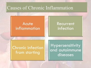
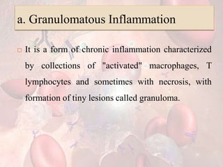
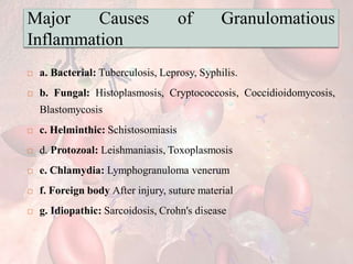
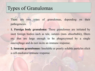
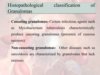
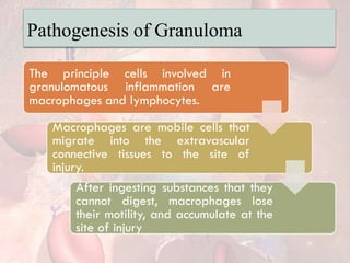
ACUTE INFLAMMATION AND
CHRONIC INFLAMMATION
Prepared by: Mayuri Gamit
M.Sc Nursing (OBG)
Nursing tutor
Definition
 Inflammation is a defensive process that a living
body initiates against local tissue damage. It takes
the form of a complex reaction of blood vessels,
certain plasma components, blood cells and
connective tissue.
Role of Inflammation
 Protection, contain and isolate the injury
 Destroy invading organisms and inactivate toxins
 Achieve healing and repair
Cardinal Signs of Inflammation
 Cornelius Celsus described four signs of
inflammation in first century BC and later Virchow
added the fifth sign in 19th century AD.
Cardinal signs of inflammation
 RUBOR
 Redness due to increased blood flow and vasodilation
 CALOR
 Heat due to increase blood flow to the periphery
 TUMOR
 Swelling from inflammatory edema
 DOLOR
 Pain from swelling and presence of inflammatory
mediators
 FUNCTIO LAESA
 Loss of function due to pain and structural necrosis
Etiology or Causes of Inflammation
• Extreme temperatures, electric shock,
radiation, surgical wound, trauma,
etc.
Physical
agents
• Products of metabolism like urea,
bilirubin, acids, alkalis, drugs, tissue
necrosis
Chemical
agents
• Microorganisms (bacteria, viruses,
fungi), parasites , immune cells and
complex
Biological
agents
Basic Patterns of Inflammation
 Acute inflammation: It is of relatively short
duration (hours to days) and is primarily
characterized by exudation(process of oozing) of
fluid and plasma proteins, as well as a neutrophilic
infiltration.
 Chronic inflammation: It is of longer duration
(days to years) and is characterized by
mononuclear cell (macrophages and lymphocytes)
infiltration, vascular proliferation and scarring.
ACUTE INFLAMMATION
Pathogenesis of Acute Inflammation:
 Though the acute inflammatory response of the
body to inciting agents is a continuous and
coordinated work of blood vessels, inflammatory
cell and the components of plasma.
Major events of inflammation
Vascular events
There are changes in ability of blood
vessels so that more blood can be
brought to the site of inflammation
and white blood cells (WBC's) can
easily move out of circulation.
Cellular events
The white blood cells move out of
blood vessels, migrate through the
tissue to the site of inflammation and
destroy the causative agent.
1. Vascular Events of Inflammation
 There is increase in the size of lumen of
microvasculature as well as the structural changes in
the microvasculature that permit plasma proteins and
leukocytes to leave the circulation.
 There are two phases of vascular events.
 a. Haemodynamic Changes
 b. Altered Vascular Permeability
a. Haemodynamic Changes
• Affects arterioles. The first response of blood vessels to
injury due to any agent is initial vasoconstriction. It is
transient and reflexive. Usually lasts from a few seconds to
upto 5 minutes.
Transient
vasoconstriction
• It results in increased blood flow due to dilatation of
microvasculature and results in increased blood volume in
affected area. It takes about 30 minutes for this process to
occur.
• This is responsible for rubor and calor at the site
Persistent
progressive
vasodilatation
• Is the outcome of vasodilatation and increased blood
volume at site of inflammation. This pushes out fluid into
the interstitial space and results in edema (tumor).
• The edema fluid at this stage is known as transudate.
(protein- poor filtrate of plasma).
Increased
hydrostatic
pressure
CONTI..
• Occurs because blood becomes
thick and there is concentration of
red blood cells (RBC's) as a result
of leakage of plasma. This is
responsible for redness or rubor.
Slowing or
stasis of blood
• As the blood flow becomes slow,
the leukocytes stick to
endothelium in vessel wall and
proceed to cellular events.
Leukocytic
margination
b. Altered Vascular Permeability
 Occurs mostly under the influence of chemicals
mediators of inflammation.
 Is seen in capillaries and small venules.
Mechanism of Altered Vascular Permeability
 The rise in hydrostatic pressure leads to increased
permeability of vessels.
 Endothelial cell contraction which is rapid and short-
lived result in widened intercellular junctions.
 Endothelial cell retraction due to reorganization of
cytoskeleton of the cells results in persistent leakage of
fluid and edema.
 Direct injury by non-specific damage to vessels
due to burns, infections, radiation and activated
leukocytes.
 Neovascularization is formation of new blood
vessels during the process of healing, which are
often leaky and may result in edema.
2. Cellular Events of Inflammation
 The purpose of inflammation is to bring the cells of
defense to the affected site and limit the effect of
injury.
 This consists of two processes:
a. Exudation of Leukocytes
b. Phagocytosis
a. Exudation of Leukocytes
 It is process by which leukocytes move from the blood
stream to tissue during inflammation.
 Escape of WBC's from circulation within blood vessels
to interstitial space is the most important event.
 Neutrophils are the most important participant cells of
acute inflammation.
Sequence of events in migration of leukocytes from
blood vessels to the site of inflammation.
b. Phagocytosis
 It is the process of engulfment of solid particulate
material by the cells.
 There are two types of phagocytic cells-
polymorphonuclear neutrophils and circulating
monocytes and fixed tissue macrophages.
 it is a cellular process by which foreign particles or infectious
agents are ingested & digested by phagocytic cells.
 The process of phagocytosis consists of 4 stages:
 a) Chemotaxis
 b) Attachment
 C) Ingestion
 d) Intracellular digestion
a) Chemotaxis:
 In this stage, the phagocytes reach the site of
infection, being attracted by chemotactic substances
produced at the site of infection.
b) Attachment:
 In this stage the infective agent gets attached or
adhered to membrane of phagocyte.
C) Ingestion:
 In this stage, the phagocytes engulf the infective
agent/particulate material into a phagosome, the
membrane of which fuses with a lysosome in the
cytoplasm forming phagolysosome.
 Lysosome contains hydrolytic enzymes & other
bactericidal substances.
d) Intracellular digestion:
 In this stage, intracellular digestion of engulfed
microbes takes place within few minutes of phagocytosis
by action of hydrolytic enzymes.
 The bacterial components are degraded in several
hours & the microorganisms which resists this
phagocytosis, get adapted to grow within these
phagocytes which later produce disease.
Chemical Mediators of Inflammation
 The group of substances that acts on blood vessels,
inflammatory cells or other cells to contribute to an
inflammatory response is known as chemical
mediators of inflammation.
 They are endogenous in origin i.e. produced in the
body.
 The mediators of inflammation comprises an extensive
network.
 Cell derived: Preformed in granules in granulocytes or
newly synthesized.
 Plasma derived: Mediators which comprise of various
products derived from activation of plasma proteins.
Most Common Mediators responsible for
various effects of inflammation
 Vasodilation: Prostaglandins, nitric oxide.
 Increased Vascular Permeability: Vasoactive
amines(histamine and serotonin), C3a and C5a
(from complement cascade), Bradykinin,
Leukotrienes C4, D4, E4 (arachadonicacid
metabolites), Platelet-activating factor.
 Chemotactic factor: C5a, Leukotriene B4,
Chemokines (e.g., IL-8)
 Fever: Interlenkin (IL-1), IL-6, tumor necrosis factor
(TNF),Prostaglandins (Arachadonic acid metabolites)
 Pain- Prostaglandins, Bradykinin (Kinin system)
 Tissue Damage: Neutrophil and macrophage products,
lysosomal enzymes, nitric oxide.
Systemic Effects of Inflammation
 Whenever acute inflammation is severe and
widespread, it is associated with systemic
manifestation or symptoms
Systemic effects of inflammation
Systemic
effects
Description
Fever The chemical mediators like prostaglandins, IL-6 and TNF-alpha have
effects on the thermoregulatory center in the hypothalamus. This causes
dermal vasoconstriction and decreased heat dissipation.
Shock Extensive vasodilatation and permeability results in hypotension and
shock
Leukocytosis Increased WBC count often accompanies acute inflammation
Lymphadenitis The lymph nodes in the region of affected area of inflammation become
enlarged and painful
Morphologic Patterns of Acute
Inflammation
 There are different morphologic patterns of acute
inflammation
Morphology of acute inflammation
Pattern Description
Serous
inflammation
This is characterized by an accumulation of a thin
fluid that is derived from either the blood or
secretion of mesothelial cells lining the peritoneal,
pleural, and pericardial cavities.
Suppurative
(Purulent)
inflammation
This type of inflammation is characterized by the
production of a large amount of pus which is a thick
creamy liquid composed of large number of living
or dead leukocytes (neutrophils) and necrotic tissue
debris.
Pattern Description
Abscess An abscess is a localized accumulation of pus with tissue destruction
and a cavity formation.
Examples:
• Pulmonary abscess.
• Cerebral abscess.
• Kidney abscess.
Fibrinous
inflammation
Outpouring of fibrinogen and conversion to fibrin over the inflamed
organ or in the body cavity example, fibrinous pericarditis seen in
Rheumatic heart disease
Catarrhal
inflammation
Discharge of mucoid material (e.g. common cold)
Hemorrhagic Accumulation of edema fluid and large amounts of blood due to
exaggerated inflammation that has broken blood vessels.
Fate of Acute Inflammation
 Acute inflammation may end up in any of the
following:
 Resolution: Complete recovery with restoration of
normal structure and function of the tissue, e.g. Lobar
pneumonia.
 Healing by fibrosis: When large amount of tissue is
destroyed, the defect is healed by formation of scar.
 Suppuration (Abscess): When the infection is
caused by pyogenic bacteria, severe liquifactive
necrosis and tissue debris form abscesses, fistula or
sinuses.
 Chronic inflammation: Persistent or recurrent
infections can progress to chronic inflammation.
CHRONIC INFLAMMATION
Definition
 Chronic inflammation is a prolonged inflammatory
process where an active inflammation, tissue
destruction and attempts to repair are proceeding
simultaneously.
Important Consideration
 It is due to the sustained inflammatory response.
 It may last for weeks, months, or even a lifetime.
 Certain chronic inflammation for Example,
chronic gastritis, autoimmune diseases, may form a
basis of development and progression to neoplasia
or cancers.
Acute
inflammation
Recurrent
infection
Chronic infection
from starting
Hypersensitivity
and autoimmune
diseases
Acute inflammation:
 When either the tissue destruction is extensive or
inflammation causing agent like bacteria or foreign
body persist in the body.
Recurrent infection:
 After repeated of acute inflammation with intervals
of healing, for example, in gall bladder after
cholelithiasis (gall stone), kidney infections, etc.
Chronic infection from starting:
 Persistent infection, prolonged exposure to non-
degradable substances.
Hypersensitivity and autoimmune
diseases:
 Rheumatoid arthritis, graves disease ect.
Morphologic features of chronic
inflammation
 Tissue destruction manifested in the form of necrosis
 Infiltration of mononuclear cells-macrophages, monocytes,
lymphocytes and plasma cells
 Granulation tissue- proliferation of small vascular channels
 Regeneration- Parenchymal tissue proliferation
 Fibrosis- Proliferation of fibroblasts and deposition of
collagen.
Cells of Chronic Inflammation
Monocytes
and
Macrophages
T-
Lymphocytes
B-lymphocytes
and Plasma
Cells
Monocytes and Macrophages
 These are the primary cells that play the role in chronic
inflammation.
 Macrophages are the scavenger cells(destroy infectious
agents) of the body.
 The mononuclear phagocyte system includes blood
monocytes, different types of tissue histiocytes and
macrophages like in the liver (Kupffer cells), spleen, lymph
nodes (sinus histiocytes), lungs (alveolar macrophages), bone
marrow, brain (microglia), skin (Langerhan's cells), etc.
 Macrophages produce inflammatory and
immunological mediators.
 They also regulate lymphocyte responses to antigens.
 They secrete other mediators that modulate the
proliferation and activities of fibroblast and endothelial
cells.
T-Lymphocytes
 These are primarily involved in cellular immunity
with lymphokine production.
 They are the key regulator and effectors cells of
the immune system.
B-lymphocytes and Plasma Cells
 These cells produce antibodies directed either
against persistent antigen in the inflammatory site
or against altered tissue components.
 They are components of humoral immunity.
Types of Chronic Inflammation
 a. Granulomatous inflammation: Characterized by
the presence of granuloma.
 b. Nonspecific chronic inflammation: This involves a
diffuse accumulation of macrophages and lymphocytes
with new fibrous tissue formations at site of injury e.g.,
Chronic cholecystitis.
a. Granulomatous Inflammation
 It is a form of chronic inflammation characterized
by collections of "activated" macrophages, T
lymphocytes and sometimes with necrosis, with
formation of tiny lesions called granuloma.
Definition
 A granuloma is a well circumscribed lesion of around 1
mm diameter which is microscopic aggregate of
modified macrophages called epithelioid cells,
surrounded by a cuff of lymphocytes and plasma cells
and a layer of fibrosis.
 Granuloma formation is a protective response to
chronic infection.
 It prevents dissemination and restricts inflammation
due to exogenous substances.
 It is seen with agents that are not effectively
digested during the acute response.
Major Causes of Granulomatious
Inflammation
 a. Bacterial: Tuberculosis, Leprosy, Syphilis.
 b. Fungal: Histoplasmosis, Cryptococcosis, Coccidioidomycosis,
Blastomycosis
 c. Helminthic: Schistosomiasis
 d. Protozoal: Leishmaniasis, Toxoplasmosis
 e. Chlamydia: Lymphogranuloma venerum
 f. Foreign body After injury, suture material
 g. Idiopathic: Sarcoidosis, Crohn's disease
Types of Granulomas
 There are two types of granulomas, depending on their
pathogenesis.
 1. Foreign body granuloma: These granulomas are initiated by
inert foreign bodies such as talc, sutures (non- absorbable), fibers
etc. that are large enough to be phagocytosed by a single
macrophage and do not incite an immune response.
 2. Immune granulomas: Insoluble or poorly soluble particles elicit
a cell-mediated immune response.
Histopathological classification of
Granulomas
 Caseating granulomas: Certain infectious agents such
as Mycobacterium tuberculosis characteristically
produce caseating granulomas (presence of caseous
necrosis)
 Non-caseating granulomas: Other diseases such as
sarcoidosis are characterized by granulomas that lack
necrosis.
Pathogenesis of Granuloma
The principle cells involved in
granulomatous inflammation are
macrophages and lymphocytes.
Macrophages are mobile cells that
migrate into the extravascular
connective tissues to the site of
injury.
After ingesting substances that they
cannot digest, macrophages lose
their motility, and accumulate at the
site of injury
They undergo transformation into nodular
collections of large sized, pale, epithelioid cells,
creating a granuloma
Epitheloid cells are so called because of their
resemblance to epithelial cells. They are
modified macrophages with a lightly staining,
slipper like nuclei and abundant cytoplasm.
Multinucleated giant cells are formed by the
cytoplasmic fusion of macrophages.
When the nuclei of such giant cells are arranged
around the periphery of the cell in a horseshoe
pattern, the cell is called as Langhans giant cell,
which is characteristic of tuberculosis.
Systemic effects of chronic inflammation
 Fever: is usually low, grade.
 Leukocytosis: Total WBC count is increased with
predominance of lymphocytes.
 ESR: Erythrocyte sedimentation rate is usually high.
 Amyloidosis: This type is seen in some chronic
infections affecting kidneys, lymph nodes and spleen
 Examples of Granulomatous Inflammation
 Three important granulomatous diseases are
tuberculosis, syphilis and leprosy.
ACUTE  AND  CHRONIC  INFLAMMATION.   pdf

ACUTE AND CHRONIC INFLAMMATION. pdf

  • 1.
    ACUTE INFLAMMATION AND CHRONICINFLAMMATION Prepared by: Mayuri Gamit M.Sc Nursing (OBG) Nursing tutor
  • 2.
    Definition  Inflammation isa defensive process that a living body initiates against local tissue damage. It takes the form of a complex reaction of blood vessels, certain plasma components, blood cells and connective tissue.
  • 3.
    Role of Inflammation Protection, contain and isolate the injury  Destroy invading organisms and inactivate toxins  Achieve healing and repair
  • 4.
    Cardinal Signs ofInflammation  Cornelius Celsus described four signs of inflammation in first century BC and later Virchow added the fifth sign in 19th century AD.
  • 5.
    Cardinal signs ofinflammation  RUBOR  Redness due to increased blood flow and vasodilation  CALOR  Heat due to increase blood flow to the periphery  TUMOR  Swelling from inflammatory edema
  • 6.
     DOLOR  Painfrom swelling and presence of inflammatory mediators  FUNCTIO LAESA  Loss of function due to pain and structural necrosis
  • 7.
    Etiology or Causesof Inflammation • Extreme temperatures, electric shock, radiation, surgical wound, trauma, etc. Physical agents • Products of metabolism like urea, bilirubin, acids, alkalis, drugs, tissue necrosis Chemical agents • Microorganisms (bacteria, viruses, fungi), parasites , immune cells and complex Biological agents
  • 8.
    Basic Patterns ofInflammation  Acute inflammation: It is of relatively short duration (hours to days) and is primarily characterized by exudation(process of oozing) of fluid and plasma proteins, as well as a neutrophilic infiltration.
  • 9.
     Chronic inflammation:It is of longer duration (days to years) and is characterized by mononuclear cell (macrophages and lymphocytes) infiltration, vascular proliferation and scarring.
  • 10.
    ACUTE INFLAMMATION Pathogenesis ofAcute Inflammation:  Though the acute inflammatory response of the body to inciting agents is a continuous and coordinated work of blood vessels, inflammatory cell and the components of plasma.
  • 11.
    Major events ofinflammation Vascular events There are changes in ability of blood vessels so that more blood can be brought to the site of inflammation and white blood cells (WBC's) can easily move out of circulation. Cellular events The white blood cells move out of blood vessels, migrate through the tissue to the site of inflammation and destroy the causative agent.
  • 12.
    1. Vascular Eventsof Inflammation  There is increase in the size of lumen of microvasculature as well as the structural changes in the microvasculature that permit plasma proteins and leukocytes to leave the circulation.  There are two phases of vascular events.  a. Haemodynamic Changes  b. Altered Vascular Permeability
  • 14.
    a. Haemodynamic Changes •Affects arterioles. The first response of blood vessels to injury due to any agent is initial vasoconstriction. It is transient and reflexive. Usually lasts from a few seconds to upto 5 minutes. Transient vasoconstriction • It results in increased blood flow due to dilatation of microvasculature and results in increased blood volume in affected area. It takes about 30 minutes for this process to occur. • This is responsible for rubor and calor at the site Persistent progressive vasodilatation • Is the outcome of vasodilatation and increased blood volume at site of inflammation. This pushes out fluid into the interstitial space and results in edema (tumor). • The edema fluid at this stage is known as transudate. (protein- poor filtrate of plasma). Increased hydrostatic pressure
  • 15.
    CONTI.. • Occurs becauseblood becomes thick and there is concentration of red blood cells (RBC's) as a result of leakage of plasma. This is responsible for redness or rubor. Slowing or stasis of blood • As the blood flow becomes slow, the leukocytes stick to endothelium in vessel wall and proceed to cellular events. Leukocytic margination
  • 17.
    b. Altered VascularPermeability  Occurs mostly under the influence of chemicals mediators of inflammation.  Is seen in capillaries and small venules.
  • 18.
    Mechanism of AlteredVascular Permeability  The rise in hydrostatic pressure leads to increased permeability of vessels.  Endothelial cell contraction which is rapid and short- lived result in widened intercellular junctions.  Endothelial cell retraction due to reorganization of cytoskeleton of the cells results in persistent leakage of fluid and edema.
  • 19.
     Direct injuryby non-specific damage to vessels due to burns, infections, radiation and activated leukocytes.  Neovascularization is formation of new blood vessels during the process of healing, which are often leaky and may result in edema.
  • 21.
    2. Cellular Eventsof Inflammation  The purpose of inflammation is to bring the cells of defense to the affected site and limit the effect of injury.  This consists of two processes: a. Exudation of Leukocytes b. Phagocytosis
  • 22.
    a. Exudation ofLeukocytes  It is process by which leukocytes move from the blood stream to tissue during inflammation.  Escape of WBC's from circulation within blood vessels to interstitial space is the most important event.  Neutrophils are the most important participant cells of acute inflammation.
  • 23.
    Sequence of eventsin migration of leukocytes from blood vessels to the site of inflammation.
  • 26.
    b. Phagocytosis  Itis the process of engulfment of solid particulate material by the cells.  There are two types of phagocytic cells- polymorphonuclear neutrophils and circulating monocytes and fixed tissue macrophages.
  • 27.
     it isa cellular process by which foreign particles or infectious agents are ingested & digested by phagocytic cells.  The process of phagocytosis consists of 4 stages:  a) Chemotaxis  b) Attachment  C) Ingestion  d) Intracellular digestion
  • 28.
    a) Chemotaxis:  Inthis stage, the phagocytes reach the site of infection, being attracted by chemotactic substances produced at the site of infection.
  • 29.
    b) Attachment:  Inthis stage the infective agent gets attached or adhered to membrane of phagocyte.
  • 30.
    C) Ingestion:  Inthis stage, the phagocytes engulf the infective agent/particulate material into a phagosome, the membrane of which fuses with a lysosome in the cytoplasm forming phagolysosome.  Lysosome contains hydrolytic enzymes & other bactericidal substances.
  • 31.
    d) Intracellular digestion: In this stage, intracellular digestion of engulfed microbes takes place within few minutes of phagocytosis by action of hydrolytic enzymes.  The bacterial components are degraded in several hours & the microorganisms which resists this phagocytosis, get adapted to grow within these phagocytes which later produce disease.
  • 35.
    Chemical Mediators ofInflammation  The group of substances that acts on blood vessels, inflammatory cells or other cells to contribute to an inflammatory response is known as chemical mediators of inflammation.  They are endogenous in origin i.e. produced in the body.
  • 36.
     The mediatorsof inflammation comprises an extensive network.  Cell derived: Preformed in granules in granulocytes or newly synthesized.  Plasma derived: Mediators which comprise of various products derived from activation of plasma proteins.
  • 37.
    Most Common Mediatorsresponsible for various effects of inflammation  Vasodilation: Prostaglandins, nitric oxide.  Increased Vascular Permeability: Vasoactive amines(histamine and serotonin), C3a and C5a (from complement cascade), Bradykinin, Leukotrienes C4, D4, E4 (arachadonicacid metabolites), Platelet-activating factor.
  • 38.
     Chemotactic factor:C5a, Leukotriene B4, Chemokines (e.g., IL-8)  Fever: Interlenkin (IL-1), IL-6, tumor necrosis factor (TNF),Prostaglandins (Arachadonic acid metabolites)  Pain- Prostaglandins, Bradykinin (Kinin system)  Tissue Damage: Neutrophil and macrophage products, lysosomal enzymes, nitric oxide.
  • 39.
    Systemic Effects ofInflammation  Whenever acute inflammation is severe and widespread, it is associated with systemic manifestation or symptoms
  • 40.
    Systemic effects ofinflammation Systemic effects Description Fever The chemical mediators like prostaglandins, IL-6 and TNF-alpha have effects on the thermoregulatory center in the hypothalamus. This causes dermal vasoconstriction and decreased heat dissipation. Shock Extensive vasodilatation and permeability results in hypotension and shock Leukocytosis Increased WBC count often accompanies acute inflammation Lymphadenitis The lymph nodes in the region of affected area of inflammation become enlarged and painful
  • 41.
    Morphologic Patterns ofAcute Inflammation  There are different morphologic patterns of acute inflammation
  • 42.
    Morphology of acuteinflammation Pattern Description Serous inflammation This is characterized by an accumulation of a thin fluid that is derived from either the blood or secretion of mesothelial cells lining the peritoneal, pleural, and pericardial cavities. Suppurative (Purulent) inflammation This type of inflammation is characterized by the production of a large amount of pus which is a thick creamy liquid composed of large number of living or dead leukocytes (neutrophils) and necrotic tissue debris.
  • 43.
    Pattern Description Abscess Anabscess is a localized accumulation of pus with tissue destruction and a cavity formation. Examples: • Pulmonary abscess. • Cerebral abscess. • Kidney abscess. Fibrinous inflammation Outpouring of fibrinogen and conversion to fibrin over the inflamed organ or in the body cavity example, fibrinous pericarditis seen in Rheumatic heart disease Catarrhal inflammation Discharge of mucoid material (e.g. common cold) Hemorrhagic Accumulation of edema fluid and large amounts of blood due to exaggerated inflammation that has broken blood vessels.
  • 44.
    Fate of AcuteInflammation  Acute inflammation may end up in any of the following:  Resolution: Complete recovery with restoration of normal structure and function of the tissue, e.g. Lobar pneumonia.  Healing by fibrosis: When large amount of tissue is destroyed, the defect is healed by formation of scar.
  • 45.
     Suppuration (Abscess):When the infection is caused by pyogenic bacteria, severe liquifactive necrosis and tissue debris form abscesses, fistula or sinuses.  Chronic inflammation: Persistent or recurrent infections can progress to chronic inflammation.
  • 46.
  • 47.
    Definition  Chronic inflammationis a prolonged inflammatory process where an active inflammation, tissue destruction and attempts to repair are proceeding simultaneously.
  • 48.
    Important Consideration  Itis due to the sustained inflammatory response.  It may last for weeks, months, or even a lifetime.  Certain chronic inflammation for Example, chronic gastritis, autoimmune diseases, may form a basis of development and progression to neoplasia or cancers.
  • 49.
  • 50.
    Acute inflammation:  Wheneither the tissue destruction is extensive or inflammation causing agent like bacteria or foreign body persist in the body.
  • 51.
    Recurrent infection:  Afterrepeated of acute inflammation with intervals of healing, for example, in gall bladder after cholelithiasis (gall stone), kidney infections, etc.
  • 52.
    Chronic infection fromstarting:  Persistent infection, prolonged exposure to non- degradable substances.
  • 53.
    Hypersensitivity and autoimmune diseases: Rheumatoid arthritis, graves disease ect.
  • 54.
    Morphologic features ofchronic inflammation  Tissue destruction manifested in the form of necrosis  Infiltration of mononuclear cells-macrophages, monocytes, lymphocytes and plasma cells  Granulation tissue- proliferation of small vascular channels  Regeneration- Parenchymal tissue proliferation  Fibrosis- Proliferation of fibroblasts and deposition of collagen.
  • 55.
    Cells of ChronicInflammation Monocytes and Macrophages T- Lymphocytes B-lymphocytes and Plasma Cells
  • 56.
    Monocytes and Macrophages These are the primary cells that play the role in chronic inflammation.  Macrophages are the scavenger cells(destroy infectious agents) of the body.  The mononuclear phagocyte system includes blood monocytes, different types of tissue histiocytes and macrophages like in the liver (Kupffer cells), spleen, lymph nodes (sinus histiocytes), lungs (alveolar macrophages), bone marrow, brain (microglia), skin (Langerhan's cells), etc.
  • 57.
     Macrophages produceinflammatory and immunological mediators.  They also regulate lymphocyte responses to antigens.  They secrete other mediators that modulate the proliferation and activities of fibroblast and endothelial cells.
  • 58.
    T-Lymphocytes  These areprimarily involved in cellular immunity with lymphokine production.  They are the key regulator and effectors cells of the immune system.
  • 59.
    B-lymphocytes and PlasmaCells  These cells produce antibodies directed either against persistent antigen in the inflammatory site or against altered tissue components.  They are components of humoral immunity.
  • 60.
    Types of ChronicInflammation  a. Granulomatous inflammation: Characterized by the presence of granuloma.  b. Nonspecific chronic inflammation: This involves a diffuse accumulation of macrophages and lymphocytes with new fibrous tissue formations at site of injury e.g., Chronic cholecystitis.
  • 62.
    a. Granulomatous Inflammation It is a form of chronic inflammation characterized by collections of "activated" macrophages, T lymphocytes and sometimes with necrosis, with formation of tiny lesions called granuloma.
  • 63.
    Definition  A granulomais a well circumscribed lesion of around 1 mm diameter which is microscopic aggregate of modified macrophages called epithelioid cells, surrounded by a cuff of lymphocytes and plasma cells and a layer of fibrosis.  Granuloma formation is a protective response to chronic infection.
  • 64.
     It preventsdissemination and restricts inflammation due to exogenous substances.  It is seen with agents that are not effectively digested during the acute response.
  • 65.
    Major Causes ofGranulomatious Inflammation  a. Bacterial: Tuberculosis, Leprosy, Syphilis.  b. Fungal: Histoplasmosis, Cryptococcosis, Coccidioidomycosis, Blastomycosis  c. Helminthic: Schistosomiasis  d. Protozoal: Leishmaniasis, Toxoplasmosis  e. Chlamydia: Lymphogranuloma venerum  f. Foreign body After injury, suture material  g. Idiopathic: Sarcoidosis, Crohn's disease
  • 66.
    Types of Granulomas There are two types of granulomas, depending on their pathogenesis.  1. Foreign body granuloma: These granulomas are initiated by inert foreign bodies such as talc, sutures (non- absorbable), fibers etc. that are large enough to be phagocytosed by a single macrophage and do not incite an immune response.  2. Immune granulomas: Insoluble or poorly soluble particles elicit a cell-mediated immune response.
  • 67.
    Histopathological classification of Granulomas Caseating granulomas: Certain infectious agents such as Mycobacterium tuberculosis characteristically produce caseating granulomas (presence of caseous necrosis)  Non-caseating granulomas: Other diseases such as sarcoidosis are characterized by granulomas that lack necrosis.
  • 68.
    Pathogenesis of Granuloma Theprinciple cells involved in granulomatous inflammation are macrophages and lymphocytes. Macrophages are mobile cells that migrate into the extravascular connective tissues to the site of injury. After ingesting substances that they cannot digest, macrophages lose their motility, and accumulate at the site of injury
  • 69.
    They undergo transformationinto nodular collections of large sized, pale, epithelioid cells, creating a granuloma Epitheloid cells are so called because of their resemblance to epithelial cells. They are modified macrophages with a lightly staining, slipper like nuclei and abundant cytoplasm. Multinucleated giant cells are formed by the cytoplasmic fusion of macrophages. When the nuclei of such giant cells are arranged around the periphery of the cell in a horseshoe pattern, the cell is called as Langhans giant cell, which is characteristic of tuberculosis.
  • 70.
    Systemic effects ofchronic inflammation  Fever: is usually low, grade.  Leukocytosis: Total WBC count is increased with predominance of lymphocytes.  ESR: Erythrocyte sedimentation rate is usually high.  Amyloidosis: This type is seen in some chronic infections affecting kidneys, lymph nodes and spleen
  • 71.
     Examples ofGranulomatous Inflammation  Three important granulomatous diseases are tuberculosis, syphilis and leprosy.