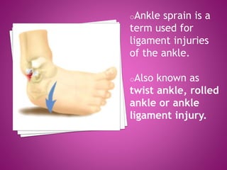
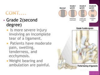
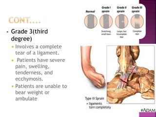
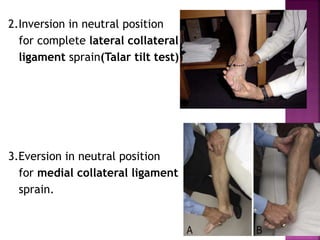
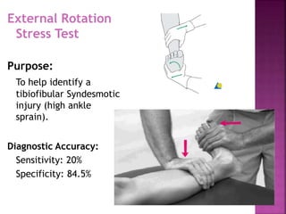
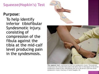
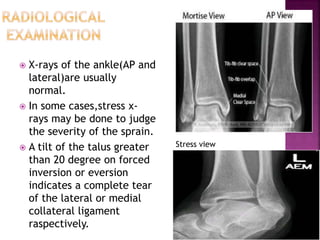
Presented by....Amit kumar
BPT 2nd year
CPRS,JMI
oAnkle sprain is a
term used for
ligament injuries
of the ankle.
oAlso known as
twist ankle, rolled
ankle or ankle
ligament injury.
 The foot is placed in forced
inversion and plantar flexion.
 It can be from an
unstable/irregular surface.
 Also caused by forced trauma.
 Swelling
 Pain
 Discoloration
 Redness
 Warmth
 Inability to walk
 Ankle Instability
 Ankle sprain classified in to:
 Low ankle sprain:
I. Lateral ankle sprain “classic sprain”- 80% to
85%
II. Medial ankle sprain- 5% to 10%
 High ankle sprain (Syndesmotic sprain) - 5% to 10%
 Lateral ankle sprain
(Inversion sprain)
o The most common mechanism of
ankle injury is inversion of the
plantar-flexed foot.
o The anterior talofibular ligament
is the first or only ligament to be
injured in the majority of ankle
sprains.
o Stronger forces lead to combined
ruptures of the anterior talofibular
ligament and the calcaneofibular
ligament.
 Medial ankle sprain
(eversion sprain)
o The medial deltoid
ligament complex is the
strongest of the ankle
ligaments and is infrequently
injured.
o Forced eversion of the
ankle can cause damage to
this structure but more
commonly results in an
avulsion fracture of the
medial malleolus because of
the strength of the deltoid
ligament.
 High ankle sprain
(Syndesmotic sprain):
o Dorsiflexion and eversion
of the ankle may cause
sprain of the syndesmotic
structures.
o There generally tends to be
less swelling with a high
ankle sprain, however there
tends to be pain that is more
severe and longer lasting.
o Syndesmotic ligament
injuries contribute to
chronic ankle instability
and are more likely to result
in recurrent ankle sprain
and the formation of
heterotopic ossification.
 Grade 1(first degree)
 It results from mild
stretching of a ligament
with microscopic tears.
 Patients have mild swelling
and tenderness.
 The patient is able to bear
weight and ambulate with
minimal pain.
 Grade 2(second
degree)
 Is more severe injury
involving an incomplete
tear of a ligament.
 Patients have moderate
pain, swelling,
tenderness, and
ecchymosis.
 Weight bearing and
ambulation are painful.
 Grade 3(third
degree)
 Involves a complete
tear of a ligament.
 Patients have severe
pain, swelling,
tenderness, and
ecchymosis.
 Patients are unable to
bear weight or
ambulate
Sign/symptom Grade I Grade II Grade III
Ligament tear None Partial Complete
Loss of
functional
ability
Minimal Some Great
Pain Minimal Moderate Severe
Swelling Minimal Moderate Severe
Ecchymosis Usually not Common Yes
Difficulty
bearing weight
None Usual Almost always
PHYSICAL EXAMINATION
 The patient gives history
of a twisting injury to the
ankle followed by pain and
swelling over the injured
ligament.
 Weight bearing gives rise
to excruciating pain.
 In case of with complete
tears,patient gives history
of feeling of ‘something
tearing’ at the time of the
injury.
 There may be swelling and tenderness localised to the site of the
torn ligament.
 If a torn ligament is subjected to stress by the following
manoeuvres the patient experiences severe pain:
1.inversion of a plantar-flexed foot for
anterior talo-fibular ligament sprain
2.Inversion in neutral position
for complete lateral collateral
ligament sprain(Talar tilt test)
3.Eversion in neutral position
for medial collateral ligament
sprain.
Anterior Draw
Test
Purpose:
To test for
ligamentous laxity or
instability in the
ankle. This test
primarily assesses the
strength of the
Anterior Talofibular
Ligament.
Diagnostic Accuracy:
Sensitivity: 71%
Specificity: 33 %
Talar Tilt test
Purpose:
The talar tilt test detects
excessive ankle inversion. If the
ligamentous tear extends
posteriorly into the
calcaneofibular portion of the
lateral ligament, the lateral
ankle is unstable and talar tilt
occurs.
Diagnostic Accuracy:
Sensitivity: 67%
Specificity: 75%
External Rotation
Stress Test
Purpose:
To help identify a
tibiofibular Syndesmotic
injury (high ankle
sprain).
Diagnostic Accuracy:
Sensitivity: 20%
Specificity: 84.5%
Squeeze(Hopkin's) Test
Purpose:
To help identify
inferior tibiofibular
Syndesmotic injury.
consisting of
compression of the
fibula against the
tibia at the mid-calf
level producing pain
in the syndesmosis.
o The Ottawa ankle rules are a set of
guidelines for clinicians to help
decide if a patient with foot
or ankle pain should be offered X-
rays to diagnose a possible bone
fracture.
o Sensitivity: 98.5%
 The rules are as follows :
۞An ankle series is only indicated for
patients who have pain in
the malleolar zone AND
 Have bone tenderness at the posterior
edge or tip of the lateral or medial
malleolus OR
 Are unable to bear weight both
immediately after the injury and for four
steps in the emergency department or
doctor's office.
۞A foot series is only indicated for patients
who have pain in the midfoot zone AND
 Have bone tenderness at the base of the
fifth metatarsal or at the navicular OR
 Are unable to bear weight both
immediately after the injury and for four
steps in the emergency department or
doctor's office.
 X-rays of the ankle(AP and
lateral)are usually
normal.
 In some cases,stress x-
rays may be done to judge
the severity of the sprain.
 A tilt of the talus greater
than 20 degree on forced
inversion or eversion
indicates a complete tear
of the lateral or medial
collateral ligament
raspectively.
Stress view
 Abnormal findings
includes:
1.Decreased tibiofibular
overlap
 normal >6 mm on AP view
 normal >1 mm on mortise view
2.Increased medial clear
space
 normal less than or equal to 4
mm
3.Increased tibiofibular clear
space
 normal <6 mm on both AP and
mortise views
1 2
3
•PRICER
•Ankle Taping/Bracing/ splints
•Gentle Mobilization
•Strengthening ex for intrinsic foot Muscles
•Proprioception training
•Balance training
•Plyometric training to regain functional level of
activity
Acute Injury/
Minor Tear
• Surgical Repair
Chronic or
recurrent Ankle
Sprain
 Conservative Management
Initial Management:
o The initial management of ankle sprain
requires the PRICER regimen;
P= Protection …. crutches, splint or brace
R= Rest ….
I= Ice …. 20 minutes every 3-4 hours
C= Compression ….
E= Elevation ….
R= Rehabilitation ….
o This is probably the single-most
important factor in treatment,
particularly with grade I and grade II
injuries.
o Pain and swelling can be reduced with
the use of electrotherapeutic modalities
o Analgesics (NSAID) may be required.
Treatment of grade III injuries:
o It requires initial conservative management
over a six-week period.
o If the patient continues to make good
progress and is able to perform sporting
activities with the aid of taping or bracing
and without persistent problems during or
following activity, surgery may not be
required.
 If, however, despite appropriate
rehabilitation and protection, the patient
complains of recurrent episodes of instability
or persistent pain, then surgical
reconstruction is indicated.
Brace on sprained ankle
 Operative Management
Indication of operation in low ankle sprain:
1. pain and instability despite extensive nonoperative management.
2. large bony avulsions.
1. severe ligamentous damage on the medial and the lateral sides
of the ankle.
2. severe recurrent injuries
Techniques of
operation in low ankle
sprain:
 Arthroscopic
reconstruction.
 Gould modification of
Brostrom anatomic
reconstruction.
o an anatomic shortening
and reinsertion of the
ATFL and CFL
 Tendon transfer and
tenodesis.
Indication of operation in high ankle
sprain:
1. syndesmotic sprain (without fracture)
with instability on stress radiographs
2. syndesmotic sprain refractory to
conservative treatment
3. syndesmotic injury with associated
fracture that remains unstable after
fixation of fracture.
Techniques of operation in high ankle
sprain:
 syndesmosis screw fixation
 syndesmosis fixation with suture button
o fiberwire suture with two buttons tensioned
around the syndesmosis screw.
 Chronic ankle instability usually develops following
an ankle sprain that has not adequately healed or
was not rehabilitated completely.
 In this case,a detailed evaluation with MRI and
arthroscopy may be necessary.
 Arthroscopy ia a good technique for diagnosis and
treatment of such cases.
SPRAINED ANKLE

SPRAINED ANKLE

  • 1.
  • 2.
    oAnkle sprain isa term used for ligament injuries of the ankle. oAlso known as twist ankle, rolled ankle or ankle ligament injury.
  • 4.
     The footis placed in forced inversion and plantar flexion.  It can be from an unstable/irregular surface.  Also caused by forced trauma.
  • 5.
     Swelling  Pain Discoloration  Redness  Warmth  Inability to walk  Ankle Instability
  • 6.
     Ankle sprainclassified in to:  Low ankle sprain: I. Lateral ankle sprain “classic sprain”- 80% to 85% II. Medial ankle sprain- 5% to 10%  High ankle sprain (Syndesmotic sprain) - 5% to 10%
  • 7.
     Lateral anklesprain (Inversion sprain) o The most common mechanism of ankle injury is inversion of the plantar-flexed foot. o The anterior talofibular ligament is the first or only ligament to be injured in the majority of ankle sprains. o Stronger forces lead to combined ruptures of the anterior talofibular ligament and the calcaneofibular ligament.
  • 8.
     Medial anklesprain (eversion sprain) o The medial deltoid ligament complex is the strongest of the ankle ligaments and is infrequently injured. o Forced eversion of the ankle can cause damage to this structure but more commonly results in an avulsion fracture of the medial malleolus because of the strength of the deltoid ligament.
  • 9.
     High anklesprain (Syndesmotic sprain): o Dorsiflexion and eversion of the ankle may cause sprain of the syndesmotic structures. o There generally tends to be less swelling with a high ankle sprain, however there tends to be pain that is more severe and longer lasting. o Syndesmotic ligament injuries contribute to chronic ankle instability and are more likely to result in recurrent ankle sprain and the formation of heterotopic ossification.
  • 10.
     Grade 1(firstdegree)  It results from mild stretching of a ligament with microscopic tears.  Patients have mild swelling and tenderness.  The patient is able to bear weight and ambulate with minimal pain.
  • 11.
     Grade 2(second degree) Is more severe injury involving an incomplete tear of a ligament.  Patients have moderate pain, swelling, tenderness, and ecchymosis.  Weight bearing and ambulation are painful.
  • 12.
     Grade 3(third degree) Involves a complete tear of a ligament.  Patients have severe pain, swelling, tenderness, and ecchymosis.  Patients are unable to bear weight or ambulate
  • 13.
    Sign/symptom Grade IGrade II Grade III Ligament tear None Partial Complete Loss of functional ability Minimal Some Great Pain Minimal Moderate Severe Swelling Minimal Moderate Severe Ecchymosis Usually not Common Yes Difficulty bearing weight None Usual Almost always
  • 14.
    PHYSICAL EXAMINATION  Thepatient gives history of a twisting injury to the ankle followed by pain and swelling over the injured ligament.  Weight bearing gives rise to excruciating pain.  In case of with complete tears,patient gives history of feeling of ‘something tearing’ at the time of the injury.
  • 15.
     There maybe swelling and tenderness localised to the site of the torn ligament.  If a torn ligament is subjected to stress by the following manoeuvres the patient experiences severe pain: 1.inversion of a plantar-flexed foot for anterior talo-fibular ligament sprain
  • 16.
    2.Inversion in neutralposition for complete lateral collateral ligament sprain(Talar tilt test) 3.Eversion in neutral position for medial collateral ligament sprain.
  • 17.
    Anterior Draw Test Purpose: To testfor ligamentous laxity or instability in the ankle. This test primarily assesses the strength of the Anterior Talofibular Ligament. Diagnostic Accuracy: Sensitivity: 71% Specificity: 33 %
  • 18.
    Talar Tilt test Purpose: Thetalar tilt test detects excessive ankle inversion. If the ligamentous tear extends posteriorly into the calcaneofibular portion of the lateral ligament, the lateral ankle is unstable and talar tilt occurs. Diagnostic Accuracy: Sensitivity: 67% Specificity: 75%
  • 19.
    External Rotation Stress Test Purpose: Tohelp identify a tibiofibular Syndesmotic injury (high ankle sprain). Diagnostic Accuracy: Sensitivity: 20% Specificity: 84.5%
  • 20.
    Squeeze(Hopkin's) Test Purpose: To helpidentify inferior tibiofibular Syndesmotic injury. consisting of compression of the fibula against the tibia at the mid-calf level producing pain in the syndesmosis.
  • 21.
    o The Ottawaankle rules are a set of guidelines for clinicians to help decide if a patient with foot or ankle pain should be offered X- rays to diagnose a possible bone fracture. o Sensitivity: 98.5%
  • 22.
     The rulesare as follows : ۞An ankle series is only indicated for patients who have pain in the malleolar zone AND  Have bone tenderness at the posterior edge or tip of the lateral or medial malleolus OR  Are unable to bear weight both immediately after the injury and for four steps in the emergency department or doctor's office. ۞A foot series is only indicated for patients who have pain in the midfoot zone AND  Have bone tenderness at the base of the fifth metatarsal or at the navicular OR  Are unable to bear weight both immediately after the injury and for four steps in the emergency department or doctor's office.
  • 23.
     X-rays ofthe ankle(AP and lateral)are usually normal.  In some cases,stress x- rays may be done to judge the severity of the sprain.  A tilt of the talus greater than 20 degree on forced inversion or eversion indicates a complete tear of the lateral or medial collateral ligament raspectively. Stress view
  • 24.
     Abnormal findings includes: 1.Decreasedtibiofibular overlap  normal >6 mm on AP view  normal >1 mm on mortise view 2.Increased medial clear space  normal less than or equal to 4 mm 3.Increased tibiofibular clear space  normal <6 mm on both AP and mortise views 1 2 3
  • 25.
    •PRICER •Ankle Taping/Bracing/ splints •GentleMobilization •Strengthening ex for intrinsic foot Muscles •Proprioception training •Balance training •Plyometric training to regain functional level of activity Acute Injury/ Minor Tear • Surgical Repair Chronic or recurrent Ankle Sprain
  • 26.
     Conservative Management InitialManagement: o The initial management of ankle sprain requires the PRICER regimen; P= Protection …. crutches, splint or brace R= Rest …. I= Ice …. 20 minutes every 3-4 hours C= Compression …. E= Elevation …. R= Rehabilitation …. o This is probably the single-most important factor in treatment, particularly with grade I and grade II injuries. o Pain and swelling can be reduced with the use of electrotherapeutic modalities o Analgesics (NSAID) may be required.
  • 28.
    Treatment of gradeIII injuries: o It requires initial conservative management over a six-week period. o If the patient continues to make good progress and is able to perform sporting activities with the aid of taping or bracing and without persistent problems during or following activity, surgery may not be required.  If, however, despite appropriate rehabilitation and protection, the patient complains of recurrent episodes of instability or persistent pain, then surgical reconstruction is indicated. Brace on sprained ankle
  • 29.
     Operative Management Indicationof operation in low ankle sprain: 1. pain and instability despite extensive nonoperative management. 2. large bony avulsions. 1. severe ligamentous damage on the medial and the lateral sides of the ankle. 2. severe recurrent injuries
  • 30.
    Techniques of operation inlow ankle sprain:  Arthroscopic reconstruction.  Gould modification of Brostrom anatomic reconstruction. o an anatomic shortening and reinsertion of the ATFL and CFL  Tendon transfer and tenodesis.
  • 31.
    Indication of operationin high ankle sprain: 1. syndesmotic sprain (without fracture) with instability on stress radiographs 2. syndesmotic sprain refractory to conservative treatment 3. syndesmotic injury with associated fracture that remains unstable after fixation of fracture. Techniques of operation in high ankle sprain:  syndesmosis screw fixation  syndesmosis fixation with suture button o fiberwire suture with two buttons tensioned around the syndesmosis screw.
  • 32.
     Chronic ankleinstability usually develops following an ankle sprain that has not adequately healed or was not rehabilitated completely.  In this case,a detailed evaluation with MRI and arthroscopy may be necessary.  Arthroscopy ia a good technique for diagnosis and treatment of such cases.