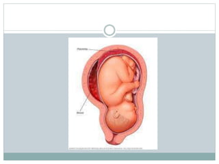
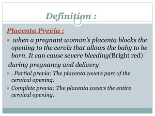
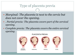
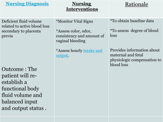
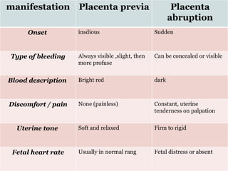
Definition :
 Placenta:
The placenta is an organ that develops in uterus
during pregnancy. This structure provides oxygen
and nutrients to growing baby and removes waste
products from baby's blood. The placenta attaches
to the wall of uterus, and baby's umbilical cord
arises from it. The organ is usually attached to the
top, side, front or back of the uterus.
Abruption
Placenta &
Placenta Previa
*Prepared By Shima maaitah
*Miss : Ghadeer Zayadeen
Objective
*At the end of this seminar we will able to Identify :
- Definetion
- Pathophysiology .
- Classification.
- Causes , risk factors and complication
-Signs and symptoms .
- Laboratory and Diagnostic Testing
-Nursing Assessment and Nursing medical
management .
- Pt education .
Abruption
Placenta
 Abruption Placenta
Placental abruption occurs when the placenta
partly or completely separates from the inner
wall of the uterus before delivery. This can
decrease or block the baby's supply of oxygen
and nutrients and cause heavy dark
bleeding in the mother.
It can occur at any time after 20 weeks of
pregnancy, but it’s most common in the third
trimester.
 the abruption start with degenerative change in the
small maternal arterioles , resulting in : thrombosis
,, and possible rupture of a vessels .
 Bleeding from the vessels forms a retroplacental clot .
The bleeding causes increase pressure behind the
placenta and result in separation .
Pathophysiology
• Mild (grade 1): minimal bleeding
(less than 500 mL), marginal
separation (10% to 20%), tender
uterus, no coagulopathy, no signs of
shock, no fetal distress
• Moderate (grade 2): moderate
bleeding (1,000 to 1,500 mL),
moderate separation (20% to 50%),
continuous abdominal pain, mild
shock, normal maternal blood
pressure , maternal tachycardia .
• Severe (grade 3):absent to moderate
bleeding (more than 1,500 ml), sever
separetion(more than 50%), profound
shock , dark vaginal bleeding , sever
abdominal pain , decreased maternal
blood pressure tachycardia and
development of dissminated
intervascular coagulopathy (DIC).
Abruptio
placentae is
classified to
the : 1. extent
of separation
2. the amount
of blood loss
from maternal
circulation
includs :
Abruptio placentae
also may be
classified as partial
or complete ,
depending on the
degree of
separation.
Causes
*The cause of placental abruption
is often unknown. *Possible
causes include : trauma or injury
to the abdomen or HTN or
smoking
Risk factors
 maternal smoking
 maternal age (over 35 years old ).
 poor nutrition.
 multiple gestation
 sever trauma ( auto accident )
 cocaine use
 alcohol ingestion
 preeclampsia
Vaginal
bleeding
Abdominal
pain
Uterine
tenderness
Back pain
Fetal
distress
Signs and symptoms
Rapid
contraction
 maternal :
 - sever hemorrage
 - need for blood transfusion
 - emergency hysterectomy

Complication
 Fetal :
 - low birth weigh .
 - preterm delivery .
 -Decreased oxygen to the baby, which could lead
to brain damage
 - stillbirth .
Complication
 Medical history
 Physical examination, including checking
the tenderness and tone of the uterus
 CBC
 Fibrinogen levels—typically are increased
in pregnancy
 (PT) , (PTT)
 ultrasound
 CT scan is more reliable method for
evaluation of placenta abruption .
Laboratory and
diagnostic testing :
Begin the health history by assessing the woman for
risk factors that may predispose her to abruption
placenta .
Assessing the pregnant woman presenting with
abdominal pain
The vaginal area is inspected for the presence
of bleeding.
Nursing Assessment
palpation of the uterus for tenderness, consistency, and
frequency and duration of uterine contractions
auscultation of fetal heart sounds and ask about fetal
movement, recent changes in activity patterns
.
Nursing Management
 Obtain maternal vital signs frequently ,even 15 min .
 Monitor the amount and characteristics of any
vaginal bleeding as frequently as every 15 to 30
minutes.
 Communicate empathy and understanding of the
client’s experience, and provide emotional support.
Medical management
 To avoid a worsening condition, these medical
procedures are implemented for both the mother
and the fetus :
 1- Intravenous therapy : Once the woman starts
to bleed, the physician would order a large gauge
catheter to replace the fluid losses.
 2- Oxygen inhalation: Delivered via face mask,
this would prevent fetal anoxia.
 3- Fibrinogen determination: This test would be
taken several times before birth to detect DIC.
 If you’re less than 34 weeks pregnant: You might have be
admitted into the hospital for monitoring -- as long as
baby’s heart rate is normal and the placental abruption
doesn’t seem to be severe. If baby appears to be doing fine
and stop bleeding in mother , you eventually might be able
to go home. You might also be given steroids to help baby’s
lungs develop faster in case you do go into labor early.
 If you’re more than 34 weeks pregnant: if the abruption
does seem severe. If it is, and it’s putting health or baby’s
health at risk, you’ll need a C-section right away. You
might also need a blood transfusion.

Health education
*urge her to seek early
and continuose prenatal
care and receive promot
health care if any signs
and symptoms occure in
future pregnancies .
*encourage the women
to avoid smoking ,or
using drugs during
pregnancy.
Nursing care plane
planning
Evaluation
Rationale
Nursing
Interventions
Nursing
Diagnosis
Goal : client
will be relief or
control of
pain .
Outcome :
Patient will
report relief or
control of pain.
.
- The patient
will be able to
feel
comfortable
and verrbalize
reduce of pain .
To help
determine the
possibility of
underlying
condition or
organ
dysfunction
requiring
treatment.
To maintain
an acceptable
level of pain.
To promote
non
pharmacologi
cal pain
management.
-Assess for
referred pain as
appropriate.
-Administer
analgesics as
indicated.
-Provide
comfort
measures, quiet
environment,
and calm
activities.
Acute Pain
RT
Sudden
separation of
placenta AMB
Sharp,
stabbing pain
high in the
uterine fundus
Uterine
tenderness
Placenta Previa
Definition :
Placenta Previa :
 when a pregnant woman's placenta blocks the
opening to the cervix that allows the baby to be
born. It can cause severe bleeding(Bright red)
during pregnancy and delivery
 . Partial previa: The placenta covers part of the
cervical opening.
 Complete previa: The placenta covers the entire
cervical opening.
Type of placenta previa
 Marginal: The placenta is next to the cervix but
does not cover the opening.
 . Partial previa: The placenta covers part of the cervical
opening.
 Complete previa: The placenta covers the entire cervical
opening.
* The exact cause of placenta previa is unknown
• It is initiated by implantation of the embryo in the
lowor uterus may be due to :
* damage in the upper segment
• Uterine endometrial scarring
• Which may incite placental growth in the
unscarred lower uterine segment ,with placenta
attachment and growth ,the cervical become
coverd by developing placenta
Pathophysiology
Risk factors
 Maternal age ( more than 35 years ) .
 Smoking .
 Previous C/S birth .
 Uterine injury .
 Multiple gestations .
 Previous induced surgical abortion .
Signs and symptoms
 Vaginal bleeding without pain
 . Contraction along with the
bleeding
Complication
 maternal :
 bleeding after delivery .
 Emergency caesarean delivery
 Shock from loss of blood
 Fetal :
 Fetal distress from lack of oxygen
 CBC
 Ultrasound
Laboratory and
diagnostic testing :
Nursing
Assessment
Rapid assessment is essential to ensure prompt,
effective interventions to prevent maternal and
fetal morbidity and mortality.
Monitor vital signs
Monitor fetal HR
Blood transfusion for the mother.
Medical management
 Depends on :
 the exent of bleeding :
If it's light, your doctor might suggest you avoid activities
including exercise. If it's heavy, you may need to go to
the emergency room.
 Whether the placenta previa is complete or
partial
 The exact location of the placenta
 The gestational age of the baby
 The position of the baby
 The health of the baby
* Rest at home
* Discharge plan
* CTG ( fetal HR )
Contraction
* dexamethazone
If the amount of
bleeding is small :
If the amount of bleeding is
large :
Management :
Blood transfusion for the
mother.
Delivary C/S
Health education
 Encourage the women to avoid smoking ,or
using drugs during pregnancy.
 Do not do any heavy activity .
Nursing Diagnosis Nursing
Interventions
Rationale
Deficient fluid volume
related to active blood loss
secondary to placenta
previa
*Monitor Vital Signs
*Assess color, odor,
consistency and amount of
vaginal bleeding
*Assess hourly intake and
output.
*To obtain baseline data
*To assess degree of blood
loss
Provides information about
maternal and fetal
physiologic compensation to
blood loss
Outcome : The
patient will re-
establish a
functional body
fluid volume and
balanced input
and output status .
summary
Placenta
abruption
Placenta previa
manifestation
Sudden
insdious
Onset
Can be concealed or visible
Always visible ,slight, then
more profuse
Type of bleeding
dark
Bright red
Blood description
Constant, uterine
tenderness on palpation
None (painless)
Discomfort / pain
Firm to rigid
Soft and relaxed
Uterine tone
Fetal distress or absent
Usually in normal rang
Fetal heart rate

Abruption Placenta & Placenta Previa (1).pptx

  • 1.
    Definition :  Placenta: Theplacenta is an organ that develops in uterus during pregnancy. This structure provides oxygen and nutrients to growing baby and removes waste products from baby's blood. The placenta attaches to the wall of uterus, and baby's umbilical cord arises from it. The organ is usually attached to the top, side, front or back of the uterus.
  • 2.
    Abruption Placenta & Placenta Previa *PreparedBy Shima maaitah *Miss : Ghadeer Zayadeen
  • 3.
    Objective *At the endof this seminar we will able to Identify : - Definetion - Pathophysiology . - Classification. - Causes , risk factors and complication -Signs and symptoms . - Laboratory and Diagnostic Testing -Nursing Assessment and Nursing medical management . - Pt education .
  • 5.
  • 6.
     Abruption Placenta Placentalabruption occurs when the placenta partly or completely separates from the inner wall of the uterus before delivery. This can decrease or block the baby's supply of oxygen and nutrients and cause heavy dark bleeding in the mother. It can occur at any time after 20 weeks of pregnancy, but it’s most common in the third trimester.
  • 7.
     the abruptionstart with degenerative change in the small maternal arterioles , resulting in : thrombosis ,, and possible rupture of a vessels .  Bleeding from the vessels forms a retroplacental clot . The bleeding causes increase pressure behind the placenta and result in separation . Pathophysiology
  • 8.
    • Mild (grade1): minimal bleeding (less than 500 mL), marginal separation (10% to 20%), tender uterus, no coagulopathy, no signs of shock, no fetal distress • Moderate (grade 2): moderate bleeding (1,000 to 1,500 mL), moderate separation (20% to 50%), continuous abdominal pain, mild shock, normal maternal blood pressure , maternal tachycardia . • Severe (grade 3):absent to moderate bleeding (more than 1,500 ml), sever separetion(more than 50%), profound shock , dark vaginal bleeding , sever abdominal pain , decreased maternal blood pressure tachycardia and development of dissminated intervascular coagulopathy (DIC). Abruptio placentae is classified to the : 1. extent of separation 2. the amount of blood loss from maternal circulation includs :
  • 9.
    Abruptio placentae also maybe classified as partial or complete , depending on the degree of separation.
  • 10.
    Causes *The cause ofplacental abruption is often unknown. *Possible causes include : trauma or injury to the abdomen or HTN or smoking
  • 11.
    Risk factors  maternalsmoking  maternal age (over 35 years old ).  poor nutrition.  multiple gestation  sever trauma ( auto accident )  cocaine use  alcohol ingestion  preeclampsia
  • 12.
  • 13.
     maternal : - sever hemorrage  - need for blood transfusion  - emergency hysterectomy  Complication
  • 14.
     Fetal : - low birth weigh .  - preterm delivery .  -Decreased oxygen to the baby, which could lead to brain damage  - stillbirth . Complication
  • 15.
     Medical history Physical examination, including checking the tenderness and tone of the uterus  CBC  Fibrinogen levels—typically are increased in pregnancy  (PT) , (PTT)  ultrasound  CT scan is more reliable method for evaluation of placenta abruption . Laboratory and diagnostic testing :
  • 16.
    Begin the healthhistory by assessing the woman for risk factors that may predispose her to abruption placenta . Assessing the pregnant woman presenting with abdominal pain The vaginal area is inspected for the presence of bleeding. Nursing Assessment
  • 17.
    palpation of theuterus for tenderness, consistency, and frequency and duration of uterine contractions auscultation of fetal heart sounds and ask about fetal movement, recent changes in activity patterns .
  • 18.
    Nursing Management  Obtainmaternal vital signs frequently ,even 15 min .  Monitor the amount and characteristics of any vaginal bleeding as frequently as every 15 to 30 minutes.  Communicate empathy and understanding of the client’s experience, and provide emotional support.
  • 19.
    Medical management  Toavoid a worsening condition, these medical procedures are implemented for both the mother and the fetus :  1- Intravenous therapy : Once the woman starts to bleed, the physician would order a large gauge catheter to replace the fluid losses.  2- Oxygen inhalation: Delivered via face mask, this would prevent fetal anoxia.  3- Fibrinogen determination: This test would be taken several times before birth to detect DIC.
  • 20.
     If you’reless than 34 weeks pregnant: You might have be admitted into the hospital for monitoring -- as long as baby’s heart rate is normal and the placental abruption doesn’t seem to be severe. If baby appears to be doing fine and stop bleeding in mother , you eventually might be able to go home. You might also be given steroids to help baby’s lungs develop faster in case you do go into labor early.  If you’re more than 34 weeks pregnant: if the abruption does seem severe. If it is, and it’s putting health or baby’s health at risk, you’ll need a C-section right away. You might also need a blood transfusion. 
  • 21.
    Health education *urge herto seek early and continuose prenatal care and receive promot health care if any signs and symptoms occure in future pregnancies . *encourage the women to avoid smoking ,or using drugs during pregnancy.
  • 22.
    Nursing care plane planning Evaluation Rationale Nursing Interventions Nursing Diagnosis Goal: client will be relief or control of pain . Outcome : Patient will report relief or control of pain. . - The patient will be able to feel comfortable and verrbalize reduce of pain . To help determine the possibility of underlying condition or organ dysfunction requiring treatment. To maintain an acceptable level of pain. To promote non pharmacologi cal pain management. -Assess for referred pain as appropriate. -Administer analgesics as indicated. -Provide comfort measures, quiet environment, and calm activities. Acute Pain RT Sudden separation of placenta AMB Sharp, stabbing pain high in the uterine fundus Uterine tenderness
  • 23.
  • 24.
    Definition : Placenta Previa:  when a pregnant woman's placenta blocks the opening to the cervix that allows the baby to be born. It can cause severe bleeding(Bright red) during pregnancy and delivery  . Partial previa: The placenta covers part of the cervical opening.  Complete previa: The placenta covers the entire cervical opening.
  • 25.
    Type of placentaprevia  Marginal: The placenta is next to the cervix but does not cover the opening.  . Partial previa: The placenta covers part of the cervical opening.  Complete previa: The placenta covers the entire cervical opening.
  • 26.
    * The exactcause of placenta previa is unknown • It is initiated by implantation of the embryo in the lowor uterus may be due to : * damage in the upper segment • Uterine endometrial scarring • Which may incite placental growth in the unscarred lower uterine segment ,with placenta attachment and growth ,the cervical become coverd by developing placenta Pathophysiology
  • 27.
    Risk factors  Maternalage ( more than 35 years ) .  Smoking .  Previous C/S birth .  Uterine injury .  Multiple gestations .  Previous induced surgical abortion .
  • 28.
    Signs and symptoms Vaginal bleeding without pain  . Contraction along with the bleeding
  • 29.
    Complication  maternal : bleeding after delivery .  Emergency caesarean delivery  Shock from loss of blood  Fetal :  Fetal distress from lack of oxygen
  • 30.
     CBC  Ultrasound Laboratoryand diagnostic testing :
  • 31.
    Nursing Assessment Rapid assessment isessential to ensure prompt, effective interventions to prevent maternal and fetal morbidity and mortality. Monitor vital signs Monitor fetal HR Blood transfusion for the mother.
  • 32.
    Medical management  Dependson :  the exent of bleeding : If it's light, your doctor might suggest you avoid activities including exercise. If it's heavy, you may need to go to the emergency room.  Whether the placenta previa is complete or partial  The exact location of the placenta  The gestational age of the baby  The position of the baby  The health of the baby
  • 33.
    * Rest athome * Discharge plan * CTG ( fetal HR ) Contraction * dexamethazone If the amount of bleeding is small : If the amount of bleeding is large : Management : Blood transfusion for the mother. Delivary C/S
  • 34.
    Health education  Encouragethe women to avoid smoking ,or using drugs during pregnancy.  Do not do any heavy activity .
  • 35.
    Nursing Diagnosis Nursing Interventions Rationale Deficientfluid volume related to active blood loss secondary to placenta previa *Monitor Vital Signs *Assess color, odor, consistency and amount of vaginal bleeding *Assess hourly intake and output. *To obtain baseline data *To assess degree of blood loss Provides information about maternal and fetal physiologic compensation to blood loss Outcome : The patient will re- establish a functional body fluid volume and balanced input and output status .
  • 36.
  • 37.
    Placenta abruption Placenta previa manifestation Sudden insdious Onset Can beconcealed or visible Always visible ,slight, then more profuse Type of bleeding dark Bright red Blood description Constant, uterine tenderness on palpation None (painless) Discomfort / pain Firm to rigid Soft and relaxed Uterine tone Fetal distress or absent Usually in normal rang Fetal heart rate