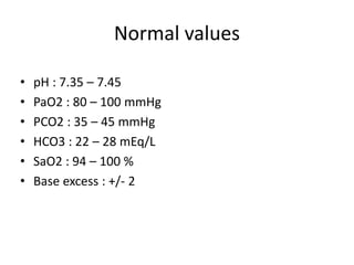
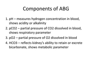
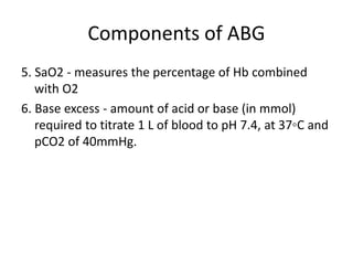
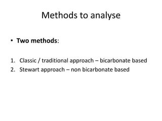
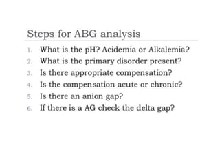
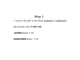
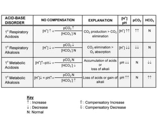
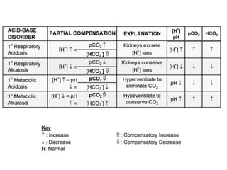
ABG
Dr. Rohit Saini
Definition
• It is a diagnostic procedure in which a blood is
obtained from an artery by direct puncture or
through indwelling arterial catheter.
Information
• Basic physiologic processes:
1. Alveolar ventilation (PaCO2)
2. Oxygenation (PaO2, SaO2)
3. Acid base balance
Indications
1. To assess ventilatory status, oxygenation, acid base
balance
2. Oxygen carrying capacity of blood
3. Aids in diagnoses / etiology
4. To assess patients response (MV, circulatory
intervention)
5. Electrolyte level
Contraindications
1. Local site infection
2. Absence of arterial pulse
3. Negative Allen’s or modified Allen’s test
4. Coagulopathies
5. Anticoagulants/thrombolytics
6. Peripheral vascular disease
7. Arterial grafts
Sites
• Radial artery (non dominant hand) – most preferred
Other sites:
• Brachial
• Femoral
• Dorsalis pedis
• Posterior tibial artery (pediatric)
Complications
• Bruise / hematoma
• Spasms
• Aneurysm
• Nerve damage
• Infection
• Pain
• Distal ischemia
• Anaphylaxis to LA
Sources of error
• Preanalytic errors (MC) – collection, storage and
transportation
• Nonarterial sample
• Air bubbles
• Inadequate or excessive anticoagulant
• Delayed analysis of uncooled sample
Normal values
• pH : 7.35 – 7.45
• PaO2 : 80 – 100 mmHg
• PCO2 : 35 – 45 mmHg
• HCO3 : 22 – 28 mEq/L
• SaO2 : 94 – 100 %
• Base excess : +/- 2
Components of ABG
1. pH – measures hydrogen concentration in blood,
shows acidity or alkalinity
2. pCO2 – partial pressure of CO2 dissolved in blood,
shows respiratory parameter
3. pO2 – partial pressure of O2 dissolved in blood
4. HCO3 – reflects kidney’s ability to retain or excrete
bicarbonate, shows metabolic parameter
Components of ABG
5. SaO2 - measures the percentage of Hb combined
with O2
6. Base excess - amount of acid or base (in mmol)
required to titrate 1 L of blood to pH 7.4, at 37◦C and
pCO2 of 40mmHg.
Abnormal values
Methods to analyse
• Two methods:
1. Classic / traditional approach – bicarbonate based
2. Stewart approach – non bicarbonate based
Classic approach
• Henderson Hasselbalch equation:
• pKa – dissociation constant (6.1)
• Conjugate base – bicarbonate
• Acid – pCO2
Classic approach
• Based on this approach – 6 primary disorders:
1. Metabolic acidosis (high or normal AG)
2. Metabolic alkalosis
3. Acute and chronic respiratory acidosis
4. Acute and chronic respiratory alkalosis
Stewart approach
• Non bicarbonate based
• Variables :
1. Respiratory – pCO2
2. Metabolic – strong ion difference (SID) and total
weak acids (ATOT)
 SID : [cations]-[anions]
 ATOT : represent all non bicarbonate buffer
Step 2
Compensation
• Maintainance of acid base balance
• Goal : to return pH back to normal
• Accomplished by buffer system (by changing H+)
• Respiratory compensation faster than metabolic
• No complete compensation (only 50 – 75%)
Expected compensation
• Metabolic acidosis : 1-1.3 pCO2↓ (1 HCO3 ↓)
• Metabolic alkalosis : 0.5-0.7 pCO2↑ (1 HCO3 ↑)
• Respiratory acidosis :
o Acute – 1 HCO3↑ (10 pCO2 ↑)
o Chronic – 3.5 HCO3 ↑ (10 pCO2 ↑)
• Respiratory alkalosis :
o Acute – 2 HCO3 ↓ (10 pCO2 ↓)
o Chronic – 5 HCO3 ↓ (10 pCO2 ↓)
Anion gap
• Difference between measured cations and measured
anions :
[Na + K] – [Cl + HCO3]
Normal value : 14-16
Or
[Na] – [Cl + HCO3]
Normal value : 8 -12
• Unmeasured anions : significant proportion e.g –
proteins, phosphates, sulfates, lactates, ketones etc.
High anion gap acidosis
Mnemonic – KULT
• Ketoacidosis : Diabetic, starvation, alcoholic
• Uremia : renal failure
• Lactic acidosis
• Toxins : ethylene glycol, salicylates
Normal anion gap acidosis
Hypokalemic :-
• GI loss HCO3 : diarrhea, ileostomy
• Renal loss HCO3 : RTA (type II)
Normokalemic / Hyperkalemic :-
• RTA (type I and IV)
• Hypoaldosteronism
• Acute tubular necrosis
Approach to normal AG acidosis
Low anion gap acidosis
• Hypoalbuminemia (MC)
• Paraproteinemia (multiple myeloma)
• Spurious hyponatremia
• Hypermagnesemia
• Hypercalcemia
Corrected anion gap :
observed AG + 2.5X(normal alb. – observed alb.)
Metabolic alkalosis
• Loss of H+ ions (vomiting, diuretics)
• Increased reabsorption of bicarbonate (low
intravascular volume)
• Alkali administration in renal failure (Ringer’s lactate)
• Hyperaldosteronism
Respiratory acidosis
Any condition causes hypoventilation:
• Airway / lung disease
• CNS depression – trauma, sedatives, narcotics
• Neuromuscular disease – spinal cord injury, NMD
residual effect, GBS etc
• Ventilation restriction – pain, obesity, chest wall
injury/deformity
Respiratory alkalosis
Any condition causes hyperventilation:
• CNS stimulation – fever, pain, CVA
• Hypoxemia – pulmonary edema, pneumonia
• Drugs – catecholamines, salicylates
• Misc. – sepsis, pregnancy
• Psychologic – anxiety, fear
Delta gap
Delta gap : to rule out co existence of 2 acid base disorders
Correlate with patient’s clinical
status is more important
Treat patient, not ABG.

ABG

  • 1.
  • 2.
    Definition • It isa diagnostic procedure in which a blood is obtained from an artery by direct puncture or through indwelling arterial catheter.
  • 3.
    Information • Basic physiologicprocesses: 1. Alveolar ventilation (PaCO2) 2. Oxygenation (PaO2, SaO2) 3. Acid base balance
  • 4.
    Indications 1. To assessventilatory status, oxygenation, acid base balance 2. Oxygen carrying capacity of blood 3. Aids in diagnoses / etiology 4. To assess patients response (MV, circulatory intervention) 5. Electrolyte level
  • 5.
    Contraindications 1. Local siteinfection 2. Absence of arterial pulse 3. Negative Allen’s or modified Allen’s test 4. Coagulopathies 5. Anticoagulants/thrombolytics 6. Peripheral vascular disease 7. Arterial grafts
  • 6.
    Sites • Radial artery(non dominant hand) – most preferred Other sites: • Brachial • Femoral • Dorsalis pedis • Posterior tibial artery (pediatric)
  • 7.
    Complications • Bruise /hematoma • Spasms • Aneurysm • Nerve damage • Infection • Pain • Distal ischemia • Anaphylaxis to LA
  • 8.
    Sources of error •Preanalytic errors (MC) – collection, storage and transportation • Nonarterial sample • Air bubbles • Inadequate or excessive anticoagulant • Delayed analysis of uncooled sample
  • 10.
    Normal values • pH: 7.35 – 7.45 • PaO2 : 80 – 100 mmHg • PCO2 : 35 – 45 mmHg • HCO3 : 22 – 28 mEq/L • SaO2 : 94 – 100 % • Base excess : +/- 2
  • 11.
    Components of ABG 1.pH – measures hydrogen concentration in blood, shows acidity or alkalinity 2. pCO2 – partial pressure of CO2 dissolved in blood, shows respiratory parameter 3. pO2 – partial pressure of O2 dissolved in blood 4. HCO3 – reflects kidney’s ability to retain or excrete bicarbonate, shows metabolic parameter
  • 12.
    Components of ABG 5.SaO2 - measures the percentage of Hb combined with O2 6. Base excess - amount of acid or base (in mmol) required to titrate 1 L of blood to pH 7.4, at 37◦C and pCO2 of 40mmHg.
  • 13.
  • 14.
    Methods to analyse •Two methods: 1. Classic / traditional approach – bicarbonate based 2. Stewart approach – non bicarbonate based
  • 15.
    Classic approach • HendersonHasselbalch equation: • pKa – dissociation constant (6.1) • Conjugate base – bicarbonate • Acid – pCO2
  • 16.
    Classic approach • Basedon this approach – 6 primary disorders: 1. Metabolic acidosis (high or normal AG) 2. Metabolic alkalosis 3. Acute and chronic respiratory acidosis 4. Acute and chronic respiratory alkalosis
  • 17.
    Stewart approach • Nonbicarbonate based • Variables : 1. Respiratory – pCO2 2. Metabolic – strong ion difference (SID) and total weak acids (ATOT)  SID : [cations]-[anions]  ATOT : represent all non bicarbonate buffer
  • 20.
  • 21.
    Compensation • Maintainance ofacid base balance • Goal : to return pH back to normal • Accomplished by buffer system (by changing H+) • Respiratory compensation faster than metabolic • No complete compensation (only 50 – 75%)
  • 25.
    Expected compensation • Metabolicacidosis : 1-1.3 pCO2↓ (1 HCO3 ↓) • Metabolic alkalosis : 0.5-0.7 pCO2↑ (1 HCO3 ↑) • Respiratory acidosis : o Acute – 1 HCO3↑ (10 pCO2 ↑) o Chronic – 3.5 HCO3 ↑ (10 pCO2 ↑) • Respiratory alkalosis : o Acute – 2 HCO3 ↓ (10 pCO2 ↓) o Chronic – 5 HCO3 ↓ (10 pCO2 ↓)
  • 26.
    Anion gap • Differencebetween measured cations and measured anions : [Na + K] – [Cl + HCO3] Normal value : 14-16 Or [Na] – [Cl + HCO3] Normal value : 8 -12 • Unmeasured anions : significant proportion e.g – proteins, phosphates, sulfates, lactates, ketones etc.
  • 27.
    High anion gapacidosis Mnemonic – KULT • Ketoacidosis : Diabetic, starvation, alcoholic • Uremia : renal failure • Lactic acidosis • Toxins : ethylene glycol, salicylates
  • 28.
    Normal anion gapacidosis Hypokalemic :- • GI loss HCO3 : diarrhea, ileostomy • Renal loss HCO3 : RTA (type II) Normokalemic / Hyperkalemic :- • RTA (type I and IV) • Hypoaldosteronism • Acute tubular necrosis
  • 29.
    Approach to normalAG acidosis
  • 30.
    Low anion gapacidosis • Hypoalbuminemia (MC) • Paraproteinemia (multiple myeloma) • Spurious hyponatremia • Hypermagnesemia • Hypercalcemia Corrected anion gap : observed AG + 2.5X(normal alb. – observed alb.)
  • 31.
    Metabolic alkalosis • Lossof H+ ions (vomiting, diuretics) • Increased reabsorption of bicarbonate (low intravascular volume) • Alkali administration in renal failure (Ringer’s lactate) • Hyperaldosteronism
  • 32.
    Respiratory acidosis Any conditioncauses hypoventilation: • Airway / lung disease • CNS depression – trauma, sedatives, narcotics • Neuromuscular disease – spinal cord injury, NMD residual effect, GBS etc • Ventilation restriction – pain, obesity, chest wall injury/deformity
  • 33.
    Respiratory alkalosis Any conditioncauses hyperventilation: • CNS stimulation – fever, pain, CVA • Hypoxemia – pulmonary edema, pneumonia • Drugs – catecholamines, salicylates • Misc. – sepsis, pregnancy • Psychologic – anxiety, fear
  • 34.
    Delta gap Delta gap: to rule out co existence of 2 acid base disorders
  • 35.
    Correlate with patient’sclinical status is more important Treat patient, not ABG.