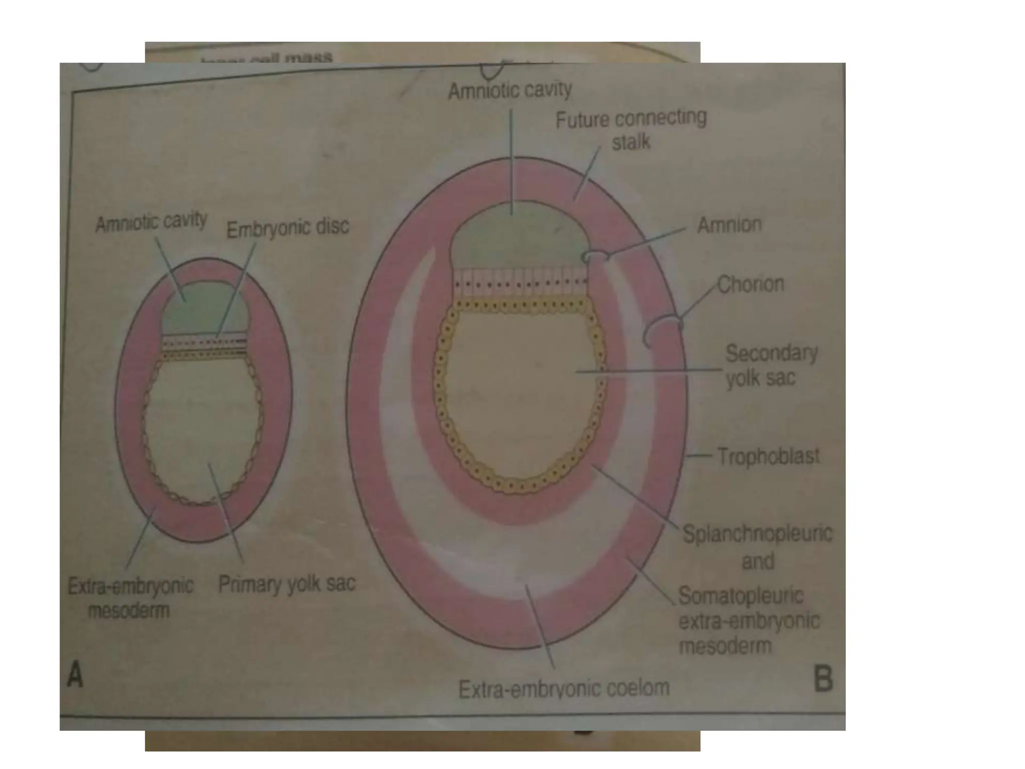
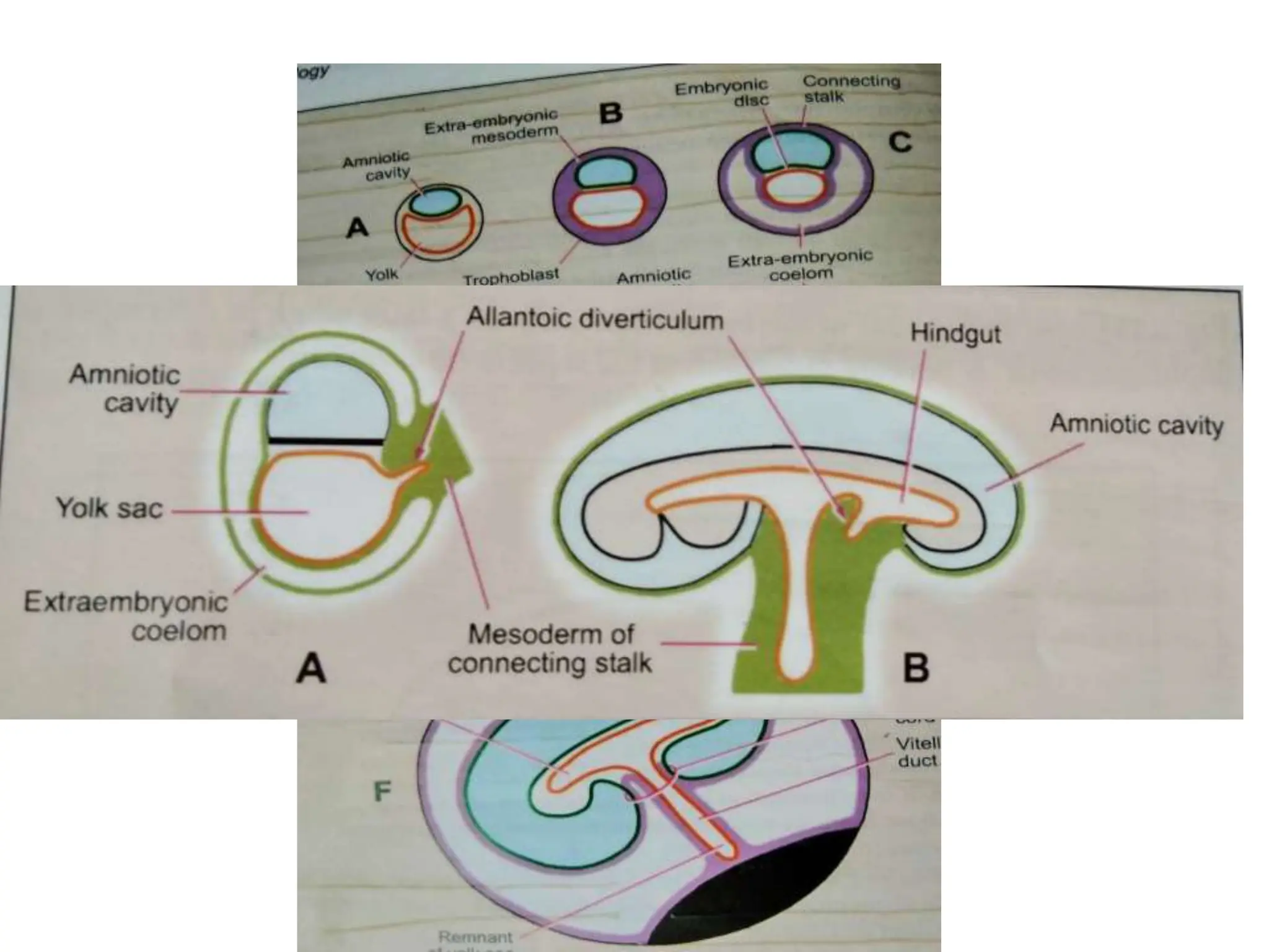
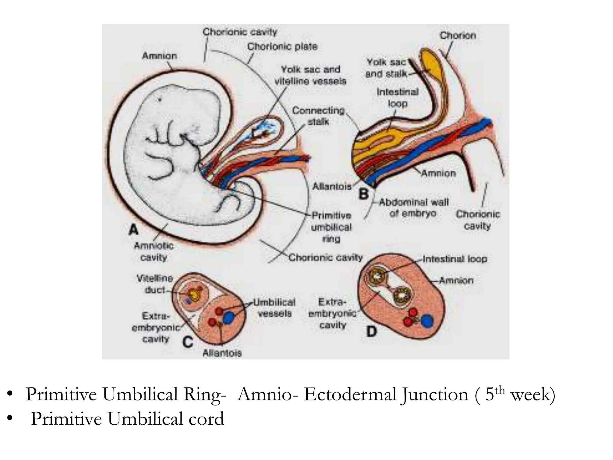
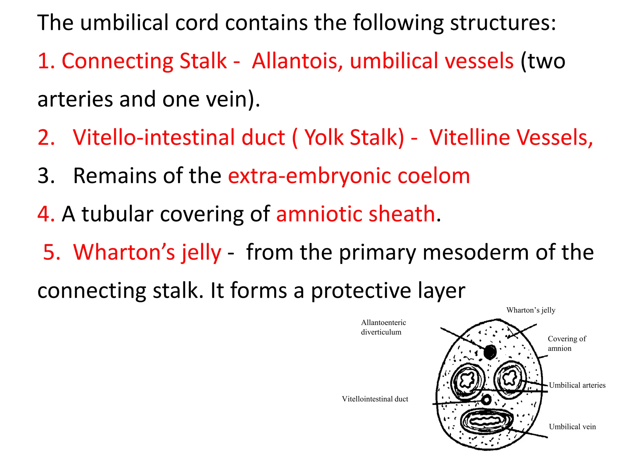
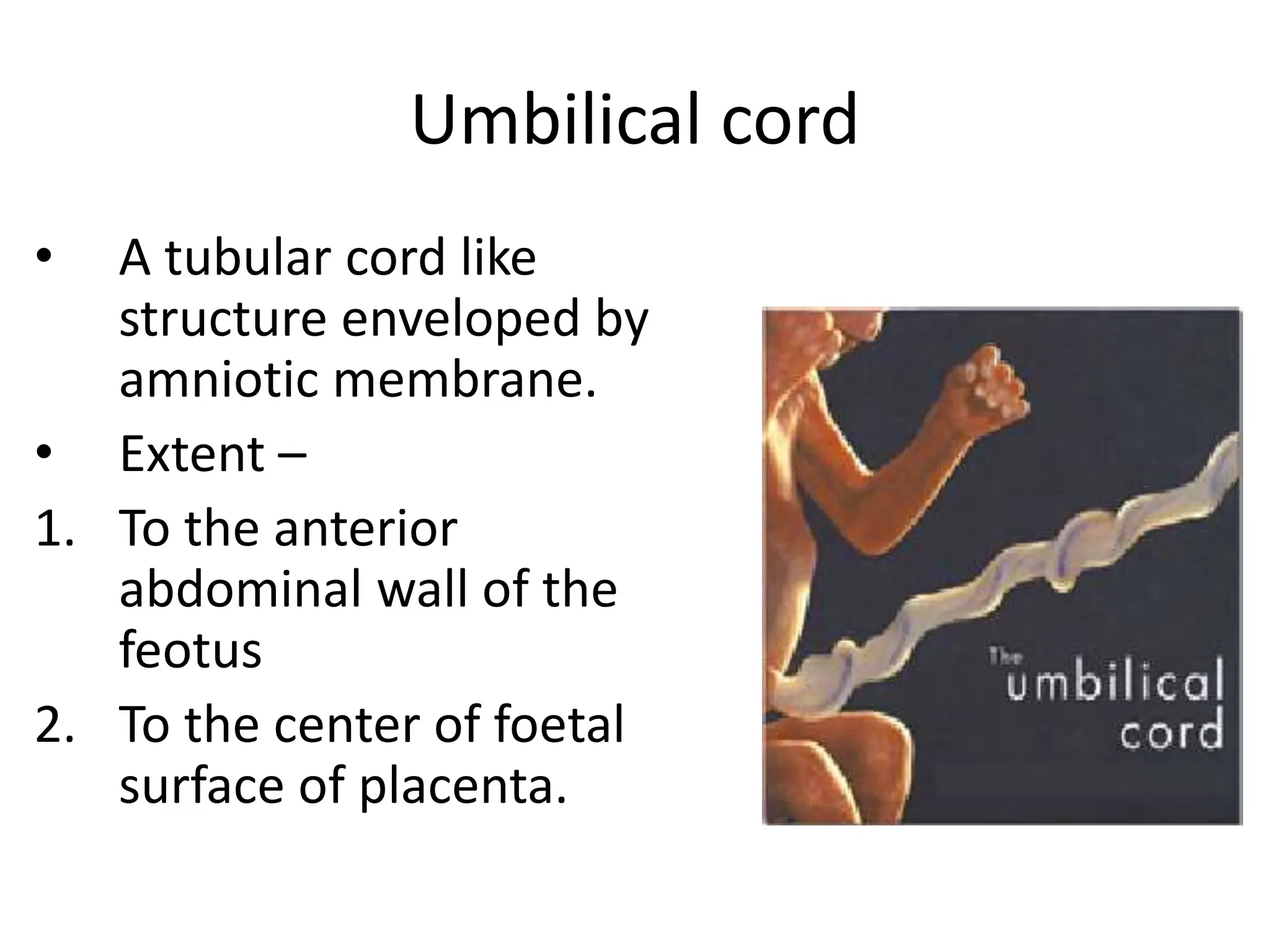
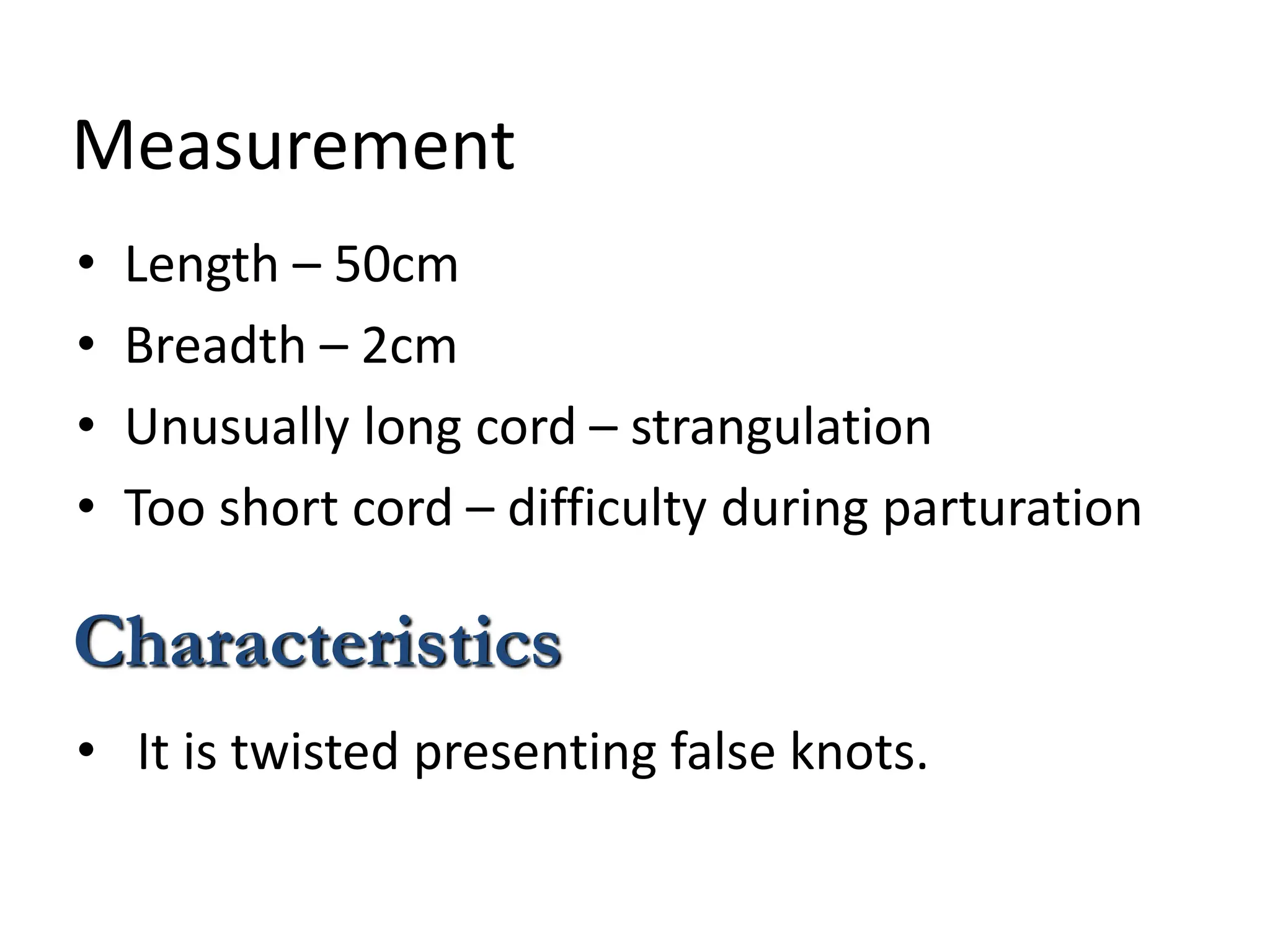
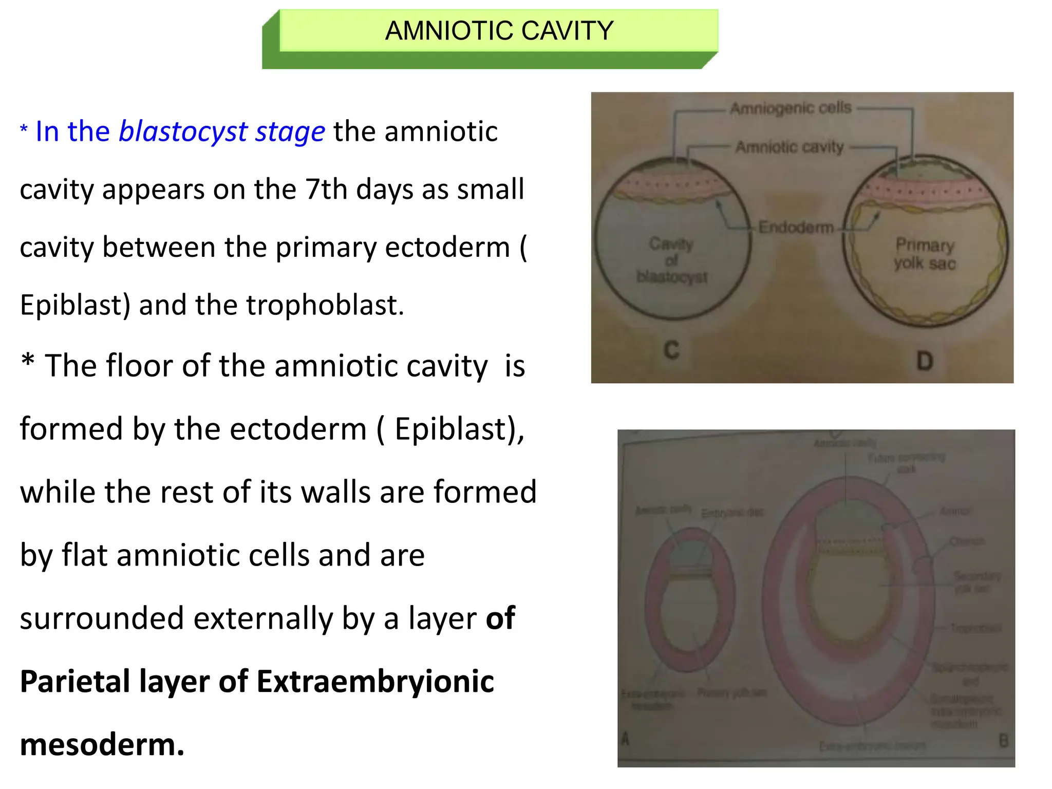
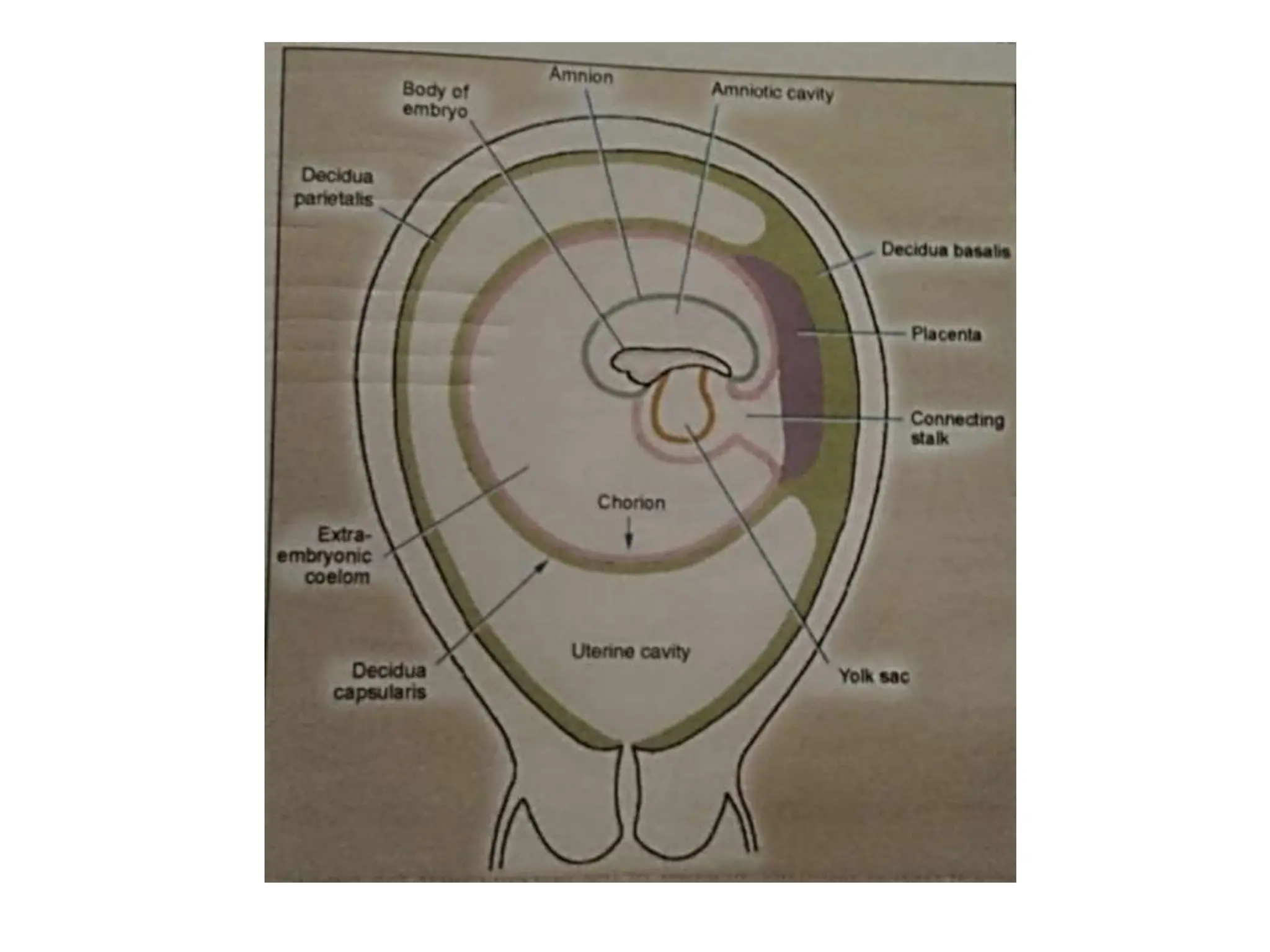
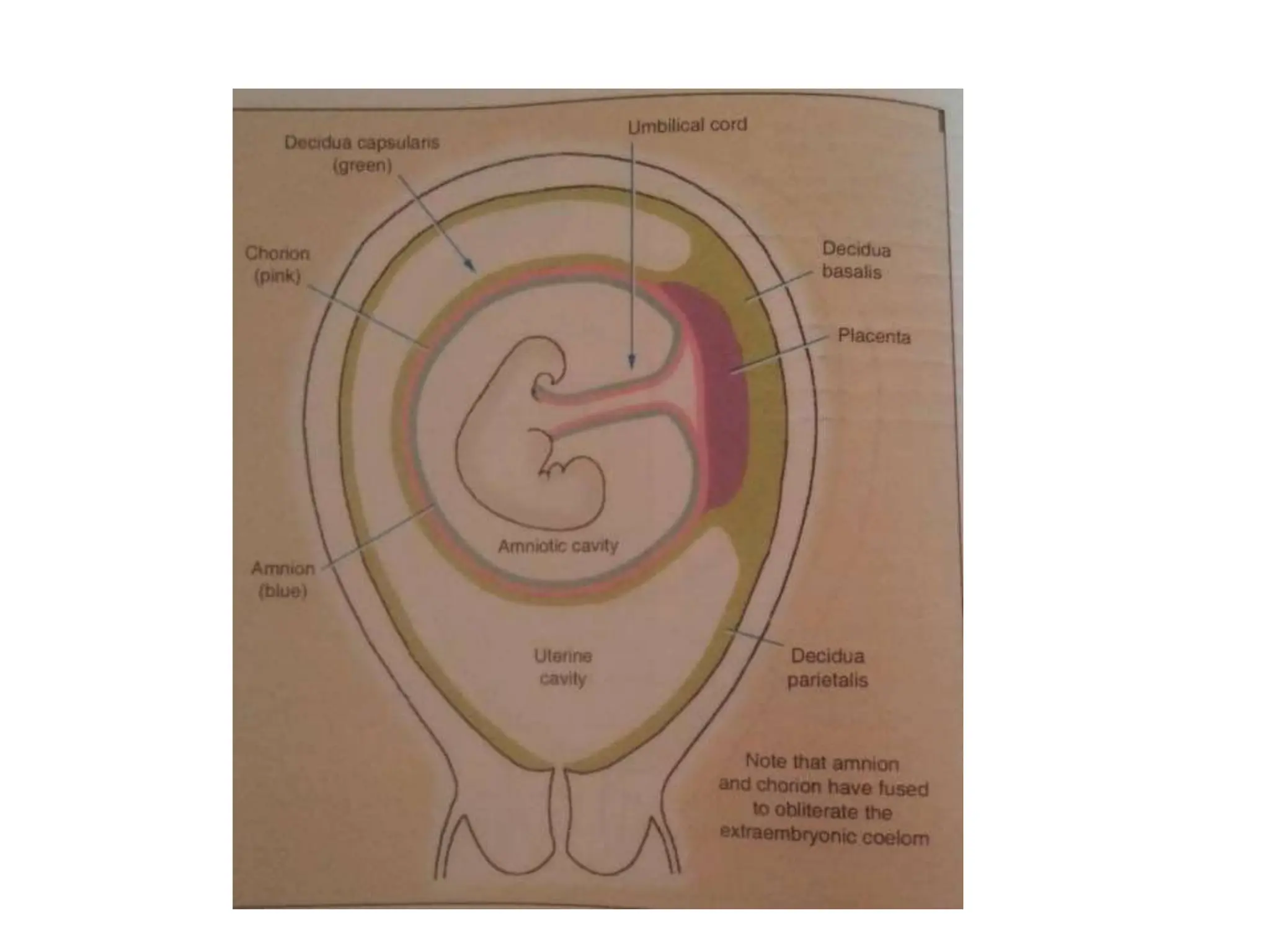
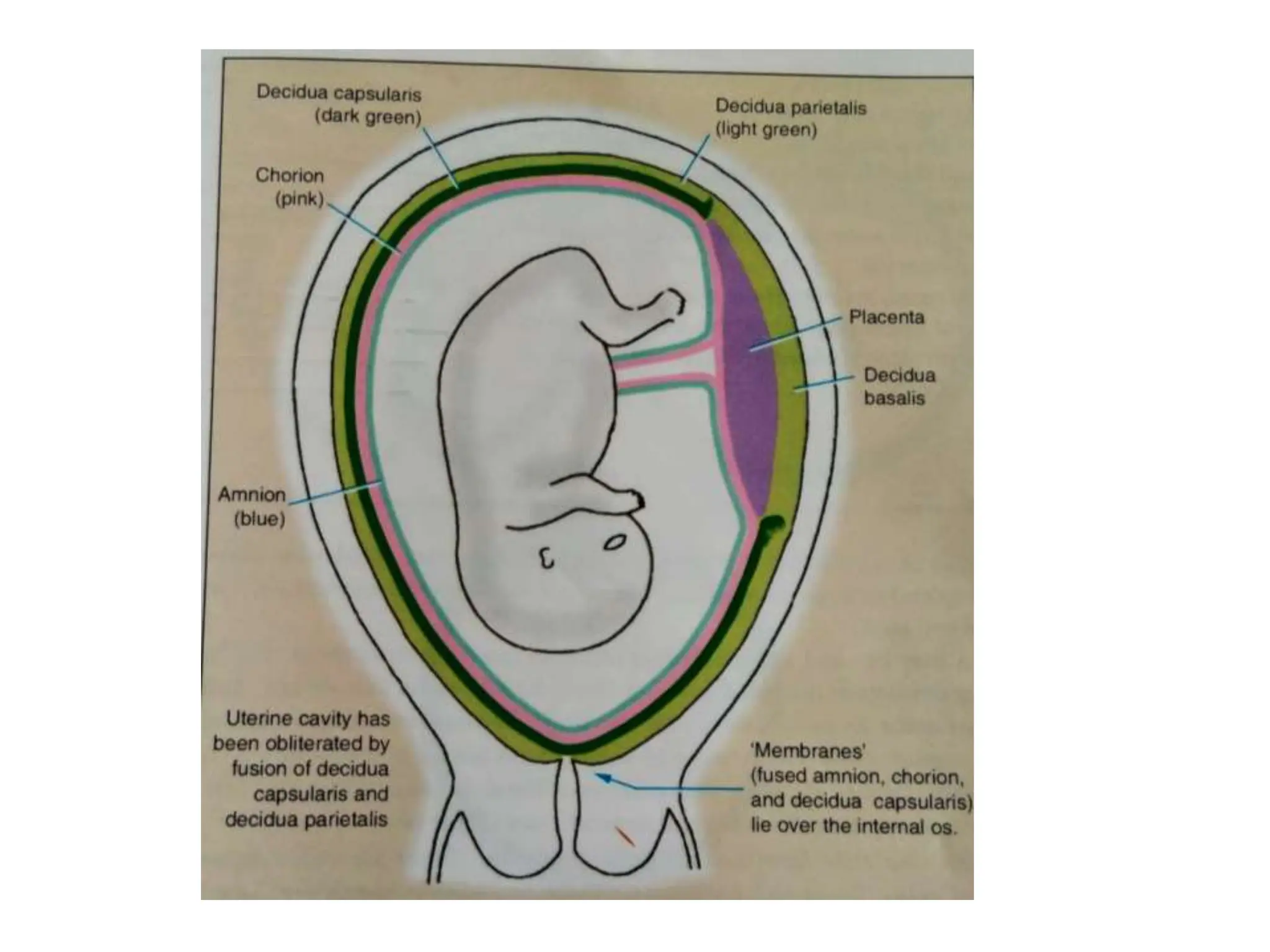
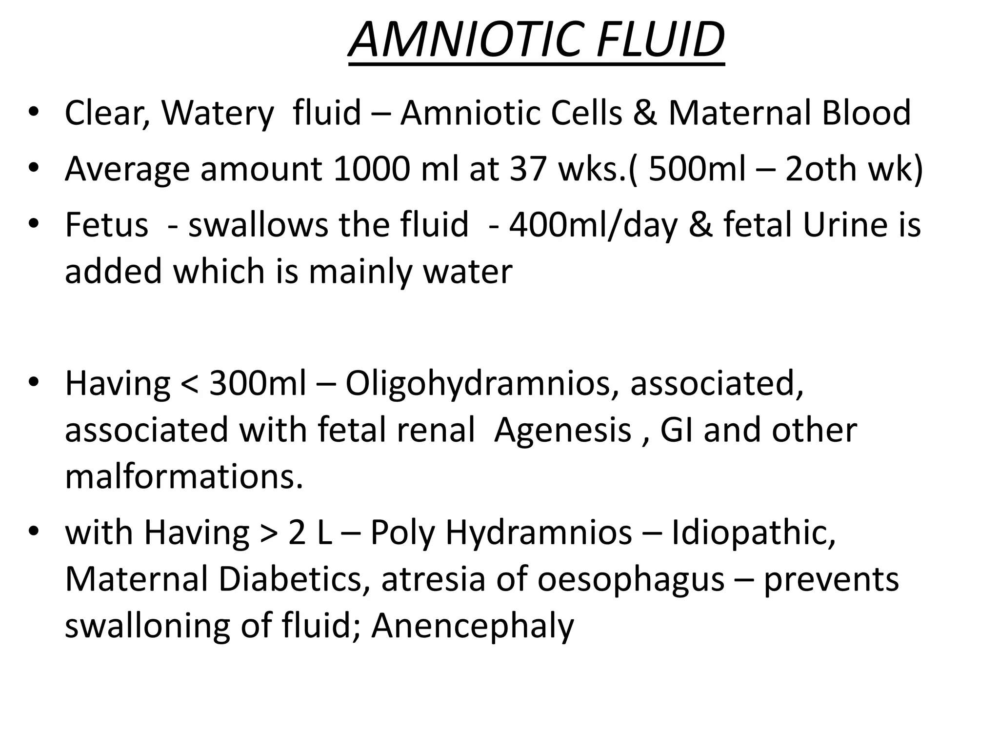
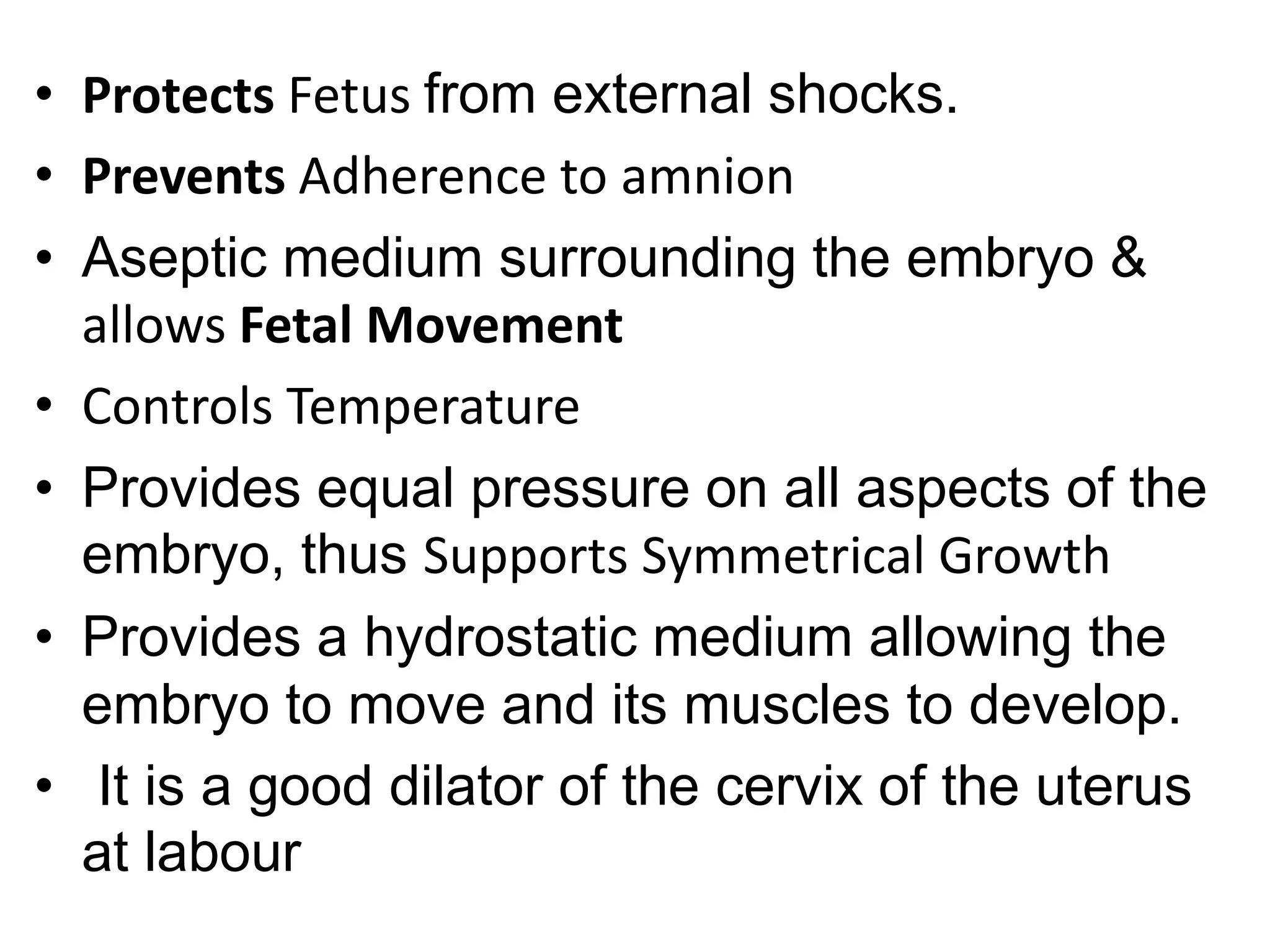
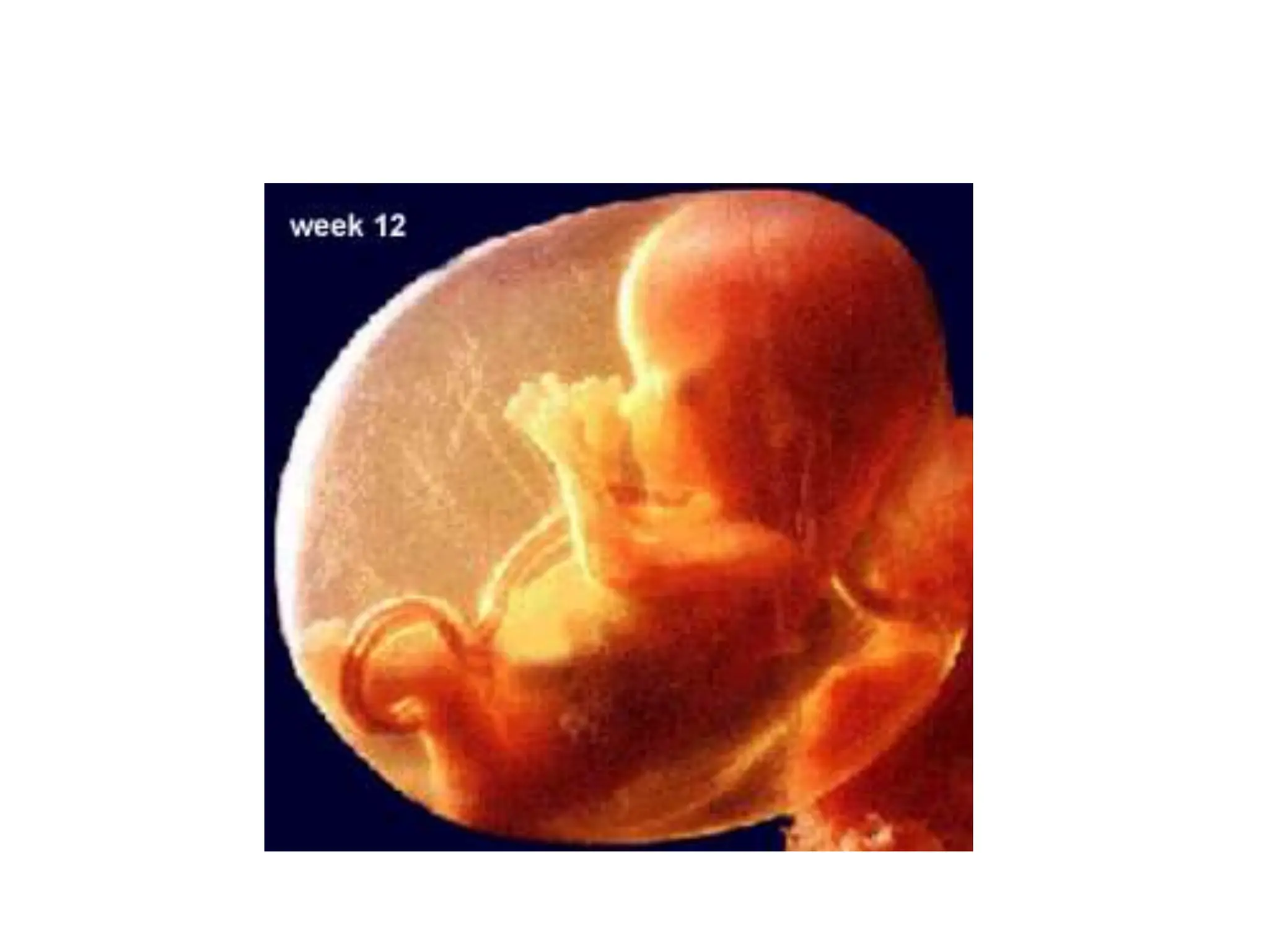
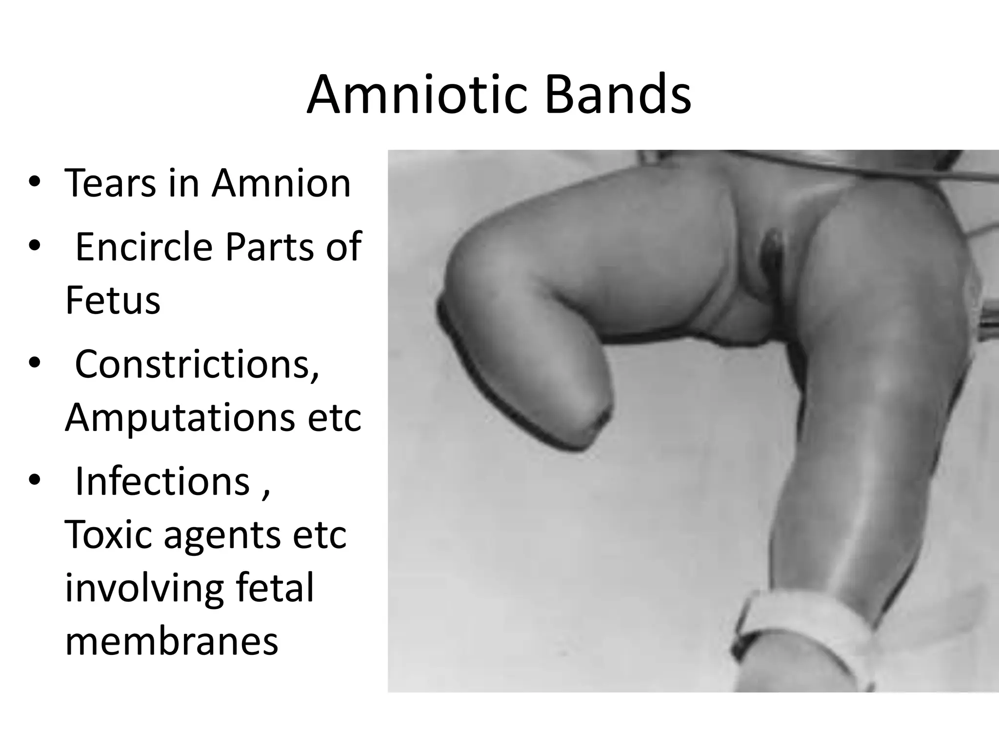
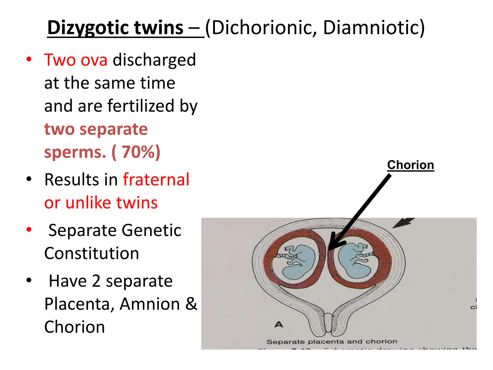
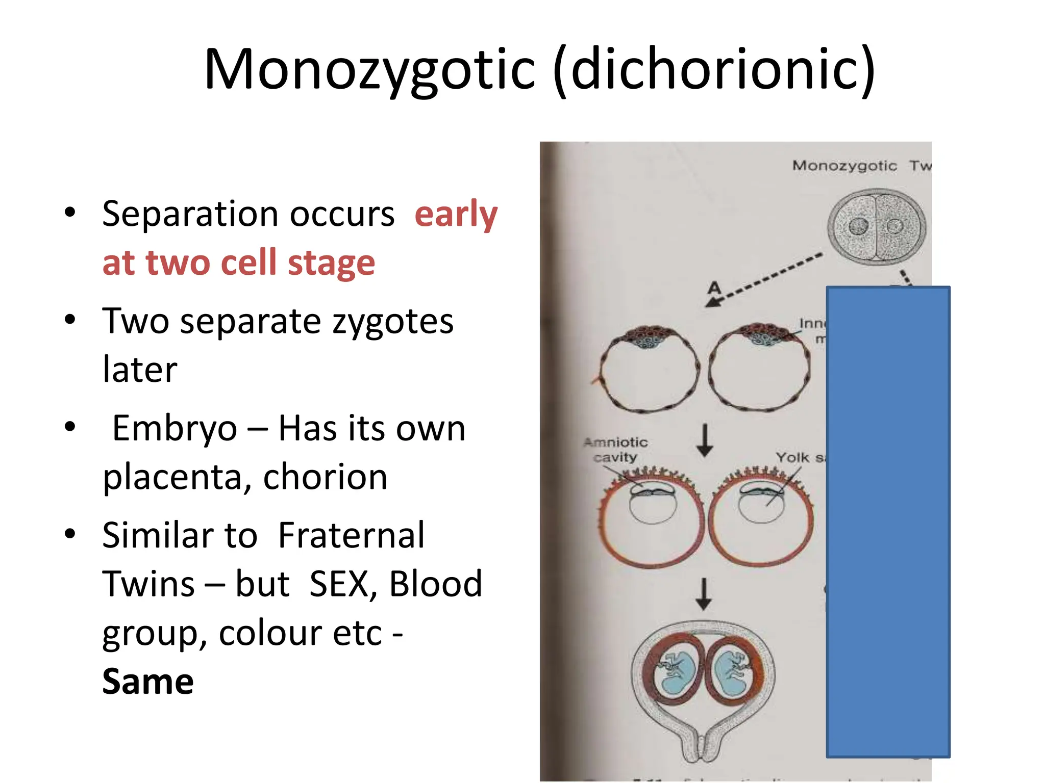
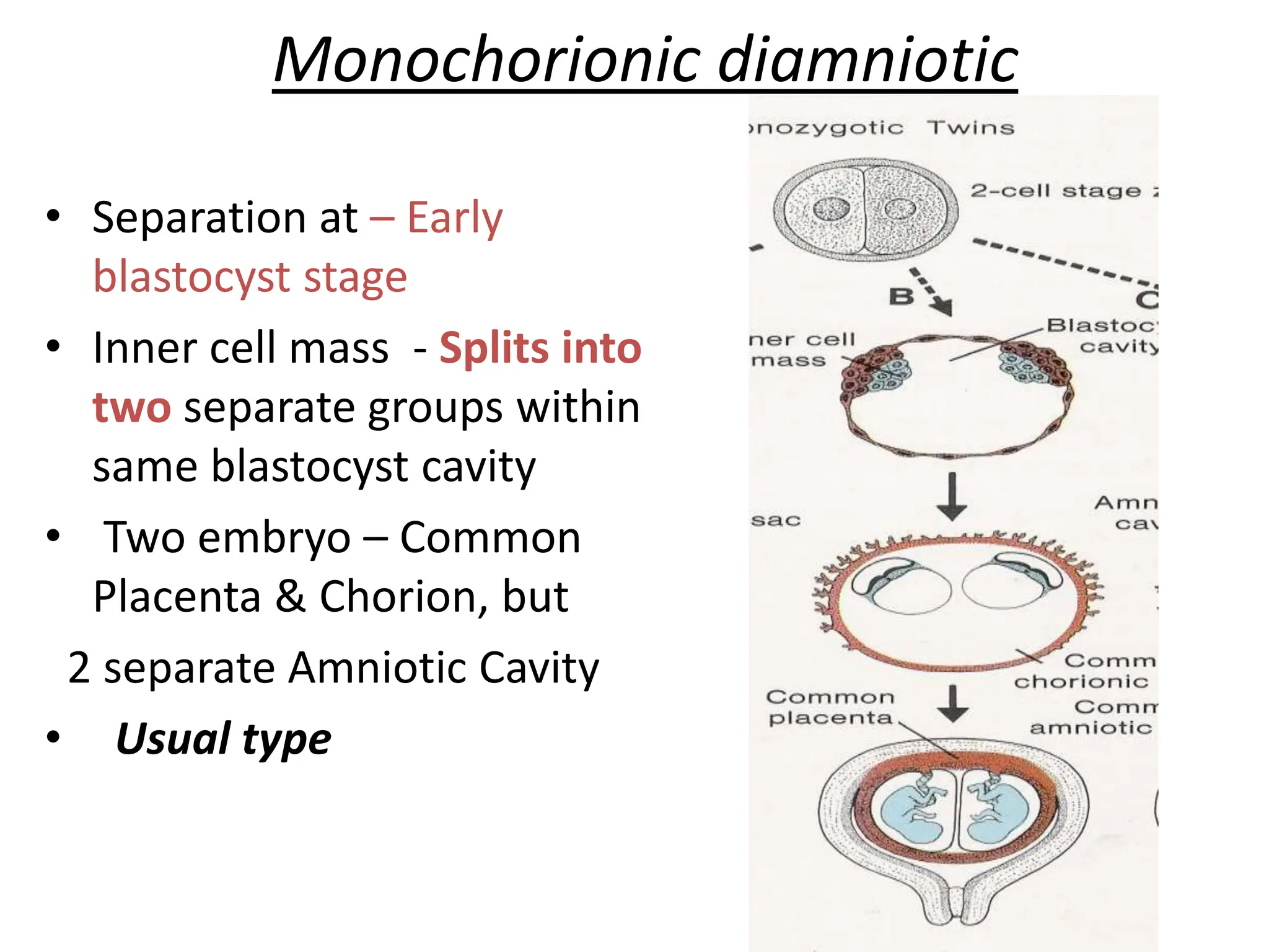
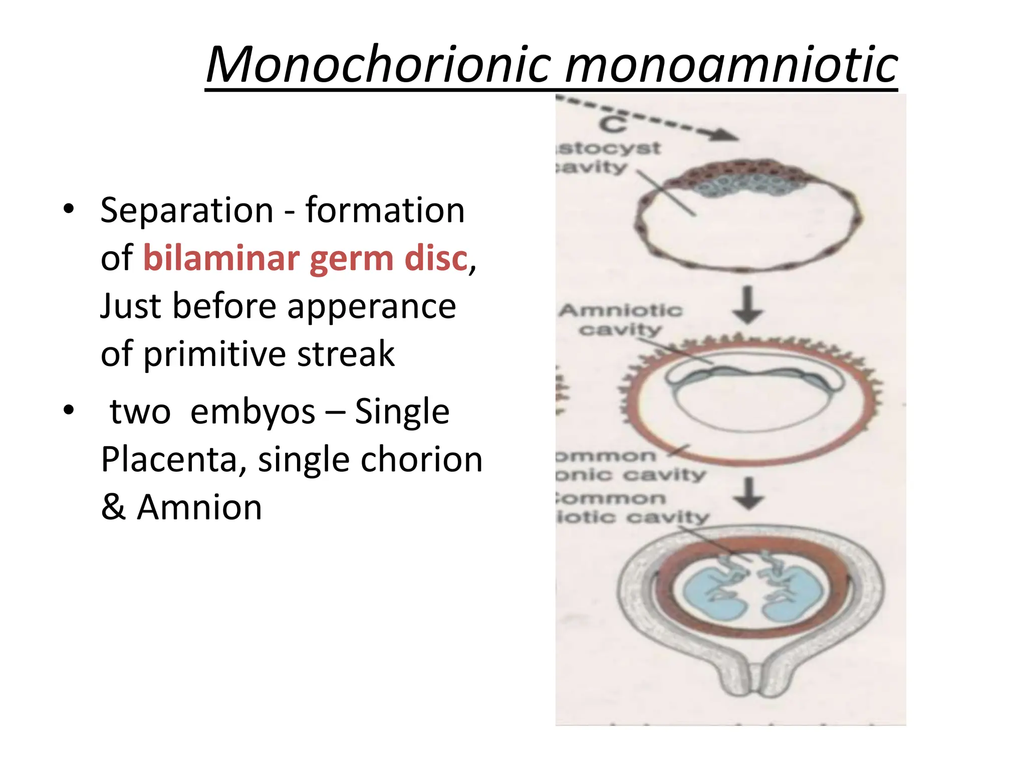
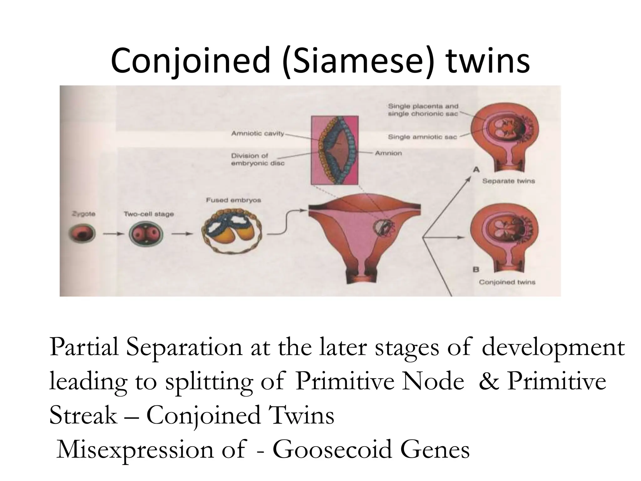
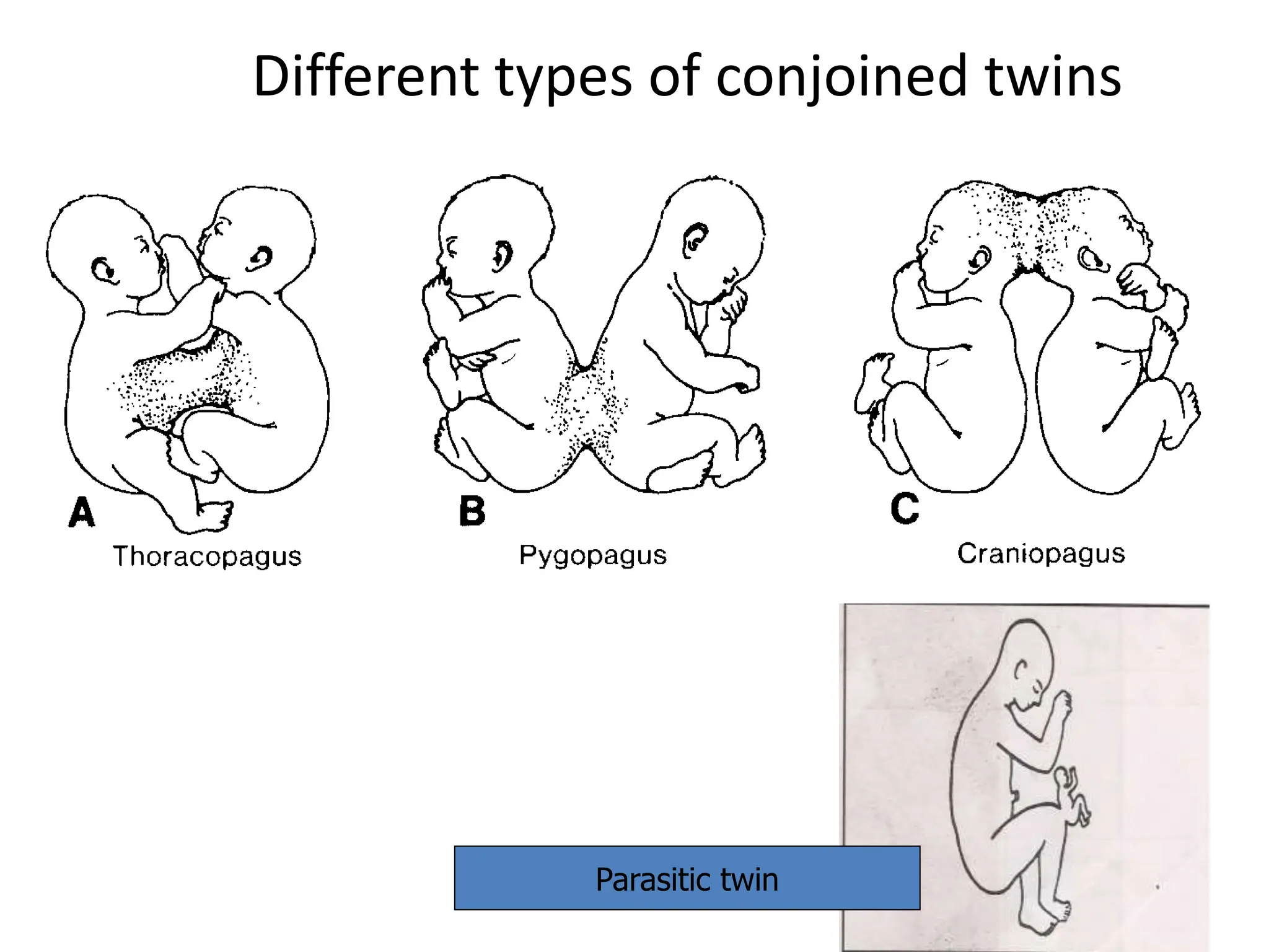
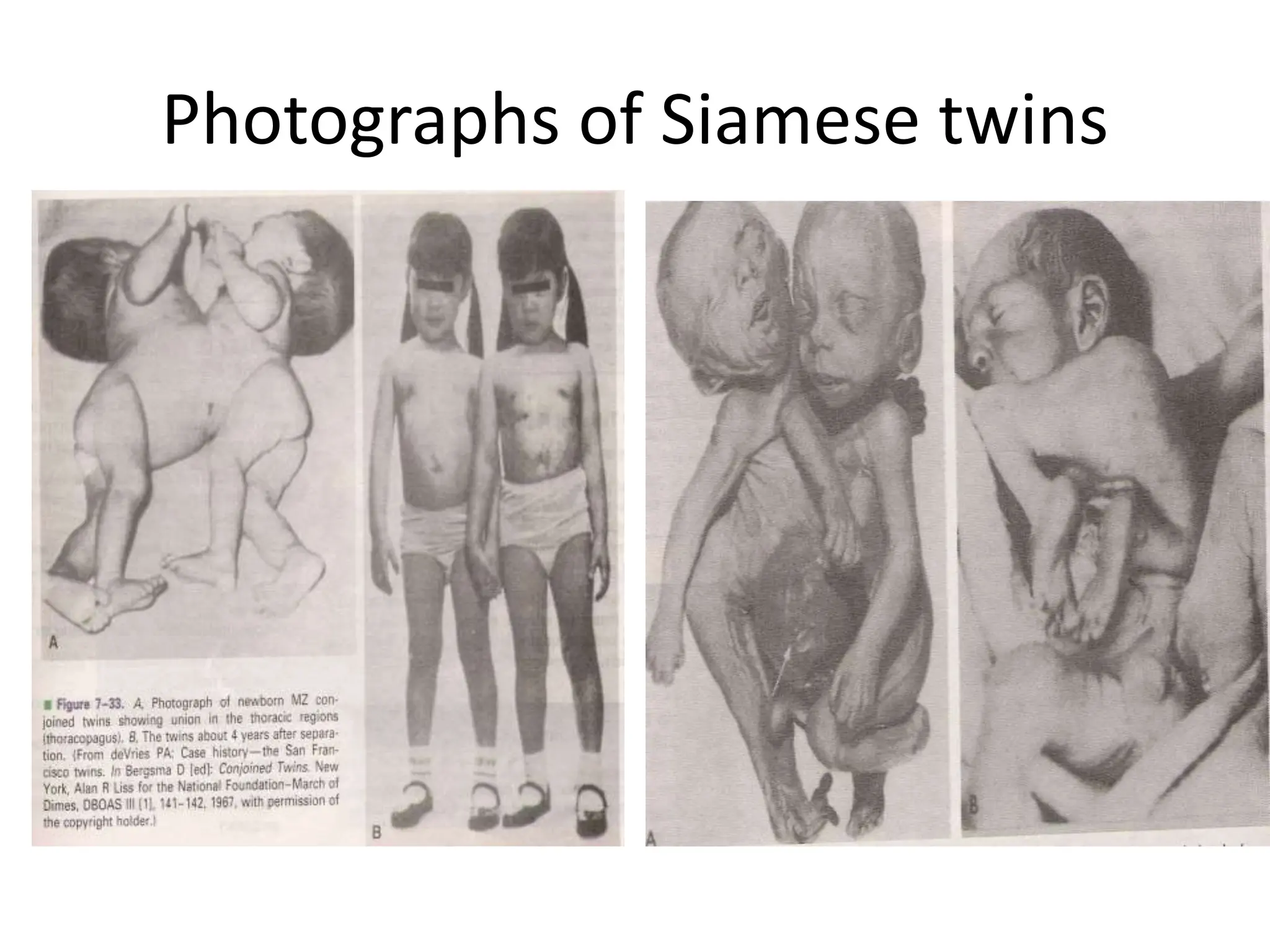
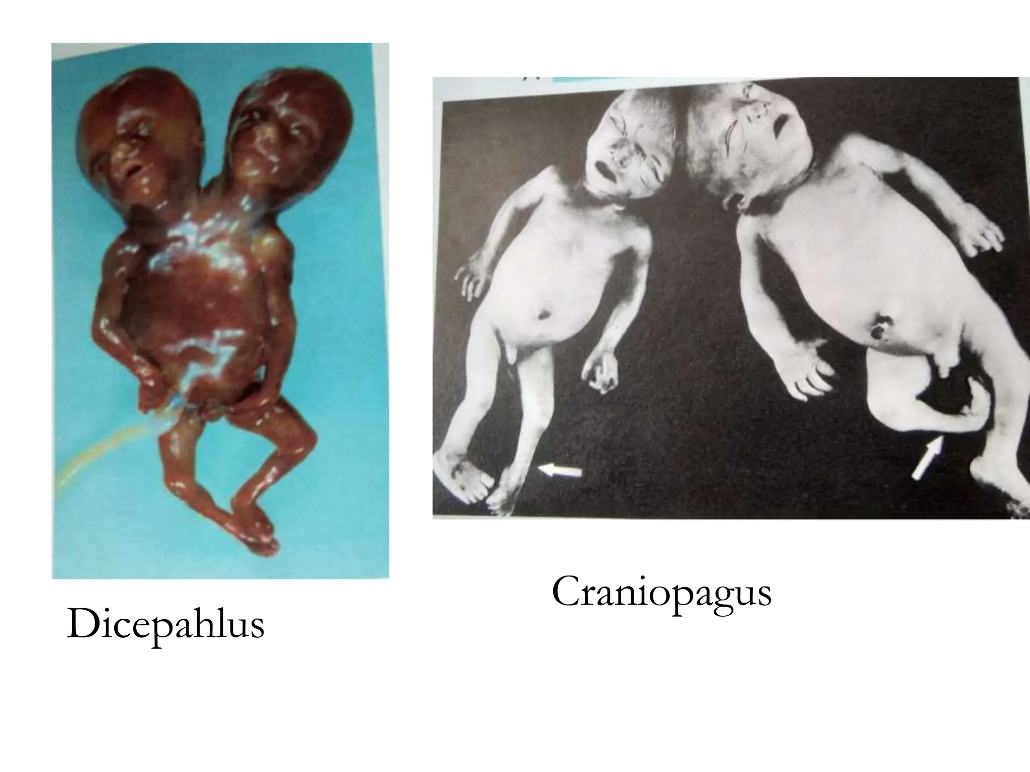
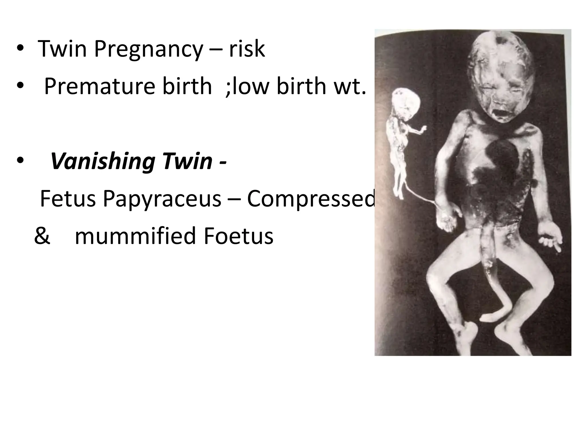
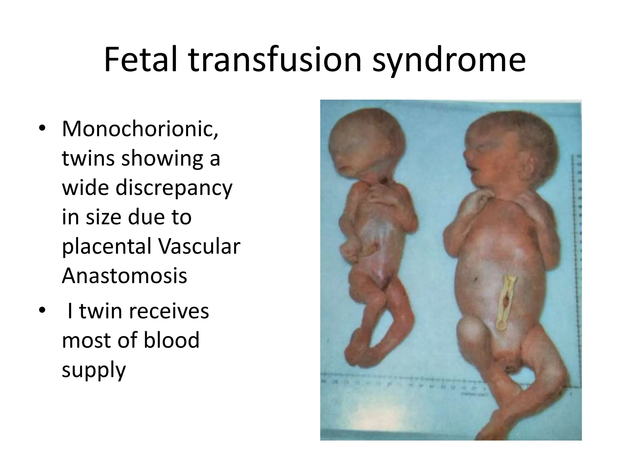
5. Allantoenteric
diverticulum
Wharton’s jelly
Covering of
amnion
Umbilical arteries
Umbilical vein
Vitellointestinal duct
The umbilical cord contains the following structures:
1. Connecting Stalk - Allantois, umbilical vessels (two
arteries and one vein).
2. Vitello-intestinal duct ( Yolk Stalk) - Vitelline Vessels,
3. Remains of the extra-embryonic coelom
4. A tubular covering of amniotic sheath.
5. Wharton’s jelly - from the primary mesoderm of the
connecting stalk. It forms a protective layer
6. Umbilical cord
• A tubular cord like
structure enveloped by
amniotic membrane.
• Extent –
1. To the anterior
abdominal wall of the
feotus
2. To the center of foetal
surface of placenta.
7. Measurement
• Length – 50cm
• Breadth – 2cm
• Unusually long cord – strangulation
• Too short cord – difficulty during parturation
• It is twisted presenting false knots.
Characteristics
8.
9.
10. Velamentous attachment of the cord
Battledore placenta
False knot True knot
ANOMAILIES OF THE CORD
1. Velamentous attachement: cord is attached to the amnion
2. Battledore placenta: cord is attached to the edge the placenta.
4. Abnormally long or short cord (normally it is 50 cm long).
5. True knot.
6. Long Cord - Encircle head of foetus
7.Short cord – Difficulty in delivery
11. AMNIOTIC CAVITY
* In the blastocyst stage the amniotic
cavity appears on the 7th days as small
cavity between the primary ectoderm (
Epiblast) and the trophoblast.
* The floor of the amniotic cavity is
formed by the ectoderm ( Epiblast),
while the rest of its walls are formed
by flat amniotic cells and are
surrounded externally by a layer of
Parietal layer of Extraembryionic
mesoderm.
15. AMNIOTIC FLUID
• Clear, Watery fluid – Amniotic Cells & Maternal Blood
• Average amount 1000 ml at 37 wks.( 500ml – 2oth wk)
• Fetus - swallows the fluid - 400ml/day & fetal Urine is
added which is mainly water
• Having < 300ml – Oligohydramnios, associated,
associated with fetal renal Agenesis , GI and other
malformations.
• with Having > 2 L – Poly Hydramnios – Idiopathic,
Maternal Diabetics, atresia of oesophagus – prevents
swalloning of fluid; Anencephaly
16. • Protects Fetus from external shocks.
• Prevents Adherence to amnion
• Aseptic medium surrounding the embryo &
allows Fetal Movement
• Controls Temperature
• Provides equal pressure on all aspects of the
embryo, thus Supports Symmetrical Growth
• Provides a hydrostatic medium allowing the
embryo to move and its muscles to develop.
• It is a good dilator of the cervix of the uterus
at labour
17.
18. Amniotic Bands
• Tears in Amnion
• Encircle Parts of
Fetus
• Constrictions,
Amputations etc
• Infections ,
Toxic agents etc
involving fetal
membranes
20. Dizygotic twins – (Dichorionic, Diamniotic)
• Two ova discharged
at the same time
and are fertilized by
two separate
sperms. ( 70%)
• Results in fraternal
or unlike twins
• Separate Genetic
Constitution
• Have 2 separate
Placenta, Amnion &
Chorion
Chorion
21. Monozygotic twins ( Identical)
• Two embryos derived from a single fertilized
ovum
• Splitting of zygote at various levels of
development
• Identical offsprings with similar genetic
constitution, appearance, structure and
belong to same sex
22. Monozygotic (dichorionic)
• Separation occurs early
at two cell stage
• Two separate zygotes
later
• Embryo – Has its own
placenta, chorion
• Similar to Fraternal
Twins – but SEX, Blood
group, colour etc -
Same
23. Monochorionic diamniotic
• Separation at – Early
blastocyst stage
• Inner cell mass - Splits into
two separate groups within
same blastocyst cavity
• Two embryo – Common
Placenta & Chorion, but
2 separate Amniotic Cavity
• Usual type
24. Monochorionic monoamniotic
• Separation - formation
of bilaminar germ disc,
Just before apperance
of primitive streak
• two embyos – Single
Placenta, single chorion
& Amnion
25. Conjoined (Siamese) twins
Partial Separation at the later stages of development
leading to splitting of Primitive Node & Primitive
Streak – Conjoined Twins
Misexpression of - Goosecoid Genes
30. Fetal transfusion syndrome
• Monochorionic,
twins showing a
wide discrepancy
in size due to
placental Vascular
Anastomosis
• I twin receives
most of blood
supply