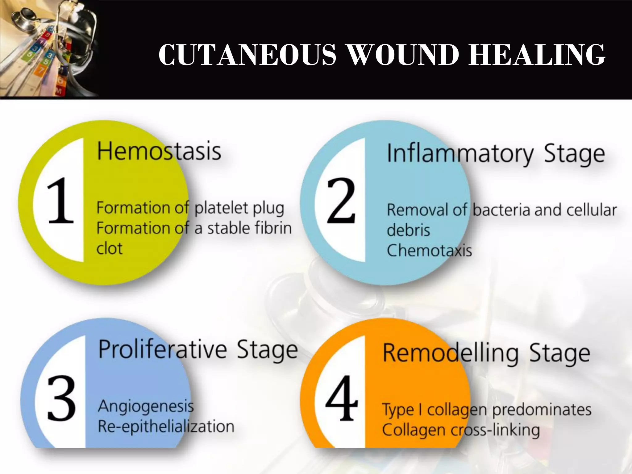
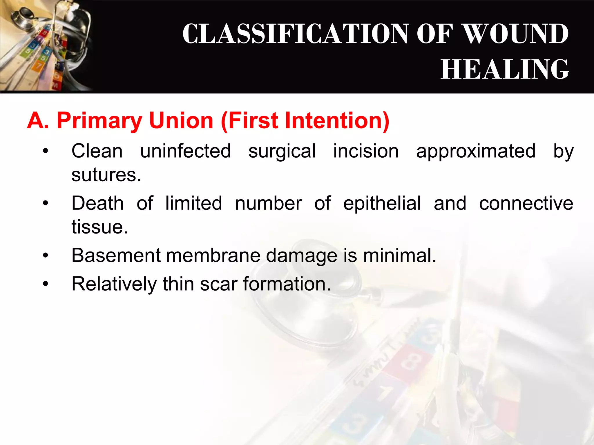
CUTANEOUS WOUND
HEALING
BY
FIRST AND SECOND
INTENTION
By:
Dr. Kinz
CUTANEOUS WOUND HEALING
STAGES OF NORMAL CUTANEOUS
WOUND HEALING
CLASSIFICATION OF WOUND
HEALING
A. Primary Union (First Intention)
• Clean uninfected surgical incision approximated by
sutures.
• Death of limited number of epithelial and connective
tissue.
• Basement membrane damage is minimal.
• Relatively thin scar formation.
B. Secondary Union (Second Intention)
• Larger defects, the edges are not attached properly,
formation of granular tissue.
• Extensive loss of cells and tissues with intense
inflammatory reaction and collagen formation.
• Fibrin clot is larger, there is more exudate and necrotic
debris.
• Granular tissue substantial scar formation which
contracts.
• Involves wound contraction.
FORMATION OF BLOOD CLOT
• Activation of coagulation pathways leading to clot
formation which prevents bleeding
• Release of Vascular Endothelial Growth Factor(VEGF)
with increased permeablity and edema
• Dehydration at the external surface of clot makes a scab
that covers the wound.
• Larger fibrin clot is seen in healing by second intention
with more exudate and necrotic debris in the wound.
• Within 24 hours, neutrophils appear at the margins of
the incision.
FORMATION OF
GRANULATION TISSUE
• Hallmark of repair.
• Occurs in the first 24 to 72 hours due to fibroblast and
vascular endothelial cell proliferation.
• Soft, pink and granular appearance on the surface of
wounds.
• The newly formed blood vessels are leaky leading to
passage of plasma proteins and fluid into extravascular
space – edematous in appearance.
• By 5 to 7 days the granulation tissue fills up the wound area
(more pronounced effect in healing by second intension).
CELL PROLIFERATION AND
COLLAGEN DEPOSITION
• Neutrophils are replaced by macrophages by 48 to 96 hours
which play role in clearing extracellular debris, fibrin and
other foreign material, promoting angiogenesis and
extracellular matrix deposition
• Fibroblast migration by chemokines and their subsequent
proliferation
• Deposition of collagen at the margins of the incision –
vertically oriented in primary intention and horizontally
oriented in secondary intention
• In 24 to 48 hours proliferation and migration of epithelial
cells adjacent to wound, migration to the margins of dermis,
depositing basement membrane components.
• Epithelial cell proliferation thickens.
• collagen fibrils (type I collagen) become more abundant and
bridges the incision.
EPITHELIZATION
SCAR FORMATION
• By second week, there is increased accumulation of
collagen with regression of vasculature.
• The granulation tissue is converted into pale, avascular scar
composed of spindle shaped fibroblasts, dense collagen,
also there is elastic tissue, and other extracellular matrix
components.
WOUND CONTRACTION
• Primarily occurs in healing by secondary intention.
• Formation of myofibroblasts from the tissue fibroblasts.
• These cells contract in the wound and produce large amount
of extracellular martrix components.
CONNECTIVE TISSUE
REMODELING
• The balance between extracellular matrix synthesis and
degradation results in remodeling of connective tissue
framework
• Matrix metalloproteinases e.g. interstitial collagenases,
gelatinases degrade the ECM, and are inhibited by Tissue
Inhibitors of metalloproteinases .
RECOVERY OF TENSILE
STRENGTH
• Tensile strength in healing wound is provided my fibrillar
collagens (type I collagen) with cross linking and increased
fibre size.
• Sutures are removed typically at first week, the wound
strength is 10% of normal.
• By 3rd month the strength plateaus upto 70 to 80% of
normal.
Cutaneous wound healing

Cutaneous wound healing

  • 1.
    CUTANEOUS WOUND HEALING BY FIRST ANDSECOND INTENTION By: Dr. Kinz
  • 2.
  • 3.
    STAGES OF NORMALCUTANEOUS WOUND HEALING
  • 4.
    CLASSIFICATION OF WOUND HEALING A.Primary Union (First Intention) • Clean uninfected surgical incision approximated by sutures. • Death of limited number of epithelial and connective tissue. • Basement membrane damage is minimal. • Relatively thin scar formation.
  • 5.
    B. Secondary Union(Second Intention) • Larger defects, the edges are not attached properly, formation of granular tissue. • Extensive loss of cells and tissues with intense inflammatory reaction and collagen formation. • Fibrin clot is larger, there is more exudate and necrotic debris. • Granular tissue substantial scar formation which contracts. • Involves wound contraction.
  • 6.
    FORMATION OF BLOODCLOT • Activation of coagulation pathways leading to clot formation which prevents bleeding • Release of Vascular Endothelial Growth Factor(VEGF) with increased permeablity and edema • Dehydration at the external surface of clot makes a scab that covers the wound. • Larger fibrin clot is seen in healing by second intention with more exudate and necrotic debris in the wound.
  • 7.
    • Within 24hours, neutrophils appear at the margins of the incision.
  • 8.
    FORMATION OF GRANULATION TISSUE •Hallmark of repair. • Occurs in the first 24 to 72 hours due to fibroblast and vascular endothelial cell proliferation. • Soft, pink and granular appearance on the surface of wounds. • The newly formed blood vessels are leaky leading to passage of plasma proteins and fluid into extravascular space – edematous in appearance. • By 5 to 7 days the granulation tissue fills up the wound area (more pronounced effect in healing by second intension).
  • 10.
    CELL PROLIFERATION AND COLLAGENDEPOSITION • Neutrophils are replaced by macrophages by 48 to 96 hours which play role in clearing extracellular debris, fibrin and other foreign material, promoting angiogenesis and extracellular matrix deposition • Fibroblast migration by chemokines and their subsequent proliferation • Deposition of collagen at the margins of the incision – vertically oriented in primary intention and horizontally oriented in secondary intention
  • 11.
    • In 24to 48 hours proliferation and migration of epithelial cells adjacent to wound, migration to the margins of dermis, depositing basement membrane components. • Epithelial cell proliferation thickens. • collagen fibrils (type I collagen) become more abundant and bridges the incision.
  • 12.
  • 13.
    SCAR FORMATION • Bysecond week, there is increased accumulation of collagen with regression of vasculature. • The granulation tissue is converted into pale, avascular scar composed of spindle shaped fibroblasts, dense collagen, also there is elastic tissue, and other extracellular matrix components.
  • 14.
    WOUND CONTRACTION • Primarilyoccurs in healing by secondary intention. • Formation of myofibroblasts from the tissue fibroblasts. • These cells contract in the wound and produce large amount of extracellular martrix components.
  • 16.
    CONNECTIVE TISSUE REMODELING • Thebalance between extracellular matrix synthesis and degradation results in remodeling of connective tissue framework • Matrix metalloproteinases e.g. interstitial collagenases, gelatinases degrade the ECM, and are inhibited by Tissue Inhibitors of metalloproteinases .
  • 17.
    RECOVERY OF TENSILE STRENGTH •Tensile strength in healing wound is provided my fibrillar collagens (type I collagen) with cross linking and increased fibre size. • Sutures are removed typically at first week, the wound strength is 10% of normal. • By 3rd month the strength plateaus upto 70 to 80% of normal.