1. Wound healing is the body's complex biological response to tissue injury. It involves regeneration and repair processes to restore tissue integrity and function.
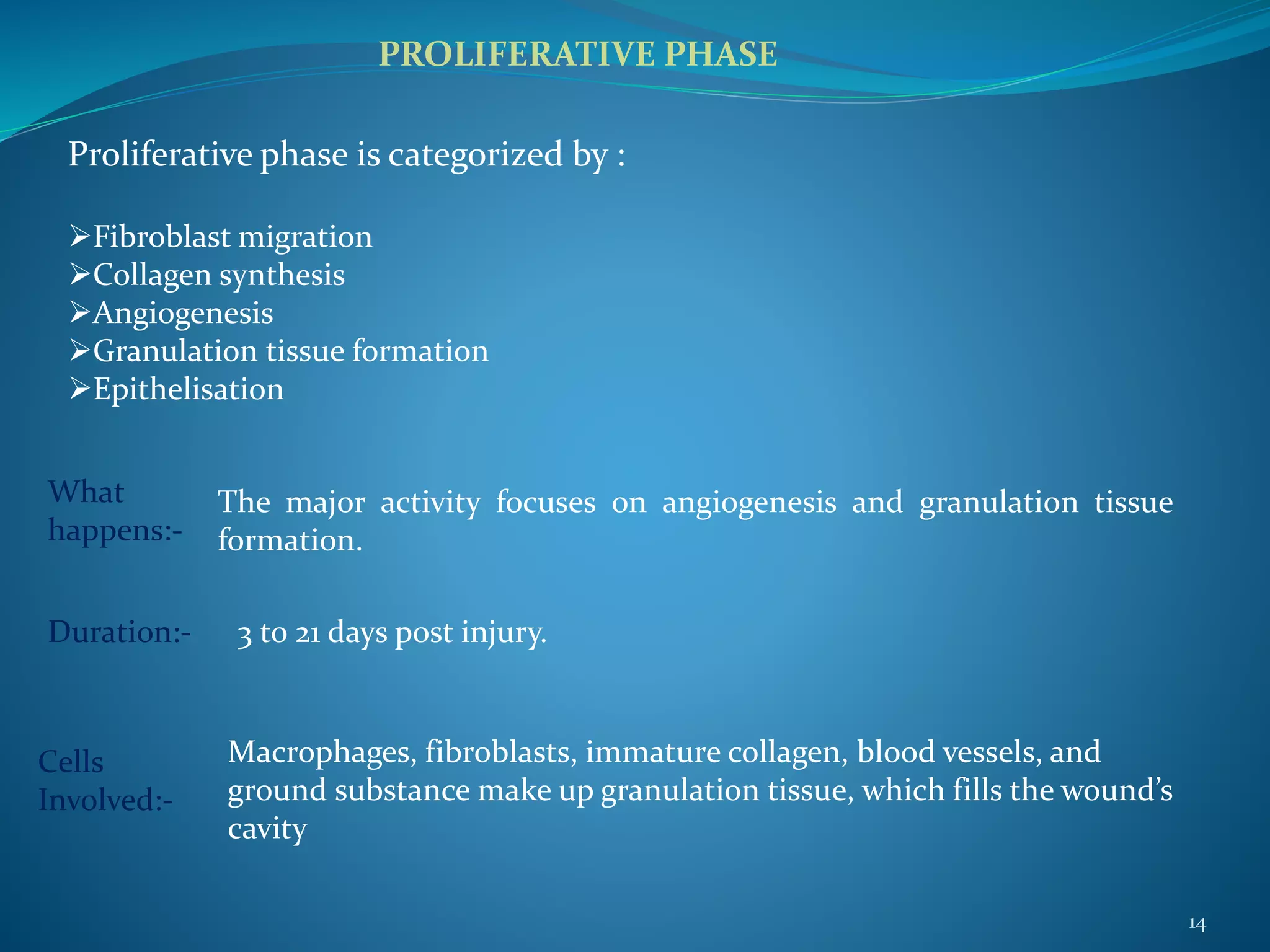
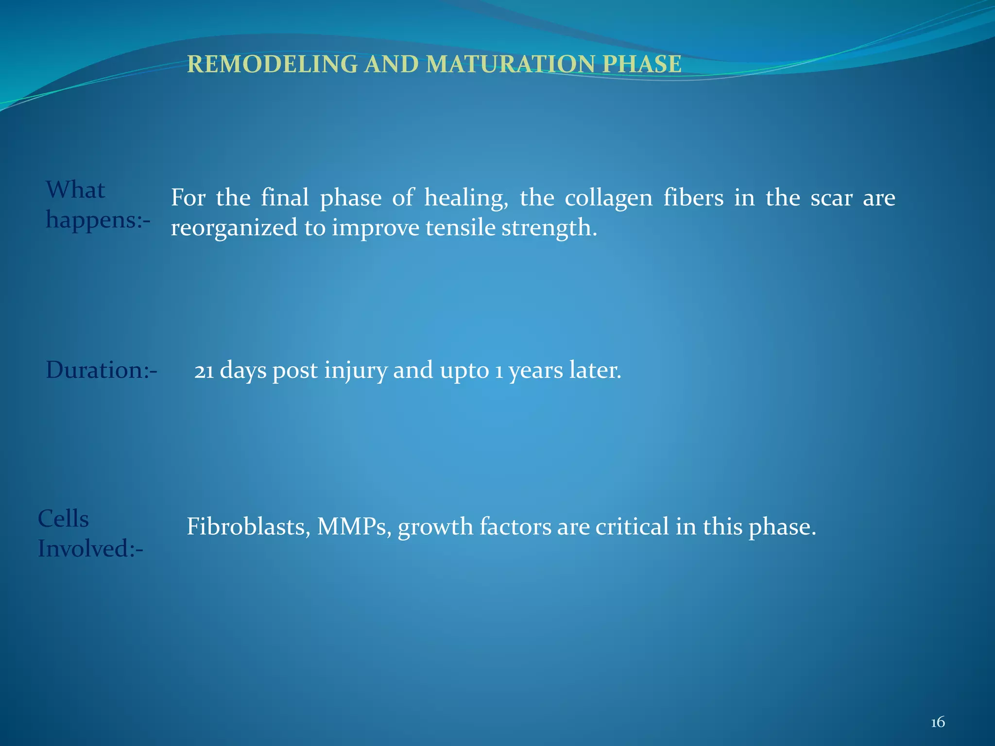
2. There are four main phases of wound healing: hemostasis, inflammation, proliferation, and remodeling. Various cell types and mediators are involved in each phase to clean the wound and promote new tissue growth.
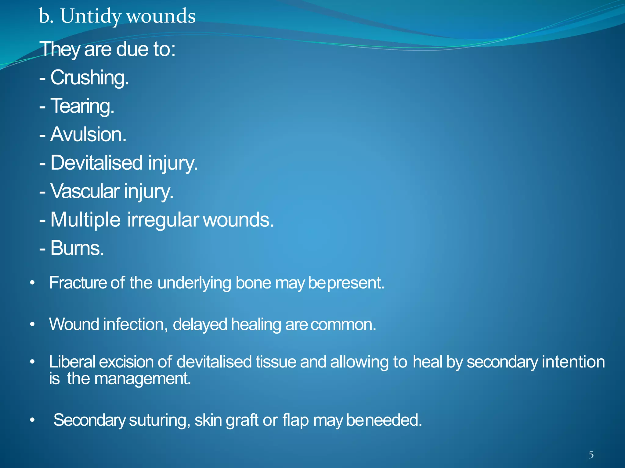
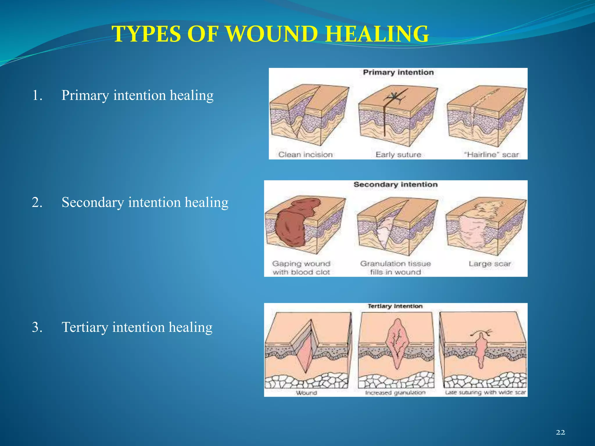
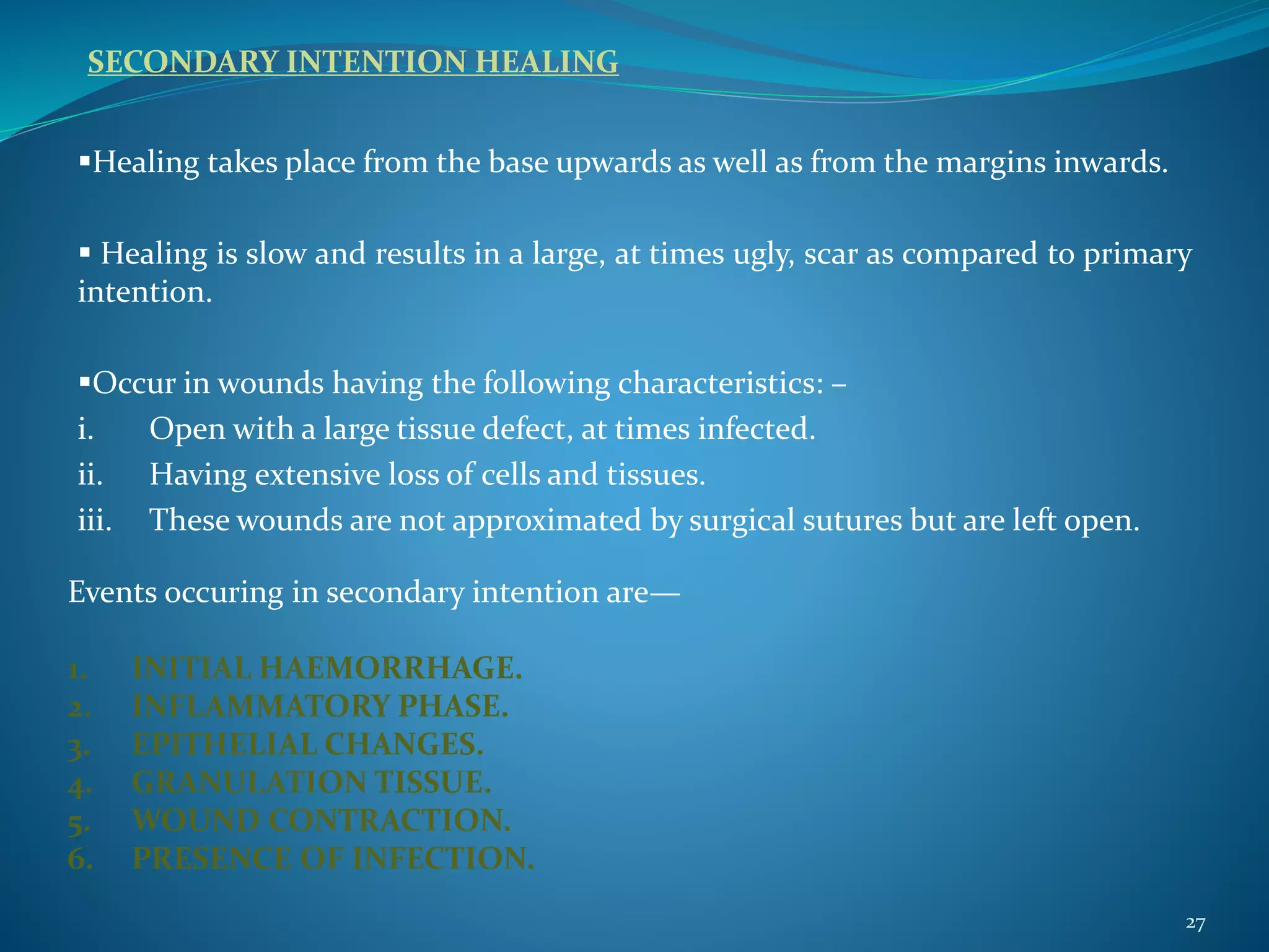
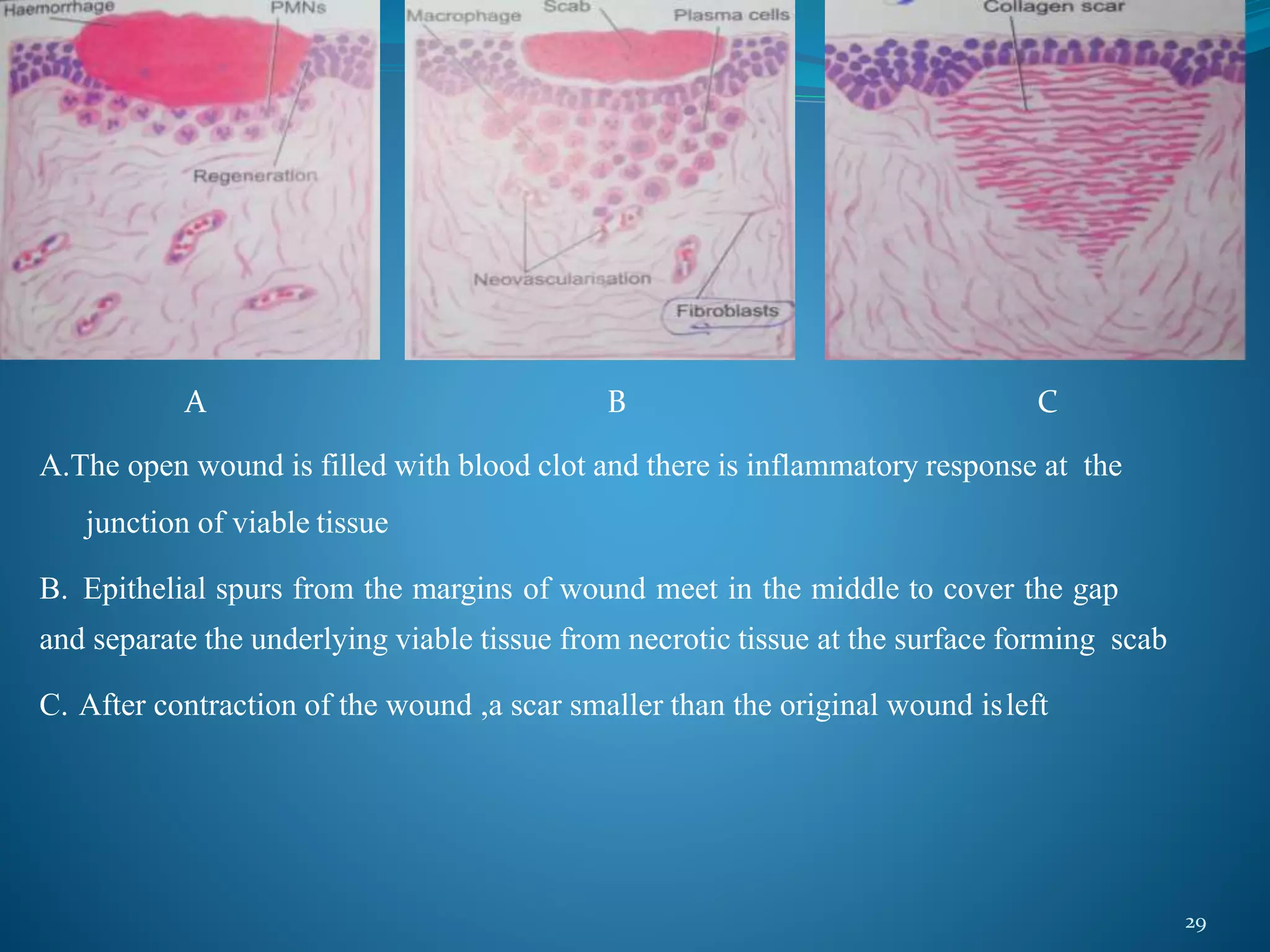
3. Wounds can heal through primary intention, secondary intention, or tertiary intention depending on factors like cleanliness and tissue loss. Primary intention involves direct wound edge approximation while secondary intention involves healing from the base up without suturing.
Introduction
Definition
Healing of skin wounds
Healing in bone
Healing of nervous tissue
Factors influencing healing
Complications of wound healing
Conclusion
References
Introduction
Definition
Healing of skin wounds
Healing in bone
Healing of nervous tissue
Factors influencing healing
Complications of wound healing
Conclusion
References
wound management briefing training course including wounds, wound healing & wound types, wound closure, wound covers, wound dressings and marketing plan for new product launch, wound assessment types and measures.
for HCP , wound care specialists, nursing, and wound care and health associations
wound management briefing training course including wounds, wound healing & wound types, wound closure, wound covers, wound dressings and marketing plan for new product launch, wound assessment types and measures.
for HCP , wound care specialists, nursing, and wound care and health associations
this a basic presentation which I got inspired to do after seeing some presentations made on slideshare. It basically covers types of wounds,wound healing process,factors affecting wound healing and wound care.
WOUND HEALING. wound healing in general, wound healing in dentistry.YasminShaik16
Wound Healing:
Wound healing is a complex and dynamic process that restores the integrity of the skin and underlying tissues after injury. This essential biological function involves a well-orchestrated interplay of cellular and molecular mechanisms. Understanding wound healing is crucial for healthcare professionals, researchers, and anyone interested in medical science. This comprehensive overview will cover the phases of wound healing, factors affecting the process, advanced wound care techniques, and current research trends. This presentation delves into the four distinct phases of wound healing: hemostasis, inflammation, proliferation, and remodeling. Each phase plays a crucial role in repairing damaged tissue and involves complex interactions between cells, growth factors, and the extracellular matrix.
Key topics include:
Hemostasis: The immediate response to injury, where blood clotting mechanisms are activated to prevent excessive bleeding.
Inflammation: The body's defense mechanism against infection, involving white blood cells, cytokines, and other inflammatory mediators.
Proliferation: The phase where new tissue forms, characterized by the proliferation of fibroblasts, collagen deposition, and angiogenesis.
Remodeling: The final phase where the newly formed tissue matures and strengthens over time, ensuring functional and aesthetic restoration.
This presentation also covers factors that influence wound healing, such as age, nutrition, underlying health conditions, and external factors like infection and mechanical stress. Additionally, it highlights advanced wound care techniques and the latest research in enhancing the healing process through innovative therapies and technologies.
Ideal for healthcare professionals, students, and anyone interested in understanding the science behind wound healing, this presentation provides valuable insights into how our bodies repair themselves and the advancements in medical science that support this vital process.
Factors Affecting Wound Healing
Wound healing can be influenced by various intrinsic and extrinsic factors. Understanding these factors is crucial for optimizing healing outcomes.
Intrinsic Factors
Age: Healing capacity declines with age due to reduced cellular proliferation, slower immune response, and decreased collagen synthesis.
Genetics: Genetic predispositions can affect the efficiency of the healing process and the likelihood of developing chronic wounds or hypertrophic scars.
Nutrition: Adequate nutrition is essential for wound healing. Proteins, vitamins (especially vitamins A and C), and minerals (like zinc) play vital roles in cell proliferation, collagen synthesis, and immune function.
Health Conditions: Chronic conditions such as diabetes, obesity, and cardiovascular diseases can impair wound healing. Diabetes, for example, can lead to poor blood circulation and reduced immune response, increasing the risk of infection and delayed healing.
Periodontics is a specialized field of dentistry that focuses on the diagnosis, treatment, and prevention of diseases that affect the gums and other supporting structures of the teeth. It plays a crucial role in maintaining overall oral health and is essential for preserving the function and aesthetics of the smile. From gum disease treatment to dental implants, periodontics encompasses a wide range of procedures and practices aimed at ensuring the health and vitality of the oral cavity. As technology continues to advance, the integration of artificial intelligence (AI) has opened up new possibilities and avenues for innovation within the field of periodontics.
Periodontal diseases have afflicted humans since the dawn of his tory. Oral hygiene is practiced since ancient times. Sushruta Samhita contains numerous descriptions of severe periodontal disease with loose teeth and purulent discharge from gingiva. Our understanding of the causes of periodontal disease have changed greatly over time. The past inabilities of generalists to pinpoint systemic causes are being overcome with the application of modern epidemiologic and clinical research approaches.
The emerging science of nanotechnology, especially within the dental and medical fields, sparked a research interest in their potential applications and benefits in comparison to conventional materials used. Therefore, a better understanding of the science behind nanotechnology is essential to appreciate how these materials can be utilized in our daily practice. Nanotechnology is the research and development of materials, devices and systems exhibiting physical, chemical and biological properties that are different from those on a large scale. Nanotechnology offers a broad range of innovations and improvement in prevention, diagnostics, and treatment of oral diseases. Periodontal disease is one of the major dental illnesses that affect millions of people around the globe. It is estimated that 90% of the world population suffers from the disease. Recent nanotechnology advancement and innovations through Nano dentistry are increasingly providing a suitable solution for the treatment of many dental disorders including periodontal disease. This review aimed to provide an overview of the role of nanotechnology in periodontics and to evaluate its applicability in prevention and treatment of oral diseases and also to provide important recent updates on the various nanotechnology-based approaches for periodontal disease therapy.
Dental indices can be considered as the main tool of epidemiological studies in dental diseases, to find out the incidence, prevalence and severity of the diseases, based on which preventive programmes are adopted for their control and prevention.
When the body is under stress, it produces more of the hormone cortisol, which acts as an anti-inflammatory agent. When cortisol is produced peripherally in the gums, it stimulates mast cells to produce more proteins, simultaneously increasing inflammation and the progression of periodontal disease.
Aggressive periodontitis is distinguished from chronic periodontitis with respect to,
Age of onset
Rapid rate of disease progression
Nature & composition of the associated subgingival micro flora
Alterations in the host’s immune response
Familial aggregation of the disease
Types of Aggressive Periodontitis
Localized Aggressive Periodontitis-LAP
Generalized Aggressive Periodontitis-GAP
Localized aggressive periodontitis
Historical background,
Diffuse atrophy of the alveolar bone (Gottlieb-1923)
Deep cementopathia (Gottlieb-1928)
Parodontitis marginalis progressiva(Wannenmacher- 1938)
Periodontosis (world workshop in periodontics -1966)
Juvenile periodontitis (Chaput etal-1971)
Localized Juvenile periodontitis (world workshop in periodontics- 1989)
Localized aggressive periodontitis (International workshop by american academy of periodontology – 1999)
Clinical characteristics LAP
LAP is localized to first molar or incisor with interproximal attachment loss on at least two permanent teeth ,one of which is a first molar & involving no more than two teeth other than first molars & incisors.
Possible reasons for limitation of the destruction
After initial colonization of the first permanent teeth( first molars & incisors) Aa evades the host defenses by different mechanisms they are –
-PMN chemotaxis inhibiting factors
-Endotoxin
-Collagenases
-Leukotoxin
After this initial attack adequate immune defenses are stimulated to produce opsonic antibodies to enhance the clearance & phagocytosis of invading bacteria & neutralize leukotoxic activity there by colonization of other sites may be prevented
Bacteria antagonistic to Aa may colonize the periodontal tissues & inhibit Aa from further colonization of periodontal sites in the mouth ,hence Aa infection & tissue destruction is localized
Aa may lose its leukotoxin producing ability for unknown reasons
A defect in cementum formation may be responsible for the localization of the lesions
Clinical features of LAP
Age of onset –puberty & around 20 years of age
It affects both male & female
There will be a lack of clinical inflammation despite the presence of deep periodontal pockets & advanced bone loss
The amount of plaque is minimal & is rarely mineralizes to calculus
Plaque Contains elevated levels of Aa & Pg
The Rate of boneloss is about 3 to 4 times faster than in chronic periodontitis
Clinical features of LAP
Distolabial migration of the maxillary incisors with concomitant diastema formation
Increasing mobility of the maxillary & mandibular incisors & first molars
Sensitivity of denuded root surfaces to thermal & tactile stimuli
Deep dull radiating pain during mastication
Robust antibody response to pathogens
Radiographs reveal ‘arc shaped loss of alveolar bone extending from distal surface of the second premolar to the mesial surface of the second molar’
Localized Aggressive periodontitis
Generalized Aggressive Periodontitis
NON SURGICAL PERIODONTAL INSTRUMENT has been designed for specific purposes such as diagnosing the periodontal disease, removing calculus, planning root surfaces, curetting the gingiva and removing diseased tissue.
More from Rama Dental College Hospital and Research Center (20)
Knee anatomy and clinical tests 2024.pdfvimalpl1234
This includes all relevant anatomy and clinical tests compiled from standard textbooks, Campbell,netter etc..It is comprehensive and best suited for orthopaedicians and orthopaedic residents.
These simplified slides by Dr. Sidra Arshad present an overview of the non-respiratory functions of the respiratory tract.
Learning objectives:
1. Enlist the non-respiratory functions of the respiratory tract
2. Briefly explain how these functions are carried out
3. Discuss the significance of dead space
4. Differentiate between minute ventilation and alveolar ventilation
5. Describe the cough and sneeze reflexes
Study Resources:
1. Chapter 39, Guyton and Hall Textbook of Medical Physiology, 14th edition
2. Chapter 34, Ganong’s Review of Medical Physiology, 26th edition
3. Chapter 17, Human Physiology by Lauralee Sherwood, 9th edition
4. Non-respiratory functions of the lungs https://academic.oup.com/bjaed/article/13/3/98/278874
New Drug Discovery and Development .....NEHA GUPTA
The "New Drug Discovery and Development" process involves the identification, design, testing, and manufacturing of novel pharmaceutical compounds with the aim of introducing new and improved treatments for various medical conditions. This comprehensive endeavor encompasses various stages, including target identification, preclinical studies, clinical trials, regulatory approval, and post-market surveillance. It involves multidisciplinary collaboration among scientists, researchers, clinicians, regulatory experts, and pharmaceutical companies to bring innovative therapies to market and address unmet medical needs.
Ozempic: Preoperative Management of Patients on GLP-1 Receptor Agonists Saeid Safari
Preoperative Management of Patients on GLP-1 Receptor Agonists like Ozempic and Semiglutide
ASA GUIDELINE
NYSORA Guideline
2 Case Reports of Gastric Ultrasound
Ethanol (CH3CH2OH), or beverage alcohol, is a two-carbon alcohol
that is rapidly distributed in the body and brain. Ethanol alters many
neurochemical systems and has rewarding and addictive properties. It
is the oldest recreational drug and likely contributes to more morbidity,
mortality, and public health costs than all illicit drugs combined. The
5th edition of the Diagnostic and Statistical Manual of Mental Disorders
(DSM-5) integrates alcohol abuse and alcohol dependence into a single
disorder called alcohol use disorder (AUD), with mild, moderate,
and severe subclassifications (American Psychiatric Association, 2013).
In the DSM-5, all types of substance abuse and dependence have been
combined into a single substance use disorder (SUD) on a continuum
from mild to severe. A diagnosis of AUD requires that at least two of
the 11 DSM-5 behaviors be present within a 12-month period (mild
AUD: 2–3 criteria; moderate AUD: 4–5 criteria; severe AUD: 6–11 criteria).
The four main behavioral effects of AUD are impaired control over
drinking, negative social consequences, risky use, and altered physiological
effects (tolerance, withdrawal). This chapter presents an overview
of the prevalence and harmful consequences of AUD in the U.S.,
the systemic nature of the disease, neurocircuitry and stages of AUD,
comorbidities, fetal alcohol spectrum disorders, genetic risk factors, and
pharmacotherapies for AUD.
micro teaching on communication m.sc nursing.pdfAnurag Sharma
Microteaching is a unique model of practice teaching. It is a viable instrument for the. desired change in the teaching behavior or the behavior potential which, in specified types of real. classroom situations, tends to facilitate the achievement of specified types of objectives.
Flu Vaccine Alert in Bangalore Karnatakaaddon Scans
As flu season approaches, health officials in Bangalore, Karnataka, are urging residents to get their flu vaccinations. The seasonal flu, while common, can lead to severe health complications, particularly for vulnerable populations such as young children, the elderly, and those with underlying health conditions.
Dr. Vidisha Kumari, a leading epidemiologist in Bangalore, emphasizes the importance of getting vaccinated. "The flu vaccine is our best defense against the influenza virus. It not only protects individuals but also helps prevent the spread of the virus in our communities," he says.
This year, the flu season is expected to coincide with a potential increase in other respiratory illnesses. The Karnataka Health Department has launched an awareness campaign highlighting the significance of flu vaccinations. They have set up multiple vaccination centers across Bangalore, making it convenient for residents to receive their shots.
To encourage widespread vaccination, the government is also collaborating with local schools, workplaces, and community centers to facilitate vaccination drives. Special attention is being given to ensuring that the vaccine is accessible to all, including marginalized communities who may have limited access to healthcare.
Residents are reminded that the flu vaccine is safe and effective. Common side effects are mild and may include soreness at the injection site, mild fever, or muscle aches. These side effects are generally short-lived and far less severe than the flu itself.
Healthcare providers are also stressing the importance of continuing COVID-19 precautions. Wearing masks, practicing good hand hygiene, and maintaining social distancing are still crucial, especially in crowded places.
Protect yourself and your loved ones by getting vaccinated. Together, we can help keep Bangalore healthy and safe this flu season. For more information on vaccination centers and schedules, residents can visit the Karnataka Health Department’s official website or follow their social media pages.
Stay informed, stay safe, and get your flu shot today!
Pulmonary Thromboembolism - etilogy, types, medical- Surgical and nursing man...VarunMahajani
Disruption of blood supply to lung alveoli due to blockage of one or more pulmonary blood vessels is called as Pulmonary thromboembolism. In this presentation we will discuss its causes, types and its management in depth.
Prix Galien International 2024 Forum ProgramLevi Shapiro
June 20, 2024, Prix Galien International and Jerusalem Ethics Forum in ROME. Detailed agenda including panels:
- ADVANCES IN CARDIOLOGY: A NEW PARADIGM IS COMING
- WOMEN’S HEALTH: FERTILITY PRESERVATION
- WHAT’S NEW IN THE TREATMENT OF INFECTIOUS,
ONCOLOGICAL AND INFLAMMATORY SKIN DISEASES?
- ARTIFICIAL INTELLIGENCE AND ETHICS
- GENE THERAPY
- BEYOND BORDERS: GLOBAL INITIATIVES FOR DEMOCRATIZING LIFE SCIENCE TECHNOLOGIES AND PROMOTING ACCESS TO HEALTHCARE
- ETHICAL CHALLENGES IN LIFE SCIENCES
- Prix Galien International Awards Ceremony
Title: Sense of Smell
Presenter: Dr. Faiza, Assistant Professor of Physiology
Qualifications:
MBBS (Best Graduate, AIMC Lahore)
FCPS Physiology
ICMT, CHPE, DHPE (STMU)
MPH (GC University, Faisalabad)
MBA (Virtual University of Pakistan)
Learning Objectives:
Describe the primary categories of smells and the concept of odor blindness.
Explain the structure and location of the olfactory membrane and mucosa, including the types and roles of cells involved in olfaction.
Describe the pathway and mechanisms of olfactory signal transmission from the olfactory receptors to the brain.
Illustrate the biochemical cascade triggered by odorant binding to olfactory receptors, including the role of G-proteins and second messengers in generating an action potential.
Identify different types of olfactory disorders such as anosmia, hyposmia, hyperosmia, and dysosmia, including their potential causes.
Key Topics:
Olfactory Genes:
3% of the human genome accounts for olfactory genes.
400 genes for odorant receptors.
Olfactory Membrane:
Located in the superior part of the nasal cavity.
Medially: Folds downward along the superior septum.
Laterally: Folds over the superior turbinate and upper surface of the middle turbinate.
Total surface area: 5-10 square centimeters.
Olfactory Mucosa:
Olfactory Cells: Bipolar nerve cells derived from the CNS (100 million), with 4-25 olfactory cilia per cell.
Sustentacular Cells: Produce mucus and maintain ionic and molecular environment.
Basal Cells: Replace worn-out olfactory cells with an average lifespan of 1-2 months.
Bowman’s Gland: Secretes mucus.
Stimulation of Olfactory Cells:
Odorant dissolves in mucus and attaches to receptors on olfactory cilia.
Involves a cascade effect through G-proteins and second messengers, leading to depolarization and action potential generation in the olfactory nerve.
Quality of a Good Odorant:
Small (3-20 Carbon atoms), volatile, water-soluble, and lipid-soluble.
Facilitated by odorant-binding proteins in mucus.
Membrane Potential and Action Potential:
Resting membrane potential: -55mV.
Action potential frequency in the olfactory nerve increases with odorant strength.
Adaptation Towards the Sense of Smell:
Rapid adaptation within the first second, with further slow adaptation.
Psychological adaptation greater than receptor adaptation, involving feedback inhibition from the central nervous system.
Primary Sensations of Smell:
Camphoraceous, Musky, Floral, Pepperminty, Ethereal, Pungent, Putrid.
Odor Detection Threshold:
Examples: Hydrogen sulfide (0.0005 ppm), Methyl-mercaptan (0.002 ppm).
Some toxic substances are odorless at lethal concentrations.
Characteristics of Smell:
Odor blindness for single substances due to lack of appropriate receptor protein.
Behavioral and emotional influences of smell.
Transmission of Olfactory Signals:
From olfactory cells to glomeruli in the olfactory bulb, involving lateral inhibition.
Primitive, less old, and new olfactory systems with different path
Evaluation of antidepressant activity of clitoris ternatea in animals
WOUND HEALING.pptx
1. 1
WOUND HEALING
DEPARTMENT OF PERIODONTOLOGY
RAMA DENTAL COLLEGE HOSPITALAND RESEARCH
CENTRE, KANPUR, UTTAR PRADESH- 208024
2. CONTENTS
Introduction
History
Regeneration and Repair
Phases of wound healing
Types of wound healing
Mediators of wound healing
Factors affecting wound healing
Wound healing in specialized tissue
Complications of wound healing
Advances in wound healing
Future strategies
Conclusion
References 2
3. INTRODUCTION
Wound is a disruption of the normal structure and
function of the skin and underlying soft tissue.
Wound healing is the body’s response to injury in an
attempt to restore normal structure and function.
Wound healing involves two distinct processes :
- Regeneration
- Repair
3
4. 4
• a. Tidy wounds
• ► Theyare wounds like surgical incisions and wounds caused
by sharpobjects.
• ► It is incised, clean, healthy wound , without any tissue loss.
• ► Usually primary suturing is done. Healing is by primary
intention.
CLASSIFICATION OF WOUNDS
I.RANK&WAKEFIELDCLASSIFICATION
5. 5
Theyare due to:
- Crushing.
- Tearing.
- Avulsion.
- Devitalised injury.
- Vascular injury.
- Multiple irregular wounds.
- Burns.
b. Untidy wounds
• Fracture of the underlying bone maybepresent.
• Wound infection, delayed healing arecommon.
• Liberal excision of devitalised tissue and allowing to heal by secondary intention
is the management.
• Secondarysuturing, skin graft or flap maybeneeded.
6. 6
CLASSIFICATION BASED ON THE THICKNESS OF WOUND
Superficial wound- involving only epidermis and dermal papillae.
Partial thickness wound- with skin loss up to deep dermis with only
deepest part of the dermis, hair follicle shafts and sweat glands are left
behind.
Full thickness wound- with loss of entire skin and subcutaneous tissue
causing spacing out of the skin edges.
Deep wound- are the one extending deeper, across deep fascia into muscles
or deeper structures.
Complicated wound- are one associated with injury to vessels or nerves.
Penetrating wound- are one which penetrates into either natural cavities
or organs.
7. 7
CLASSIFICATION BASED ON THE INVOLVEMENT OF STRUCTURES
Simple wounds- are one involving only one organ or tissue.
Combined wounds- are one involving mixed tissues.
8. 8
Acute wounds in normal, healthy individuals heal through
an orderly sequence of physiological events that include
hemostasis, inflammation, epithelization, fibroplasia, and
maturation. Acute wound is upto 8 hours of trauma.
When this process is altered, chronic wound may develop
and is more likely to occur in patients with underlying
disorders . Chronic wounds are generally associated with
physiological impairments that slow or prevent wound
healing. Chronic wound develops after 8 hours from the
trauma.
CLASSIFICATION BASED ON THE TIME ELAPSED
9. HISTORY
9
The earliest accounts of wound healing dates back to about 2000 B.C
Galen of Pergamum emphasized the importance of maintaining a moist
environment to ensure adequate healing.
Ambriose Paré found that simply dressed gunshot wounds heal faster and are
less painful than when treated with boiling oil, the previously accepted method.
Ignaz Philipp Semmelweis advocated need for washing hands before any
treatment.
Joseph Lister began soaking his instruments in phenol and spraying the
operating rooms, reducing the mortality rates close to 50%.
10. REGENERATION
10
It is the natural renewal of a structure, produced by growth
and differentiation of new cells and intercellular substances
to form new tissues or parts.
It takes place by proliferation of parenchymal cells and
usually results in complete restoration of the original tissue.
In order to maintain proper structure of tissues ,these cells
are under constant regulatory control of their cell-cycle.
11. REPAIR
11
Repair simply restores the continuity of the diseased tissue
and re-establishes normal margins. This process is also called
“Healing by Scar”.
It takes place by proliferation of connective tissue elements
resulting in fibrosis and scarring.
2 processes are involved in repair:
a) Granulation tissue formation.
b) Contraction of wounds.
12. 12
INFLAMMATORY PHASE
Objective:- Leukocytes and macrophages destroy bacteria, cleaning the
wound of cellular debris.
Duration:-
Immediately following the Hemostasis Phase from 0 to 3 days
post injury.
Cells
Involved:-
Host cells infiltrate the wound site, such as leukocytes and
marcophages. Bacteria are destroyed by leukocytes. Macrophages
cleanse the wound of cellular debris.
Signs &
Symptoms:-
Swelling, Increased fluid, profusion of blood, Redness, Release of
Epinephrine, Histamine response, Heat, Pain.
PHASES OF WOUND HEALING
14. PROLIFERATIVE PHASE
14
Proliferative phase is categorized by :
Fibroblast migration
Collagen synthesis
Angiogenesis
Granulation tissue formation
Epithelisation
What
happens:-
The major activity focuses on angiogenesis and granulation tissue
formation.
Duration:- 3 to 21 days post injury.
Cells
Involved:-
Macrophages, fibroblasts, immature collagen, blood vessels, and
ground substance make up granulation tissue, which fills the wound’s
cavity
16. 16
REMODELING AND MATURATION PHASE
What
happens:-
For the final phase of healing, the collagen fibers in the scar are
reorganized to improve tensile strength.
Duration:- 21 days post injury and upto 1 years later.
Cells
Involved:-
Fibroblasts, MMPs, growth factors are critical in this phase.
18. 18
GRANULATION TISSUE FORMATION:
Has 3 phases—
A. PHASE OF INFLAMMATION–
-Following trauma, blood clots at the site of injury. There is acute
inflammatory response within 24 hours.
B. PHASE OF CLEARANCE–
-Neutrophils and phagocytic activity of macrophages clear off the necrotic
tissues , debris and RBC.
C. PHASE OF INGROWTH OF GRANULATION TISSUE—
-Consist of 2 main process:-- I)Angiogenesis
II)Fibrogenesis
19. 19
ANGIOGENESIS–
-Formation of new blood vessels at the site of
injury takes place by proliferation of
endothelial cells.
-Newly formed blood vessels are more leaky,
accounting for the oedematous appearance of
new granulation tissue.
-Soon, these blood vessels differentiate into
muscular arterioles, thin-walled venules and
true capillaries.
20. 20
FIBROGENESIS–
-The newly formed blood vessels are present in an amorphous ground substance
or matrix.
-The new fibroblasts originate from fibrocytes or by mitotic division of
fibroblasts.
-Collagen fibrils begin to appear by about 6th day.
-As maturation proceeds, more collagen is formed while the number of active
fibroblasts and new blood vessels decreases and results in inactive looking scar
known as cicatrisation.
21. 21
CONTRACTION OF WOUNDS: -
-It starts after 2-3 days and the process is completed by the 14th day.
-The wound is reduced by approximately 80% of its original size.
-Contracted wound results in rapid healing since lesser surface area of the
injured tissue has to be replaced.
23. 23
PRIMARY INTENTION HEALING
Wound healing by primary intention is typical for non-complicated surgical
wounds.
It can be characterized by—
i. Cleaned and uninfected.
ii. Surgically incised.
iii. Without much loss of cells and tissues.
iv. Edges of wound are approximated by surgical sutures.
Events occuring in primary intention are—
1. INITIAL HAEMORRHAGE.
2. ACUTE INFLAMMATORY RESPONSE.
3. EPITHELIAL CHANGES.
4. ORGANIZATION.
5. SUTURE TRACKS.
24. 24
•Each suture track is a separate wound and have same
phenomenon as in healing of primary wound .
•On removal of suture around 7th day, much of epithelialized
suture track is avulsed and remaining epithelial tissue gets
absorbed.
•Sometimes suture tract gets infected and resulting in STITCH
ABSCESS or when epithelial cells may persists in the tract it
causes- IMPLANTATION or EPIDERMAL CYST.
Suture Tracks
27. 27
SECONDARY INTENTION HEALING
Healing takes place from the base upwards as well as from the margins inwards.
Healing is slow and results in a large, at times ugly, scar as compared to primary
intention.
Occur in wounds having the following characteristics: –
i. Open with a large tissue defect, at times infected.
ii. Having extensive loss of cells and tissues.
iii. These wounds are not approximated by surgical sutures but are left open.
Events occuring in secondary intention are—
1. INITIAL HAEMORRHAGE.
2. INFLAMMATORY PHASE.
3. EPITHELIAL CHANGES.
4. GRANULATION TISSUE.
5. WOUND CONTRACTION.
6. PRESENCE OF INFECTION.
29. 29
A.The open wound is filled with blood clot and there is inflammatory response at the
junction of viable tissue
B. Epithelial spurs from the margins of wound meet in the middle to cover the gap
and separate the underlying viable tissue from necrotic tissue at the surface forming scab
C. After contraction of the wound ,a scar smaller than the original wound isleft
A B C
30. Difference between 1˚ & 2˚ union of
wound
FEATURES PRIMARY SECONDARY
CLEANLINESS CLEAN NOT CLEAN
INFECTION NOT INFECTED INFECTED
MARGINS SURGICALLY CLEAN IRREGULAR
SUTURES USED NOT USED
HEALING SMALL GRANULATION
TISSUE
LARGE GRANULATION
TISSUE
OUT COME LINEAR SCAR IRREGULAR WOUND
COMPLICATION NOT FREQUENT FREQUENT
31. 31
TERTIARY INTENTION HEALING
Wounds that are too heavily contaminated for primary closure but start
appear clean and well vascularized after 4-5 days of open observation
can be dealt with tertiary intention healing.
When resolution has occurred, the wound edges can be brought
together (approximated) and the wound proceeds to heal.
Most commonly indicated in:-
-infected wounds with high bacterial content,
-wounds with a long time lapse since injury, or
-wounds with a severe crush component with significant tissue
devitalization.
36. 36
WOUND HEALING IN SPECIALIZED TISSUES
FRACTURE HEALING
Healing of fracture by callus formation depends upon some clinical considerations
whether the fracture is-
Traumatic or pathologic
Complete or incomplete
Simple or comminuted or compound
However, basic events in healing of any type of fracture are similar and resemble
healing of skin wound to some extent .
37. 37
Primary Union of Fractures
Occurs in a few special situations when the ends of fracture are approximated.
Bony union takes place with formation of medullary callus without periosteal callus
formation.
Patient can be made ambulatory early but there is more extensive bone necrosis
and slow healing.
Secondary union of Fractures
Is a more common process of fracture healing.
Though it is a continuous process, secondary bone union is described under the
following 3 headings:
1. Procallus formation
2. Osseous callus formation
3. Remodelling
38. A. PROCALLUS FORMATION: Steps involved are:
The osteoid undergoes calcification and is called woven bone callus.
Cells of inner layer of the periosteum lay down collagen and osteoid
matrix in the granulation tissue.
Callus composed of woven bone and cartilage starts forming within
first few days
Ingrowth of granulation tissue occurs and with it a soft tissue callus is
formed which joins the ends of fractured bone
Hematoma formation and Local inflammatory response occurs by
exudation of fibrin, polymorphs and macrophages
38
39. .
B. OSSEOUS CALLUS FORMATION:
1. Procallus act as scaffolding on which osseous callus
composed of lamellar bone is formed.
2. Woven bone is cleared away by osteoclasts and calcified
cartilage disintegrates.
3. Newly-formed blood vessels and osteoblasts invade, laying
down osteoid and lamellar bone is formed by developing
haversian system concentrically around blood vessels.
C. REMODELLING:
During the formation of lamellar bone, osteoblastic
laying and osteoclastic removal take place remodelling the
united bone ends, which become indistinguishable from normal
bone.
39
40. 40
Complications Of Fracture Wound Healing:
1. Fibrous union- when immobilization of fractured bone is not done. False
joint develop at fracture site.(pseudo-arthrosis)
2. Non-union may result if some soft tissue is interposed between fractured
ends.
3. Delayed union may occur from to delayed wound healing due to infection,
inadequate blood supply, poor nutrition, movement and old age.
Pseudo-arthrosis of Tibia
41. 41
Periodontalwoundhealing
HEALING FOLLOWING SCALING & ROOT PLANING
• Immediately after Scalingof Teeththe epithelial attachment willbe severed,
junctional & crevicular epithelium partiallyremoved
• Numerous polymorphonuclear leucocytes canbe seenbetween residual epithelial
cells & crevicular surface inabout 2 hrs
• There is dilation of blood vessels, oedema & necrosis in the lateral wall of the
pocket
• Theremaining epithelial cells show very little pre-mitotic activity at that time. 24
hrs after scaling awidespread & intense labeling of the cells have been observed, in
all areas of the remaining epithelium & in 2 days the entire pocket isepithlialized.
42. 42
• In 4-5 daysanew epithelial attachment mayappear at bottom of sulcus.
Depending on the severity of inflammation & the depth of the gingival
crevice, complete epithelial healing occursin 1-2weeks
• Immature collagen fibers occur within 21days.Following scaling,root
planning & curettage procedure healing occurs with the formation of a
long thin junctional epithelium with no connective tissueattachment.
43. 43
• A blood clot formsbetween the root surface & the lateral wall of the
pocket,soonafter the curettage
- Largenumber of polymorphonuclear leucocytesappearin the
areashortly after the procedure
- Thisisfollowed by rapid proliferation of granulationtissue.
- Epithelial cells proliferate along thesulcus.
HEALING FOLLOWING CURETTAGE
- Epithelisation of the inner surfaceof the lateral wall is completed in 2-7
days
- Thejunctional epithelium is alsoformed in about 5 days
- Healing results in the formation of a long junctional epithelium
adherent to the root surface.
44. 44
HEALING AFTER SURGICAL GINGIVECTOMY
• Initial responseafter gingivetomy isthe formation of a
protective surfaceclot
• Underlying tissue becomesacutely inflamed withsome necrosis
• Clot is then replaced by granulationtissue
• By24 hours there is anincreasein new connective tissuecells, mainly
angioblastsjust beneath the surfacelayer of inflammation and necrosis
45. 45
• By the 3rd daynumerous young fibroblast are located In the area
• Thehighly vasculargranulation tissue grows coronally, creating a new free
gingival margin andsulcus
• Capillaries derived from the blood vesselsof the periodontal ligament migratein
to the granulation tissueand within 2 weeks they connect with gingival
vessels.
• After 12 to 24hours ,epithelial cells at the marginsof the wound start tomigrate
over the granulation tissue, seperating it fromthe contaminated surfacelayer of
theclot
• Epithelial activity at the marginsreaches peakin 24to 33 hours
• Thenew epithelial cells arise from the basaland deeper spinous layersof the
wound edgeepithelium andmigrate over the wound over afibrin layer that is
later resorbed and replaced by aconnective tissuebed
• Theepithelial cells advancesby tumbling action,with cell becoming fixed to the
substrate by heidesmosomes andanew basement lamina
46. 46
• After 5 to 14 days,surface epithelization isgenerallycompleted
• During the first 4 weeks after gingivectomy ,keratinization isless
than it wasbeforesurgery
• Complete epithelial repair takesabout onemonth
• Vasodilation and vascularity begin to decreaseafter fourth dayof healing and
appear to be almost normal by the sixteenthday
• Completerepair of the connective tissuetakesabout 7weeks
• Theflow of gingival fluid in humansisinitiallyincreased after gingivectomy and
diminishes ashealingprogresses
• Maximal flow isreachedafter 1week,coinciding with thetime of maximal
inflammation
• In patientswith physiologic gingivalmelanosisthe pigmentation isdiminished in the
healedgingiva.
47. 47
Thereappearsto be little difference in the results obtained after shallow
gingival resection withelectrosurgery and that with periodontal knives.
However,when usedfor deep resection closeto bone, electrosurgery can
produce gingival recession,bone necrosisand sequestration, lossof bone
height, furcation exposure, and tooth mobility, which do not occur with the
useofperiodontal knives.
HEALING FOLLOWING ELECTROSURGICAL GINGIVECTOMY
48. 48
HEALING FOLLWING DEPIGMENTATION OF GINGIVA
Healingafter surgicaldepigmentation:
• After surgery it wasfound necessaryto cover the exposed lamina propria with
periodontal packsfor 7 to 10 days.Thewound healed uneventfully.After 6 weeks the
attached gingiva regenerated by only adelicate scarpresent. Thenewly formed
gingiva wasclinically non-pigmented.
Healingfollowingcryosurgical depigmentation:
• At 2nd to 3rd day: superficial necrosis becomes apparent and a whitish slough
could be separated from the underlyingtissue, leaving aclean pink surface.
• In 1-2 weeks: normal gingiva
• In 3-4 weeks: keratinization completed.
• Nopostoperative pain, hemorrhage, infection or scarring seen in patients.
50. 50
HEALING FOLLOWING FLAP SURGERY
• Immediately after suturing of the flap againsttooth surface a clot forms between the
2 tissues
• Theclot consistsof fibrin reticulum with many polymorphonuclear leukocytes,
erythrocytes & remnentsof injured clots
• At edgeof flap numerous capillaries areseen
• 1-3days after surgery spacebetween flap & tooth surface & bone appears reduced
& the epithelial cells along border of the flap start migrating
By1week after surgery
• epithelial cells havemigrated & established anattachmentto root surfaceby means
of hemidesmosomes
51. 51
• The blood clot isreplaced by granulation tissueproliferating from the gingival
connective tissue, alveolar bone and periodontal ligament
• By2ndweek collagenfibers beginsto appear.Collagenfibers getsarranged parallel to
root surfacerather than at right angles.Theattachment between soft tissue & tooth
surfaceis weak
• Byend of one month following surgerythe epithelial attachmentiswell formed &
the gingival creviceisalsowell epithealised
• Thereisbeginning functional arrangement ofsupracrestal fibres.
In caseswhereMucoperiostealFlap
• hasbeen reflected, superficial bone necrosishavebeen observed during first 3
days
• OsteoclasticResorption occursin that areawhich reachesits peakat 4-6days
• Osteoblastic Remodelling occurssubsequently
• Lossof alveolar bone height by about 1mm may be expected
after healing.
52. 52
HEALING OF PEDICLE AUTOGRAFTS
ADAPTATION SATAGE(0-4 days): Clot & thin fibrinous exudate between flap and
root surface
-PMNLs in clot and connective tissue
-Epithelium at margins of flap starts to
proliferate-may contact tooth surface
PROLIFERATION STAGE(4-21 days): Connective tissue invades the fibrin layer
- 6-10 days- fibroblasts apposed against root
surface
-Collagen within the flap- oriented parallel to
root surface
-Thin collagen fibers adjacent to root(no
fibrous union)
-Apical proliferation of epithelium-peaks at 10-
14 days.
-Osteoclastic resorption(peaks at 6th day)-
decreases by 14th day.
- Slight cemental resorption
53. 53
ATTACHMENT STAGE(21-28 days): Collagen fibers insert into new cementum
- Cementoid deposition(by 28th day- along
the entire root)
-Connective tissue attachment
- New gingival margin, sulcus and epithelial
attachment
- Osteoblastic activity
MATURATION STAGE(28-90 days): Continuous formation of collagen fibers
-Completely formed gingival sulcus and
epithelial attachment
-Bone apposition at alveolar crest
54. 54
HEALING OF FREE GINGIVAL GRAFT
INITIAL PHASE(0-3 days): Thin layer of exudate between graft and recipient bed
- Avascular plasmatic circulation(Forman 1960; Reese &
Stark 1961)
- Epithelium of free graft gets desquamated
REVASCULARISATION(2-11 days): Anastomosis between graft and recipient site
blood vessels
- Capillaries proliferate in the graft tissue
- Fibrous union between graft and connective
tissue bed
- Re- epithelization of the graft
TISSUE MATURATION(11-42 days): Decrease in the no. of blood vessel to normal
by the 14th day
- Epithelium maturation- formation of
keratin layer
-Functional integration-by 17th day
- Morphologically distinguishable for several
months
55. 55
DONOR SITE
-Granulation tissue fills the donor site.
-Initial healing is usually complete within 2-3 weeks after the removal of
a 4to 5mm thick graft.
-Patients should wear the surgical stent for about 2 weeks to protect the
healing wound.
-Palate returns to its pre surgical contour after about 3 months.
56. 56
RECIPIENT SITE
First week- Postoperative swelling
The epithelium of the graft will slough
Epithelial cells together with fibrin form a white film on the surface of
the grafts
The “white stage” passes in 4-7 days.
Epithelium will not cover the grafted tissue until a functional capillary circulation
has been re-established.
Therefore- Red stage(2nd & 3rd week)
-Normal colour as epithelialization is completed
- Epithelium thickens via stratification.
The graft reverts to its original volume or remains slightly larger as the swelling
subsides.
Tissue form is usually stable after 2 months, but some shrinkage may occur
between the 2nd and 4th months after surgery.
Final restorative measures should be initiated until after 4-6 months.
57. 57
HEALING OF CONNECTIVE TISSUE GRAFTS
Healing is similar to Free Gingival Graft.
2nd day- Epithelialization commences
7-10 days- Initial epithelialization completed
4 weeks- Keratinization commences
HEALING FOLLOWING FRENECTOMY
-Initial hemorrhage
-Acute inflammatory response- within 24 hours
-Epithelial changes- completes by 48 hours
-Organization of fibroblasts- starts around 3rd day
-Wound maturation- starts after 1 week and completes around 4 weeks.
58. 58
HEALING FOLLOWING OSSEOUS RESECTION
• Osseoussurgery initiates ainflammatory response
• Elevation of Mucoperiosteal Flapresults in temporary lossof
nutrient supply to the bone
• In additition surgicalresection of bone alsocontributes to inflammatory changes.
Necrosisof the alveolar crest & osteoclastic resorption of the bonetakesplace
initially
• Theosteoclastic resorption isfollowed by bone deposition& remodeling.
59. 59
• Theinitial lossin bone height iscompensatedtosomeextent by the repair and
remodeling.
• Thusfinal lossin bone height isclinicallyinsignificant
• Osteoblastic activity isevenseenafter 1yr.post-operatively
• As mucoperiosteum issutured backon to alveolar process the osteoclastic activity
doesn’t last for long
60. 60
HEALING AFTER IMPLANT PLACEMENT
• The interface areaconsistsof bone, marrow tissue, and a hematoma mixed with
bone fragments from the drillingprocess.
• In the early phaseof healing, woven bone is formed by osteoblasts at the surfaces
of trabecular and endosteal cortical bone surrounding the implant.
• Thenewly formed bone approaching the implant surface leads to bone
condensation into both, the implant threads and towards the implant surface.
• Consequently, the amount of bone in the threads and the degree of bone-implant
contact increasewithtime.
• In the late phasesof healing, lamellar bone replaceswoven bonein
aprocessof creeping substitution.
61. 61
STAGES OF HEALING OF IMPLANTS
.WovenBoneFormation: When bone matrix isexposed to extra-cellular fluid, non-
collagenous proteins &growth factors are set free & initiaterepair.
• Wovenbone isfirst formed & bridge agap within afew days.
• Wovenbone formation dominates the first 4-6 weeks
b. Lamellar BoneFormation : From2ndmonth post-operatively the microscopic
structure of bone changestolamellar or parallel fibered bone
c. BoneRemodelling : It begins around 3rd month post- operatively.Initially rapid
remodeling occurswhichslows down & continuos for rest of the life
Thuscomplete healing probably takeslonger than 3to6months
62. 62
HEALING IN NERVOUS TISSUES
1. Central Nervous System: The nerve cells of brain, spinal cord
and ganglia once destroyed are not replaced. Damaged neuroglial cells,
however, show proliferation of astrocytes called gliosis.
2.. Peripheral Nervous System: Cells of peripheral nervous system
show regeneration, mainly by proliferation of Schwann cells and fibrils from
distal end.
63. 63
HEALING OF MUSCLE
All 3 types of muscle have limited capacity to regenerate:
SKELETAL MUSCLE : Regeneration is similar to peripheral nerves. On injury cut
ends retract but are held together by stromal connective tissue injured site is
filled with fibrinous material, polymorphs and macrophages. It is then regenerated
either:
If muscle is intact- sarcolemmal tubes containing histiocytes appear and in about
3 month time restores properly.
If muscle sheath is damaged- it forms a disorganized multinucleate mass and scar
composed of fibrovascular tissue.
SMOOTH MUSCLE : Non-striated muscle has limited regenerative capacity e.g.,
appearance of smooth muscle in the arterioles in granulation tissue. However, in
large destructive lesions, the smooth muscle is replaced by permanent scar tissue.
CARDIAC MUSCLE : Destruction of heart muscle is replaced by fibrous tissue.
However, in cases where endomysium of individual cardiac fiber is intact
regeneration of cardiac fibers may occur in young patients
64. 64
HEALING OF MUCOSAL SURFACES
The cells of mucosal surfaces have very good regeneration and are normally
being lost and replaced continuously.
This occurs by proliferation from margins, migration, multilayering and
differentiation of epithelial cells in the same way as in epidermal cells in
healing of skin wounds.
65. 65
COMPLICATIONS OF WOUND HEALING
1. Wound infection
Wound infection result from gross bacterial contamination of susceptible wounds, where
the bacterial burden of replicating microorganisms actually impair healing.
Continual presence of a bacterial infection stimulates the host immune defenses leading
to production of inflammatory mediators, such as prostaglandins and thromboxane.
These cause wound hypoxia , leading to enhanced bacterial proliferation and continued
tissue damage.
Neutrophil proteases and endotoxins released by the breakdown of bacteria by the host
defense mechanism cause further destruction of newly formed cells and their collagen
matrix, resulting in impaired wound healing.
Clinical manifestation of wound infection include : erythema, warmth, swelling, pain,
odor, and pus.
66. 66
2. Deficient scar formation–
This may occur due to inadequate formation of granulation tissue .
3. Wound dehiscence-
Dehiscence result from tissue failure rather than improper suturing techniques.
The dehisced wound may be closed again or left to heal by secondary intention,
depending upon the extent of the disruption and the surgeon’s assessment of
the clinical situation.
4. Implantation (epidermal) cyst-
formation may occur due to persistence of epithelial cells
in the wound after healing.
67. 67
5. Proliferative scarring-
The two common forms of hyperproliferative healing are hypertrophied scars
and keloids.
These are characterized by hypervascularity and hypercellularity.
It results from altered apoptotic behaviour, which leads to scarring , persistent
inflammation, and an overproduction of extracellular matrix components,
including glycosaminoglycans and collagen type I.
Keloid
68. 68
6. Pigmentation- Healed wounds may at times have rust like colour
due to staining with haemosiderin .
7. Excessive contraction– An exaggeration of wound contraction may
result in formation of contractures or cicatrisation.
69. 69
ADVANCES IN WOUND HEALING
Advances in wound care include :
1. Growth factors :
• Currently, PDGF-BB is used for the treatment of cutaneous ulcers, specifically
diabetic foot ulcers.
• Recombinant KGF-2 enhance the formation of granulation tissues in rabbits and
wound closure in human meshed skin grafts.
• rhBMP-2 and rhBMP-7 induce undifferentiated mesenchymal cells to
differentiate into osteoblasts
• Platelet-rich fibrin (PRF) is a second-generation PRP where
autologous platelets and leucocytes are present in a complex fibrin matrix to
accelerate the healing of soft and hard tissue and is used as a tissue-engineering
scaffold for filling the intrabony defects and furcation defects.
70. 70
2. Dermal and Mucosal substitutes :
• Immediate coverage protects the wound from water loss, drying and
mechanical injury and so accelerates wound healing .
• Autologous grafts remains standard for replacing dermal mucosal surfaces , a
no. of bioengineered substitutes can be used in surgical practice.
Engineered skin contains all the components necessary to modulate healing and
allow healing that replicates native tissue and limits scar formation .
71. 71
Bioengineered Skin
• Living bioengineered skin equivalents provide a
living supply of growth factors and cytokine and a
collagen matrix to build upon.
• The mechanism of action is not fully understood
on how these skin equivalents initiates wound healing,
but it has been studied extensively with the Apligraf product.
• The cells contained in the Apligraf grow and proliferate,
producing growth factors, collagens, and extracellular matrix
proteins, which stimulate re-epithelization, formation of
granulation tissue, angiogenesis, and neutrophil and
monocyte chemotaxis.
• Apligraf acts as a potent cellular remedy that can provide a
different and adaptable response in acute and chronic
wounds.
72. 72
3. Hydrotherapy
• Hydrotherapy supports wound healing by debriding the wound, warming the
injured extremity, and providing gentle limb resistance for physical therapy.
• New forms used are pulsed lavage and the VersaJet.
• Pulsed lavage delivers an irrigating solution under pressure (4 to 15 psi) and
the rate of granulation tissue formation was greater in patients receiving
pulse lavage compared with those receiving conventional Hydrotherapy.
• The VersaJet Hydrosurgery System is a water jet–powered surgical tool
designed to efficiently debride a wound by removing damaged tissues and
contaminants precisely, without the collateral trauma associated with
traditional surgical modalities .
73. 73
4. Hyperbaric Oxygen:
-Dividing cells in a wound require an oxygen tension of at least 30
mmHg.
-Tissues in wounds that are not healing have partial pressure of
oxygen values of 5 to 20 mmHg
- Many reports in the literature have demonstrated benefit from
hyperbaric oxygen treatment for a variety of conditions, including
amputations, osteoradionecrosis, surgical flaps, and skin grafts.
In addition to simply providing more oxygen to the wound site,
hyperbaric oxygen therapy also increases expression of nitric oxide,
which is crucial for wound healing.
74. 74
5. Lasers
• Lasers for open wound management are low-energy lasers capable of raising
tissue temperature 0.1°C to 0.5°C. This is called as bio-stimulation.
• It is caused by the stimulation of ascorbic acid uptake by cells, stimulation of
photoreceptors in the mitochondria respiratory chain, changes in cellular ATP
and cell membrane stabilization.
• It increase healing (especially when combined with hyperbaric oxygen
treatments) of ischemic, hypoxic, and infected wounds.
The 2 most common lasers used clinically are helium-neon lasers and gallium-
arsenide lasers.
75. 75
6. Electrostimulation
In 1982, Barker described the “skin battery.” He found that the skin surface was
always negatively charged (compared with the deeper skin layers), and he
measured transcutaneous voltages up to 40mV.
Electrostimulation is believed to restart or accelerate the wound-healing process
by imitating the natural electrical current that occurs in skin when it is injured.
Electrical current applied to wounded tissue increases the migration of cells vital
to the wound-healing process (i.e., neutrophils, macrophages, and fibroblasts).
Electrostimulation may also play a role in wound healing through improved blood
flow.
76. 76
7. Negative Pressure Therapy
• Negative pressure therapy (or vacuum-assisted closure), uses a sub-atmospheric
pressure dressing to convert an open wound into a controlled closed wound.
The negative pressure removes interstitial fluid and edema to improve tissue
oxygenation. It also removes inflammatory mediators that suppress the normal
progression of wound healing.
Granulation tissue forms more rapidly and 5 days of therapy decreases the wound
bacterial count.
77. 77
FUTURE STRATEGIES
Gene Therapy :
Gene therapy is the future for wound-healing strategies. Current research is
aimed at inserting growth factor genomes into the wound.
Inserting the desired genome can be accomplished through several
different vehicles:
biologic (viral) techniques,
chemical methods via cationic liposomes, and
physical insertion:
hypodermic needle,
microseeding,
particle-mediated transfer (using a gene gun), and
electroporation
78. 78
CONCLUSION
The variety of wounds present challenges to the physician to select the most
appropriate management to facilitate healing.
A complete wound history along with knowledge of the healing potential of the
wound, as it relates to the specific medical and environmental considerations for
each patient, provides the basis of decision making for wound management.
It is essential to consider each wound individually in order to create the optimal
conditions for wound healing.
Understanding of wound healing is as important as knowing the pathogenesis
of disease, because satisfactory wound healing is the ultimate goal of treatment.
If we are able to understand the mechanism of wound healing, we can design
treatment approaches that maximize favorable conditions for wound healing to
occur.
79. 79
REFERENCES
Pathology By Harsh Mohan - 4th edition.
Peterson's Principles of Oral and Maxillofacial Surgery, Third Edition 3rd Edition
by Michael Miloro , GE Ghali , Peter Larsen , Peter Waite .
Carranza’s Clinical Periodontology- By Newman, Takei, Klokkevold, Carranza 10th
Edition.
Wound Healing: An Overview. George Broughton.Plast. Reconstr. Surg. 117: 1e-S,
2006
Biology of wound healing. Ikramuddin Aukhil. Perio 2000, vol. 22. 2000 -44-50.
Polypeptide growth factors and attachment proteins in periodontal wound healing
and regeneration. Raul G. Caffesse & Carlos R. Quinones.
-Perio 2000, vol.1 1993- 69-79
-Perio 2000, vol 24:connective tissue of periodontium (cell biology of woundhealing)
• Journel of dental rsearch2010 march,89(3),219-229
(factors affecting wound healing)