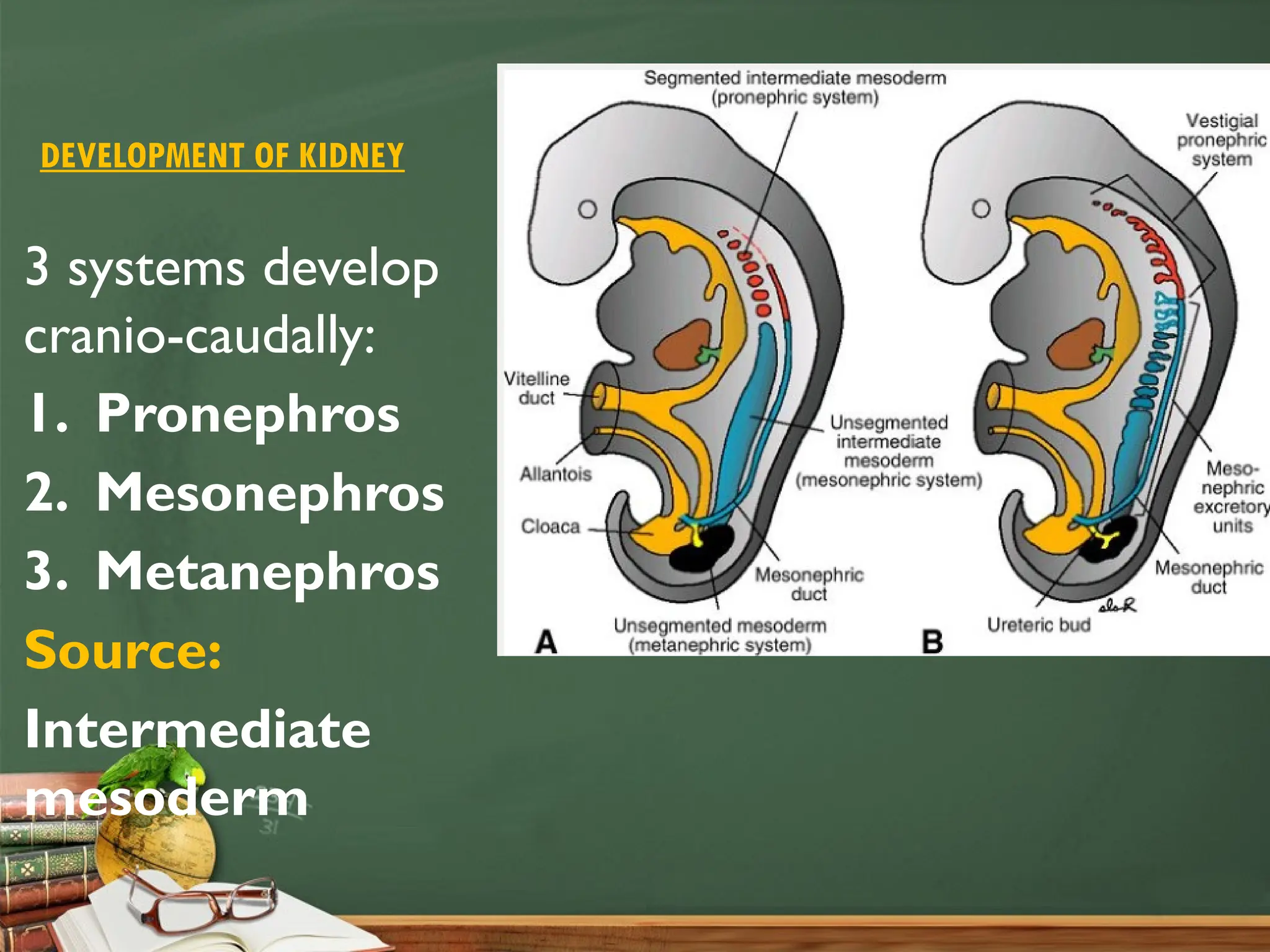
DEVELOPMENT OF RENAL SYSTEM
By:
Phanindra Prasad Poudel
Assistant Professor
Department of Anatomy
MCOMS, Pokhara
DEVELOPMENT OF KIDNEY
3 systems develop
cranio-caudally:
1. Pronephros
2. Mesonephros
3. Metanephros
Source:
Intermediate
mesoderm
PRONEPHROS
 4th
week
 Cervical and upper thoracic regions
 7-10 segmentation- solid cell groups- vestigial excretory
units- Nephrotome/ Pronephric tubule
 By the end of 4th
week- pronephric system disappears
MESONEPHROS
 Lower thoracic and
upper lumbar region
(L3)
 After disappearance of
pronephros
 70-80 segmentation
appear- each segment-
nephrotome/
mesonephric tubule
Mesonephric tubule:
S- shaped loop
 Medial end
 forms glomerulus
enclosed by bowman’s
capsule- together k/a-
renal corpuscle
 Lateral end
 terminates in the
longitudinal collecting
duct k/a mesonephric
duct/ wolffian duct
 Opens distally in cloaca
(urogenital sinus)
 5th
week- proximal
mesonephric tubules
disappear
 By the end of 1st
trimester
 26 mesonephric tubules
& mesonephric duct
persist
In female- disappears
In male- persist- efferent
ductules of epididymis,
vasa deferens
METANEPHROS
 3rd
urinary organ
 Appears lastly in
lumbosacral segments
 Starts to function by 5th
weeks
 Develops from
metanephric mesoderm
 Persists as permanent
kidney
 Contains 2 parts:
i. Collecting part
ii. Excreting part
Collecting system:
 In 5th
week
 Ureteric bud appears
 Outgrowth from the
dorsomedial side of caudal
part of mesonephric duct just
before its entrance into cloaca
 Penetrates the metanephric
tissue
 Covered by cap like investment
k/a metanephric blastema
 Distal end dilates- primordial
renal pelvis
 Splits into cranial & caudal
portions- major calyces
 Ureteric bud further undergoes 12 or
more generations of division until the
end of 5th
month
 Terminal branches form ampullated
dilated extremities
 Branches of 2nd
, 3rd
and 4th
orders–
absorbed– to form minor calyces
 All other branches from 5th
&
subsequent orders– persist as– 1-3
millions collecting tubules, renal pyramid
Excretory system
 Developed from
metanephric blastema
(metanephros)
 Induction by cells of
collecting tubules to
the metanephric
blastema form small
vesicles- renal
vesicles/metanephric
tubules
Renal vesicle
 S-shaped tubule
 Proximal end– bowman’s
capsule
 Capillaries/glomeruli grow into
the bowman’s capsule– renal
corpuscle
 Distal end– connects with
collecting tubule
 Distal end of S-shaped tubule-
lengthens- forms- proximal
convoluted tubule, loop of
henle, distal convoluted tubule
 Tubules + capillaries form
nephron/ excretory unit
 Capillaries and mesangial cells
derived from mesenchyme
Fetal kidney
• Subdivided into lobes
• Lobulation disappears during infancy:
 due to growth and increase in size of
nephrons
• Glomerular filtration begins at 9/10th
week
• Urine production begins at 10th
week
After birth
• Kidney increases in size
 elongation of the PCT
 increase of interstitial tissue
• Uriniferous tubule consists of two
embryologically different parts:
 nephron is derived from metanephric
blastema
 collecting tubule is derived from ureteric
bud
ASCENDS OF KIDNEY
• Initial- metanephric kidneys- closer to
pelvis, ventral to the sacrum
• Later- in abdomen- 9th
week
 Due to more growth of embryo
caudal to the kidneys
 Too small pelvic cavity
 Search of better nutrition
 Continuous lengthening of ureteric
bud
Rotation of kidneys:
 Initially hilum of kidney- ventrally
 As kidney ascends, it rotates
medially almost 900
 At 9th
week- hilum directs- antero-
medially
DEVELOPMENT OF URETER
• Ureteric bud-
origins from
mesonephric duct
• Elongates
• Forms the ureter
• Mucosa + muscular
layer + adventitia-
mesoderm from
mesonephric ducts
II CLASS
CLOACA
• Expanded terminal part of
hind gut lined by endoderm
• Divided into dorsal and
ventral parts by urorectal
septum
• Ventral part- urogenital sinus-
part of urinary and
reproductive system
• Dorsal part- rectum and anal
canal
• 7th
week- urorectal septum
fuses with cloacal membrane
& divides it into– ventral
urogenital membrane &
dorsal anal membrane
• Urogenital membrane-
ruptures- openings of urethra
(M&F) & vagina (F)
• Anal membrane- ruptures-
anus
• Area of fusion of urorectal
septum with cloacal
membrane- perineal body
UROGENITAL SINUS
Primitive urogenital
sinus- receives-
mesonephric ducts- 5th
week- divides it into
two parts:
a. Cephalic part- k/a
vesicourethral part
b. Caudal part- k/a
definitive urogenital
sinus
VESICOURETHRAL PART
Dilated upper part
Communicates
with allantois
DEFINITIVE UROGENITAL SINUS
Subdivided into:
a. Upper:
 Pelvic part
 Receives opening of
mesonephric ducts
b. Lower:
 Phallic part
 Closed below by cloacal
membrane
 Receives paramesonephric
ducts forming mullerian
tubercle
DEVELOPMENT OF URINARY BLADDER
 Vesicourethral part of urogenital
sinus except internal trigone
 Apex of bladder- proximal part of
allatois, distal part- obliterates-
urachus/ median umbilical
ligament
 Internal trigone- incorporation of
caudal part of mesonephric ducts,
later- overgrown by endoderm
from surrounding bladder wall
 Lamina propria, submucosa,
muscularis externa-
splanchnopleuric layer of lateral
plate mesoderm
DEVELOPMENT OF MALE URETHRA a. Prostatic part:
Above the ejaculatory ducts
 Dorsal wall-
mesonephric ducts
 Ventral wall-
vesicourethral part of
urogenital sinus
 Seminal colliculus-
mullerian eminence
 Prostratic utricle-
remnant of
paramesonephric/
mullerian ducts
Below the ejaculatory ducts:
 pelvic part of
urogenital sinus
b. Membranous
part: pelvic part
of urogenital sinus
c. Spongy part:
 Urethral groove
 At 4th
month- ectodermal
cells- tip of glans penis-
epithelial cord- later
acquire lumen- external
urethral meatus
DEVELOPMENT OF FEMALE URETHRA
Upper part-
vesicourethral
part of
urogenital sinus
Lower part- pelvic
and phallic parts
of urogenital
sinus
CONGENITAL ANOMALIES
Renal agenesis
• Ureteric bud fail to
develop
• Congenital absence of
kidney
• Two types:
a. Unilateral
b. Bilateral
Multiple kidneys
• More than one kidney
• Early splitting of ureteric bud
& metanephric blastema
Ectopic kidney/ Pelvic kidney
• Kidney fails to ascend
Fused kidney
• Sometimes kidney of one side
fuses with kidney of other
side
Lobulated kidney
• Persistent of fetal lobulations
Accessory renal arteries & veins
• Supernumerary renal arteries
above & below renal arteries
Horseshoe shaped kidney
• Lower poles of both kidneys are
connected by isthmus of kidney
• Ascent of kidneys are arrested
by inferior mesenteric artery
• At lower lumbar vertebra
Pancake kidney
• Also k/a disc kidney
• Complete fusion of both
kidneys across the midline
• Abnormal ascend of kidneys
with fusion
Floating kidney
• Suspended by a fold of
peritoneum from the posterior
abdominal wall
Mal-rotated kidney
• Failure of rotation or
abnormal rotation
• Hilum- faces- posterior,
anterior
Duplication of ureter
• Results from early
splitting of ureteric bud
• Splitting may be partial
or complete
Extrophy of bladder/
Ectopia vesicae
 Anterior wall of
bladder is deficient
 Interior of bladder is
exposed to the
infraumbilical part of
anterior abdominal
wall
URACHAL ANOMALIES
a. Urachal cyst:
 Obliteration of
allantois in proximal &
distal part leaving
central as a cyst
b. Urachal fistula:
 Persistent of allantois
 Urine appears through
umbilicus
c. Urachal sinus:
 Persitent of distal
part of allantois and
enlarge as a sinus
HYPOSPADIASIS
Opening of penile part of
urethra along the
undersurface of penis,
perineum
a. Balanic/ glandular
hypospadias:
 Urethra opens on the
undersurface of base of
glans penis
b. Penile type:
 Urethra opens on the
undersurface of body of
penis
c. Penoscrotal hypospadias:
 Urethra opens on the
undersurface of penis
and scrotum
d. Complete perineal
hypospadias:
 Complete failure of
fusion of genital folds
 Urethra opens on the
undersurface of whole
length of penis &
scrotum, perineum
EPISPADIAS
 Urethra opens on the
dorsal surface of
penis, close to the
anterior abdominal
wall
THANK YOU

Development of renal system- PPP.pptx anatomy

  • 1.
    DEVELOPMENT OF RENALSYSTEM By: Phanindra Prasad Poudel Assistant Professor Department of Anatomy MCOMS, Pokhara
  • 2.
    DEVELOPMENT OF KIDNEY 3systems develop cranio-caudally: 1. Pronephros 2. Mesonephros 3. Metanephros Source: Intermediate mesoderm
  • 3.
    PRONEPHROS  4th week  Cervicaland upper thoracic regions  7-10 segmentation- solid cell groups- vestigial excretory units- Nephrotome/ Pronephric tubule  By the end of 4th week- pronephric system disappears
  • 4.
    MESONEPHROS  Lower thoracicand upper lumbar region (L3)  After disappearance of pronephros  70-80 segmentation appear- each segment- nephrotome/ mesonephric tubule
  • 5.
    Mesonephric tubule: S- shapedloop  Medial end  forms glomerulus enclosed by bowman’s capsule- together k/a- renal corpuscle  Lateral end  terminates in the longitudinal collecting duct k/a mesonephric duct/ wolffian duct  Opens distally in cloaca (urogenital sinus)
  • 6.
     5th week- proximal mesonephrictubules disappear  By the end of 1st trimester  26 mesonephric tubules & mesonephric duct persist In female- disappears In male- persist- efferent ductules of epididymis, vasa deferens
  • 7.
    METANEPHROS  3rd urinary organ Appears lastly in lumbosacral segments  Starts to function by 5th weeks  Develops from metanephric mesoderm  Persists as permanent kidney  Contains 2 parts: i. Collecting part ii. Excreting part
  • 8.
    Collecting system:  In5th week  Ureteric bud appears  Outgrowth from the dorsomedial side of caudal part of mesonephric duct just before its entrance into cloaca  Penetrates the metanephric tissue  Covered by cap like investment k/a metanephric blastema  Distal end dilates- primordial renal pelvis  Splits into cranial & caudal portions- major calyces
  • 9.
     Ureteric budfurther undergoes 12 or more generations of division until the end of 5th month  Terminal branches form ampullated dilated extremities  Branches of 2nd , 3rd and 4th orders– absorbed– to form minor calyces  All other branches from 5th & subsequent orders– persist as– 1-3 millions collecting tubules, renal pyramid
  • 10.
    Excretory system  Developedfrom metanephric blastema (metanephros)  Induction by cells of collecting tubules to the metanephric blastema form small vesicles- renal vesicles/metanephric tubules
  • 11.
    Renal vesicle  S-shapedtubule  Proximal end– bowman’s capsule  Capillaries/glomeruli grow into the bowman’s capsule– renal corpuscle  Distal end– connects with collecting tubule  Distal end of S-shaped tubule- lengthens- forms- proximal convoluted tubule, loop of henle, distal convoluted tubule  Tubules + capillaries form nephron/ excretory unit  Capillaries and mesangial cells derived from mesenchyme
  • 12.
    Fetal kidney • Subdividedinto lobes • Lobulation disappears during infancy:  due to growth and increase in size of nephrons • Glomerular filtration begins at 9/10th week • Urine production begins at 10th week After birth • Kidney increases in size  elongation of the PCT  increase of interstitial tissue • Uriniferous tubule consists of two embryologically different parts:  nephron is derived from metanephric blastema  collecting tubule is derived from ureteric bud
  • 13.
    ASCENDS OF KIDNEY •Initial- metanephric kidneys- closer to pelvis, ventral to the sacrum • Later- in abdomen- 9th week  Due to more growth of embryo caudal to the kidneys  Too small pelvic cavity  Search of better nutrition  Continuous lengthening of ureteric bud Rotation of kidneys:  Initially hilum of kidney- ventrally  As kidney ascends, it rotates medially almost 900  At 9th week- hilum directs- antero- medially
  • 14.
    DEVELOPMENT OF URETER •Ureteric bud- origins from mesonephric duct • Elongates • Forms the ureter • Mucosa + muscular layer + adventitia- mesoderm from mesonephric ducts
  • 15.
  • 16.
    CLOACA • Expanded terminalpart of hind gut lined by endoderm • Divided into dorsal and ventral parts by urorectal septum • Ventral part- urogenital sinus- part of urinary and reproductive system • Dorsal part- rectum and anal canal
  • 17.
    • 7th week- urorectalseptum fuses with cloacal membrane & divides it into– ventral urogenital membrane & dorsal anal membrane • Urogenital membrane- ruptures- openings of urethra (M&F) & vagina (F) • Anal membrane- ruptures- anus • Area of fusion of urorectal septum with cloacal membrane- perineal body
  • 18.
    UROGENITAL SINUS Primitive urogenital sinus-receives- mesonephric ducts- 5th week- divides it into two parts: a. Cephalic part- k/a vesicourethral part b. Caudal part- k/a definitive urogenital sinus
  • 19.
    VESICOURETHRAL PART Dilated upperpart Communicates with allantois
  • 20.
    DEFINITIVE UROGENITAL SINUS Subdividedinto: a. Upper:  Pelvic part  Receives opening of mesonephric ducts b. Lower:  Phallic part  Closed below by cloacal membrane  Receives paramesonephric ducts forming mullerian tubercle
  • 21.
    DEVELOPMENT OF URINARYBLADDER  Vesicourethral part of urogenital sinus except internal trigone  Apex of bladder- proximal part of allatois, distal part- obliterates- urachus/ median umbilical ligament  Internal trigone- incorporation of caudal part of mesonephric ducts, later- overgrown by endoderm from surrounding bladder wall  Lamina propria, submucosa, muscularis externa- splanchnopleuric layer of lateral plate mesoderm
  • 22.
    DEVELOPMENT OF MALEURETHRA a. Prostatic part: Above the ejaculatory ducts  Dorsal wall- mesonephric ducts  Ventral wall- vesicourethral part of urogenital sinus  Seminal colliculus- mullerian eminence  Prostratic utricle- remnant of paramesonephric/ mullerian ducts Below the ejaculatory ducts:  pelvic part of urogenital sinus
  • 23.
    b. Membranous part: pelvicpart of urogenital sinus
  • 24.
    c. Spongy part: Urethral groove  At 4th month- ectodermal cells- tip of glans penis- epithelial cord- later acquire lumen- external urethral meatus
  • 25.
    DEVELOPMENT OF FEMALEURETHRA Upper part- vesicourethral part of urogenital sinus Lower part- pelvic and phallic parts of urogenital sinus
  • 26.
  • 27.
    Renal agenesis • Uretericbud fail to develop • Congenital absence of kidney • Two types: a. Unilateral b. Bilateral
  • 28.
    Multiple kidneys • Morethan one kidney • Early splitting of ureteric bud & metanephric blastema Ectopic kidney/ Pelvic kidney • Kidney fails to ascend Fused kidney • Sometimes kidney of one side fuses with kidney of other side Lobulated kidney • Persistent of fetal lobulations Accessory renal arteries & veins • Supernumerary renal arteries above & below renal arteries
  • 29.
    Horseshoe shaped kidney •Lower poles of both kidneys are connected by isthmus of kidney • Ascent of kidneys are arrested by inferior mesenteric artery • At lower lumbar vertebra Pancake kidney • Also k/a disc kidney • Complete fusion of both kidneys across the midline • Abnormal ascend of kidneys with fusion Floating kidney • Suspended by a fold of peritoneum from the posterior abdominal wall
  • 30.
    Mal-rotated kidney • Failureof rotation or abnormal rotation • Hilum- faces- posterior, anterior Duplication of ureter • Results from early splitting of ureteric bud • Splitting may be partial or complete
  • 31.
    Extrophy of bladder/ Ectopiavesicae  Anterior wall of bladder is deficient  Interior of bladder is exposed to the infraumbilical part of anterior abdominal wall
  • 32.
    URACHAL ANOMALIES a. Urachalcyst:  Obliteration of allantois in proximal & distal part leaving central as a cyst b. Urachal fistula:  Persistent of allantois  Urine appears through umbilicus c. Urachal sinus:  Persitent of distal part of allantois and enlarge as a sinus
  • 33.
    HYPOSPADIASIS Opening of penilepart of urethra along the undersurface of penis, perineum a. Balanic/ glandular hypospadias:  Urethra opens on the undersurface of base of glans penis b. Penile type:  Urethra opens on the undersurface of body of penis
  • 34.
    c. Penoscrotal hypospadias: Urethra opens on the undersurface of penis and scrotum d. Complete perineal hypospadias:  Complete failure of fusion of genital folds  Urethra opens on the undersurface of whole length of penis & scrotum, perineum
  • 35.
    EPISPADIAS  Urethra openson the dorsal surface of penis, close to the anterior abdominal wall
  • 36.