
More Related Content
Similar to 3.METABOLISM OF LIPIDS.pptx
Similar to 3.METABOLISM OF LIPIDS.pptx (20)
More from RoopeshGupta5
More from RoopeshGupta5 (17)
Recently uploaded
Recently uploaded (20)
3.METABOLISM OF LIPIDS.pptx
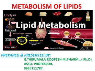
- 1. METABOLISM OF LIPIDS G.THIRUMALA ROOPESH M.PHARM .,( Ph.D) ASSO. PROFESSOR, 9985111787. PREPARED & PRESENTED BY:
- 2. METABOLISM OF LIPIDS •β-Oxidation of saturated fatty acid (Palmitic acid); •Formation and utilization of ketone bodies; ketoacidosis; • De novo synthesis of fatty acids (Palmitic acid); •Biological significance of cholesterol and conversion of cholesterol into; bile acids, steroid hormone and vitamin D; •Disorders of lipid metabolism: Hypercholesterolemia, atherosclerosis, fatty liver and obesity.
- 3. β-Oxidation of saturated fatty acid (Palmitic acid) The fatty acids in the body are mostly oxidized by -oxidation. -Oxidation may be defined as the oxidation of fatty acids on the BETA carbon atom. This results in the sequential removal of a two carbon fragment, acetyl CoA. Fatty acid oxidation —stages and tissues…………………….. The beta oxidation of fatty acids involves three stages:………. I. Activation of fatty acids occurring in the cytosol II. Transport of fatty acids into mitochondria III. Oxidation proper in the mitochondrial matrix. Fatty acids are oxidized by most of the tissues in the body. However, brain, erythrocytes and adrenal medulla cannot utilize fatty acids for energy requirement. I.Fatty acid activation: Fatty acids are activated to acyl CoA by thiokinases or acyl CoA synthetases. The reaction occurs in two steps and requires ATP, coenzyme A and Mg2+. Fatty acid reacts with ATP to form acyladenylate which then combines with coenzyme A to produce acyl CoA (Fig.14.5). In the activation, two high energy phosphates are utilized, since ATP is converted to pyrophosphate (PPi). The enzyme inorganic pyrophosphatase hydrolyses PPi to phosphate(Pi). The immediate elimination of PPi makes this reaction totally irreversible. Three different thiokinases, to activate long chain (10-20 carbon), medium chain (4-12 carbon) and short chain (< 4 carbon) fatty acids have been identified.
- 4. β-Oxidation of saturated fatty acid CARNITINE SHUTTLE FOR TRANSPORT OF ACTIVATED FATTY ACID (ACYL Co A) INTO MITOCHONDRIA
- 5. II. Transport of acyl CoA into mitochondria: The inner mitochondrial membrane is impermeable to fatty acids. A specialized carnitine carrier system (carnitine shuttle) operates to transport activated fatty acids from cytosol to the mitochondria. This occurs in four steps (Fig.14.6). 1. Acyl group of acyl CoA is transferred to carnitine (beta-hydroxyl gamma-trimethyl aminobutyrate), catalysed by carnitine acyltransferase I (present on the outer surface of inner mitochondrial membrane). 2. The acyl-carnitine is transported across the membrane to mitochondrial matrix by a specific carrier protein. 3. Carnitine acyl transferase II (found on the inner surface of inner mitochondrial membrane) converts acyl-carnitine to acyl CoA. 4. The carnitine released returns to cytosol for reuse. It should be noted that the coenzyme A used for activation is different from the one that finally combines with fatty acid in the mitochondria to form acyl CoA. Thus, the cell has two separate pools (cytosolic and mitochondrial) of coenzyme A. Inhibitor of carnitine shuttle : Carnitine acyl transferase I is inhibited by malonyl CoA, a key metabolite involved in fatty acid synthesis that occurs in cytosol details given later). In other words, while the fatty acid synthesis is in progress (reflected by high concentration of malonyl CoA), their oxidation does not occur, since carnitine shuttle is impaired.
- 6. III. Beta Oxidation proper: Each cycle of BETA-oxidation, liberating a two carbon unit- acetyl CoA, occurs in a sequence of four reactions (Fig.14.7). 1. Oxidation : Acyl CoA undergoes dehydrogenation by an FAD-dependent flavoenzyme, acyl CoA dehydrogenase. A double bond is formed between ALPHA and BETA carbons (i.e., 2 and 3 carbons). 2. Hydration : Enoyl CoA hydratase brings about the hydration of the double bond to form - hydroxyacyl CoA. 3. Oxidation : BETA -Hydroxyacyl CoA dehydrogenase catalyses the second oxidation and generates NADH. The product formed is BETA - ketoacyl CoA. 4. Cleavage : The final reaction in beta oxidation is the liberation of a 2 carbon fragment, acetyl CoA from acyl CoA. This occurs by a thiolytic cleavage catalysed by BETA ketoacyl CoA thiolase (or simply thiolase).The new acyl CoA, containing two carbons less than the original, reenters the BETA-oxidation cycle. The process continues till the fatty acid is completely oxidized. The overall reaction for each cycle of BETA -oxidation Cn Acyl CoA + FAD + NAD+ + H2O +CoASH C(n–2) Acyl CoA + Acetyl CoA + FADH2 + NADH + H+. The scheme of fatty acid oxidation discussed above corresponds to saturated (no double bond) and even carbon fatty acids. This occurs most predominantly in biological system.
- 7. Oxidation of palmitoyl CoA: The summary of -oxidation of palmitoyl CoA is shown below Palmitoyl CoA + 7 CoASH + 7 FAD + 7 NAD+ + 7H2O 8 Acetyl CoA + 7 FADH2 + 7 NADH + 7H+ Palmitoyl CoA undergoes 7 cycles of BETA oxidation to yield 8 acetyl CoA. Acetyl CoA can enter citric acid cycle and get completely oxidized to CO2 and H2O. Energetics of BETA-oxidation : The ultimate aim of fatty acid oxidation is to generate energy. The energy obtained from the complete oxidation of palmitic acid (16 carbon) is given in Table 14.2 and Fig.14.8.
- 8. DISORDERS OF α,β, ᵞ & ᵟ-Oxidation of fatty acidS: ALPHA BETA GAMMA OMEGA REFSUMS DISEASE (PHYTANIC ACID ) •SUDDEN INFANT DEATH SYNDROME (SIDS) DUE TO DEFICIENCY OF ( ACYL COA DEHYDROGENASE (MCAD) •PEROXISOMES --- ZELLWEGER SYNDROME DUE TO LONG CHAIN FA NOT OXIDIZED) •JAMAICAN VOMING SICKNESS (Caused due to eating of unripe ackee fruit an unusual toxic amino acid hypoglycin-A) NOT KNOWN HYDROXYLATION REQUIRED PRIOR OXIDATION (DEPENDS ON NADPH, CYT P450.)
- 9. Formation and utilization of ketone bodies The compounds namely acetone, acetoacetate and ᵦ-hydroxybutyrate (or 3-hydroxybutyrate) are known as ketone bodies (Fig.14.10). Only the first two are true ketones while ᵦ-hydroxybutyrate does not possess a keto (CO) group. Ketone bodies are water-soluble and energy yielding. Acetone, however, is an exception, since it cannot be metabolized. Ketogenesis The synthesis of ketone bodies occurs in the liver. The enzymes for ketone body synthesis are located in the mitochondrial matrix. Acetyl CoA, formed by oxidation of fatty acids, pyruvate or some amino acids, is the precursor for ketone bodies. Ketogenesis occurs through the following reactions (Fig.14.11). 1. Two moles of acetyl CoA condense to form acetoacetyl CoA. This reaction is catalysed by thiolase, an enzyme involved in the final step of ᵦ-oxidation. Hence, acetoacetate synthesis is appropriately regarded as the reversal of thiolase reaction of fatty acid oxidation. 2. Acetoacetyl CoA combines with another molecule of acetyl CoA to produce ᵦ-hydroxy ᵦ-methyl glutaryl CoA (HMG CoA). HMG CoA synthase, catalysing this reaction, regulates the synthesis of ketone bodies. 3. HMG CoA lyase cleaves HMG CoA to produce acetoacetate and acetyl CoA. 4. Acetoacetate can undergo spontaneous decarboxylation to form acetone. 5. Acetoacetate can be reduced by a dehydrogenease to ᵦ-hydroxybutyrate. The carbon skeleton of some amino acids (ketogenic) is degraded to acetoacetate or acetyl CoA and, therefore, to ketone bodies, e.g. leucine, lysine, phenylalanine etc.
- 10. Metabolism & Utilization of ketone bodies
- 11. Utilization of ketone bodies: The ketone bodies, being water-soluble, are easily transported from the liver to various tissues. The two ketone bodies—acetoacetate and ᵦ-hydroxybutyrate serve as important sources of energy for the peripheral tissues such as skeletal muscle, cardiac muscle, renal cortex etc. The tissues which lack mitochondria (e.g. erythrocytes) however, cannot utilize ketone bodies. The production of ketone bodies and their utilization become more significant when glucose is in short supply to the tissues, as observed in starvation, and diabetes mellitus. Reactions of ketone bodies : ᵦ-Hydroxybutyrate is first converted to acetoacetate (reversal of synthesis) and metabolized. Acetoacetate is activated to acetoacetyl CoA by a mitochondrial enzyme thiophorase (succinyl CoA acetoacetate CoA transferase). The coenzyme A is donated by succinyl CoA, an intermediate in citric acid cycle. Thiophorase is absent in liver, hence ketone bodies are not utilized by the liver. Thiolase cleaves acetoacetyl CoA to two moles of acetyl CoA (Fig.14.12). The summary of ketone body synthesis, utilization and excretion is depicted in Fig.14.13. Overproduction of ketone bodies : In normal individuals, there is a constant production of ketone bodies by liver and their utilization by extrahepatic tissues. The concentration of ketone bodies in blood is maintained around 1 mg/dl. Their excretion in urine is very low and undetectable by routine tests (Rothera’s test). Regulation of ketogenesis: The ketone body formation (particularly overproduction) occurs primarily due to nonavailability of carbohydrates to the tissues. This is an outcome of excessive utilization of fatty acids to meet the energy requirements of the cells. The hormone glucagon stimulates ketogenesis whereas insulin inhibits.
- 12. Utilization of ketone bodies: DISORDERS: KETONURIA, KETONEMIA, KETOSIS( UNCONTROLLED DIABETES & STARVATION), KETO ACIDOSIS (INSULIN)
- 13. De novo synthesis of fatty acids (Palmitic acid) The dietary carbohydrates and amino acids,when consumed in excess, can be converted to fatty acids and stored as triacylglycerols. Denovo (new) synthesis of fatty acids occurs predominantly in liver, kidney, adipose tissue and lactating mammary glands. The enzyme machinery for fatty acid production is located in the cytosomal fraction of the cell. Acetyl CoA is the source of carbon atoms while NADPH provides the reducing equivalents and ATP supplies energy for fatty acid formation. The fatty acid synthesis may be learnt in 3 stages I. Production of acetyl CoA and NADPH II. Conversion of acetyl CoA to malonyl CoA III. Reactions of fatty acid synthase complex. I. Production of acetyl CoA and NADPH Acetyl CoA and NADPH are the prerequisites for fatty acid synthesis. Acetyl CoA is produced in the mitochondria by the oxidation of pyruvate and fatty acids, degradation of carbon skeleton of certain amino acids, and from ketone bodies. Mitochondria, however, are not permeable to acetyl CoA. An alternate or a bypass arrangement is made for the transfer of acetyl CoA to cytosol. Acetyl CoA condenses with oxaloacetate in mitochondria to form citrate.Citrate is freely transported to cytosol where it is cleaved by citrate lyase to liberate acetyl CoA and xaloacetate. Oxaloacetate in the cytosol is converted to malate (Fig.14.14). Malic enzyme converts malate to pyruvate. NADPH and CO2 are generated in this reaction. Both of them are utilized for fatty acid synthesis. Advantages of coupled transport of acetyl CoA and NADPH : The transport of acetyl CoA from mitochondria to cytosol is coupled with the cytosomal production of NADPH and CO2 which is highly advantageous to the cell for optimum synthesis of fatty acids.
- 14. 1. Production of acetyl CoA and NADPH
- 15. II. Formation of malonyl CoA Acetyl CoA is carboxylated to malonyl CoA by the enzyme acetyl CoA carboxylase (Fig.14.15). This is an ATP- dependent reaction and requires biotin for CO2 fixation. The mechanism of action of acetyl CoA carboxylase is similar to that of pyruvate carboxylase. Acetyl CoA carboxylase is a regulatory enzyme in fatty acid synthesis.
- 16. III. Reactions of fatty acid synthase complex The remaining reactions of fatty acid synthesis are catalysed by a multifunctional enzyme known as fatty acid synthase (FAS) complex. In eukaryotic cells, including man, the fatty acid synthase exists as a dimer with two identical units. Each monomer possesses the activities of seven different enzymes and an acyl carrier protein (ACP) bound to 4’- phosphopantetheine. Fatty acid synthase functions as a single unit catalysing all the seven reactions. Dissociation of the synthase complex results in loss of the enzyme activities. In the lower organisms (prokaryotes), the fatty acid synthesis is carried out by a multienzyme complex in association with a separate acyl carrier protein. This is in contrast to eukaryotes where ACP is a part of fatty acid synthase. The sequence of reactions of the extra— mitochondrial synthesis of fatty acids (palmitate)is depicted in Fig.14.16.
- 17. III. Reactions of fatty acid synthase complex
- 18. 1. The two carbon fragment of acetyl CoA is transferred to ACP of fatty acid synthase, catalysed by the enzyme, acetyl CoA-ACP transacylase. The acetyl unit is then transferred from ACP to cysteine residue of the enzyme. Thus ACP site falls vacant. 2. The enzyme malonyl CoA-ACP transacylase transfers malonate from malonyl CoA to bind to ACP. 3. The acetyl unit attached to cysteine is transferred to malonyl group (bound to ACP). The malonyl moiety loses CO2 which was added by acetyl CoA carboxylase. Thus, CO2 is never incorporated into fatty acid carbon chain. The decarboxylation is accompanied by loss of free energy which allows the reaction to proceed forward. This reaction is catalyzed by ᵦ-ketoacyl ACP synthase. 4. BETA-Ketoacyl ACP reductase reduces ketoacyl group to hydroxyacyl group. The reducing equivalents are supplied by NADPH. 5. BETA-Hydroxyacyl ACP undergoes dehydration. A molecule of water is eliminated and a double bond is introduced between ALPHA and BETA carbons. 6. A second NADPH-dependent reduction, catalysed by enoyl-ACP reductase occurs to produce acyl-ACP. The four-carbon unit attached to ACP is butyryl group. The carbon chain attached to ACP is transferred to cysteine residue and the reactions 2-6 are repeated 6 more times. Each time, the fatty acid chain is lengthened by a two-carbon unit (obtained from malonyl CoA). At the end of 7 cycles, the fatty acid synthesis is complete and a 16-carbon fully saturated fatty acid—namely palmitate—bound to ACP is produced. 7. The enzyme palmitoyl thioesterase separates palmitate from fatty acid synthase. This completes the synthesis of palmitate. Fatty acid synthase (FAS) functions as a single unit catalysing all the seven reactions.
- 19. Summary of palmitate synthesis Of the 16 carbons present in palmitate, only two come from acetyl CoA directly. The remaining 14 are from malonyl CoA which, in turn, is produced by acetyl CoA. The overall reaction of palmitate synthesis is summarized . 8 Acetyl CoA + 7 ATP + 14 NADPH + 14 H+ Palmitate + 8 CoA + 7 ADP + 7 Pi + 6H2O Regulation of fatty acid synthesis Fatty acid production is controlled by enzymes, metabolites, end products, hormones and dietary manipulations. Some of the important regulatory mechanisms are discussed hereunder. Acetyl CoA carboxylase : This enzyme controls a committed step in fatty acid synthesis. Acetyl CoA carboxylase exists as an inactive protomer (monomer) or an active polymer. Citrate promotes polymer formation, hence increases fatty acid synthesis. On the other hand, palmitoyl CoA and malonyl CoA cause depolymerization of the enzyme and, therefore, inhibit fatty acid synthesis. Hormonal influence : Hormones regulate acetyl CoA carboxylase by a separate mechanism— phosphorylation (inactive form) and dephosphorylation (active form) of the enzyme. Glucagon, epinephrine and norepinephrine inactivate the enzyme by cAMPdependent phosphorylation. Insulin, on the other hand, dephosphorylates and activates the enzyme. Thus, insulin promotes fatty acid synthesis while glucagon inhibits. Insulin stimulates tissue uptake of glucose, and conversion of pyruvate to acetyl CoA. This also facilitates fatty acid formation. Dietary regulation : Consumption of high carbohydrate or fat-free diet increases the synthesis of acetyl CoA carboxylase and fatty acid synthase, which promote fatty acid formation. On the other hand, fasting or high fat diet decreases fatty acid production by reducing the synthesis of these two enzymes. Availability of NADPH : The reducing equivalents for fatty acid synthesis are provided by NADPH which come either from citrate (acetyl CoA) transport or hexose monophosphate shunt. About 50-60% of required NADPH is obtained from HMP shunt, which significantly influences fatty acid synthesis.
- 20. Cholesterol is found exclusively in animals, hence it is often called as animal sterol. The total body content of cholesterol in an adult man weighing 70 kg is about 140 g i.e., around 2 g/kg body weight. Cholesterol is amphipathic in nature, since it possesses both hydrophilic and hydrophobic regions in the structure. Cholesterol Functions of cholesterol Cholesterol is essential to life, as it performs a number of important functions 1. It is a structural component of cell membrane. 2. Cholesterol is the precursor for the synthesis of all other steroids in the body. These include steroid hormones, vitamin D and bile acids. 3. It is an essential ingredient in the structure of lipoproteins in which form the lipids in the body are transported. 4. Fatty acids are transported to liver as cholesteryl esters for oxidation.
- 21. CHOLESTEROL BIOSYNTHESIS About 1 g of cholesterol is synthesized per day in adults. Almost all the tissues of the body participate in cholesterol biosynthesis. The largest contribution is made by liver (50%),intestine (15%), skin, adrenal cortex, reproductive tissue etc. The enzymes involved in cholesterol synthesis are found in the cytosol and microsomal fractions of the cell. Acetate of acetyl CoA provides all the carbon atoms in cholesterol. The reducing equivalents are supplied by NADPH while ATP provides energy. For the production of one mole of cholesterol, 18 moles of acetyl CoA, 36 moles of ATP and 16 moles of NADPH are required. By administering acetate with 14C isotope label either on the methyl ( CH3) group or carboxyl ( COO) group, the origin of carbon atoms in the entire molecule of cholesterol has been established. The sources of carbon atoms and the key intermediates of cholesterol formation are depicted in Fig.14.26, and the detailed reactions are given in Fig.14.27. The synthesis of cholesterol may be learnt in 5 stages 1. Synthesis of HMG CoA 2. Formation of mevalonate (6C) 3. Production of isoprenoid units (5C) 4. Synthesis of squalene (30C) 5. Conversion of squalene to cholesterol (27C).
- 22. 1. Synthesis of BETA-hydroxy BETA-methylglutaryl CoA (HMG CoA) : Two moles of acetyl CoA condense to form acetoacetyl CoA. Another molecule of acetyl CoA is then added to produce HMG CoA. These reactions are similar to that of ketone body synthesis. However, the two pathways are distinct, since ketone bodies are produced in mitochondria while cholesterol synthesis occurs in cytosol. Thus, there exist two pools of HMG CoA in the cell. Further, two isoenzymes of HMG CoA synthase are known. The cytosomal enzyme is involved in cholesterol synthesis whereas the mitochondrial HMG CoA synthase participates in ketone body formation. 2. Formation of mevalonate : HMG CoA reductase is the rate limiting enzyme in cholesterol biosynthesis. This enzyme is present in endoplasmic reticulum and catalyses the reduction of HMG CoA to mevalonate. The reducing equivalents are supplied by NADPH. SYNTHESIS OF CHOLESTEROL
- 23. 3. Production of isoprenoid units : In a three step reaction catalysed by kinases, mevalonate is converted to 3-phospho 5-pyrophosphomevalonate which on decarboxylation forms isopentenyl pyrophosphate (IPP). The latter isomerizes to dimethylallyl pyrophosphate (DPP). Both IPP and DPP are 5-carbon isoprenoid units. 4. Synthesis of squalene : IPP and DPP condense to produce a 10-carbon geranyl pyrophosphate (GPP). Another molecule of IPP condenses with GPP to form a 15-carbon farnesyl pyrophosphate (FPP). Two units of farnesyl pyrophosphate unite and get reduced to produce a 30-carbon squalene. 5. Conversion of squalene to cholesterol : Squalene undergoes hydroxylation and cyclization utilizing O2 and NADPH and gets converted to lanosterol. The formation of cholesterol from lanosterol is a multistep process with a series of about 19 enzymatic reactions. The following are the most important reactions…………………… Reducing the carbon atoms from 30 to 27. Removal of two methyl groups from C4 and one methyl group from C14. Shift of double bond from C8 to C5. Reduction in the double bond present between C24 and C25. The enzymes (about 19?) involved in the conversion of lanosterol to cholesterol are associated with endoplasmic reticulum. 14- Desmethyl lanosterol, zymosterol, cholestadienol and desmosterol are among the intermediates in the cholesterol biosynthesis. The penultimate product is 7-dehydrocholesterol which, on reduction, finally yields cholesterol. Cholesterol biosynthesis is now believed to be a part of a major metabolic pathway concerned with the synthesis of several other isoprenoid compounds. These include ubiquinone (coenzyme Q of electron transport chain) and dolichol (found in glycoprotein). Both of them are derived from farnesyl pyrophosphate.
- 24. SYNTHESIS OF CHOLESTEROL 1. Synthesis of HMG CoA 2. Formation of mevalonate (6C) 3. Production of isoprenoid units (5C) 4. Synthesis of squalene (30C) 5. Conversion of squalene to cholesterol (27C).
- 25. Regulation of cholesterol synthesis Cholesterol biosynthesis is controlled by the rate limiting enzyme HMG CoA reductase, at the beginning of the pathway (Fig.14.28). HMG CoA reductase is found in association with endoplasmic reticulum, and is subjected to different metabolic controls. 1. Feedback control : 2. Hormonal regulation : 3. Inhibition by drugs : 4. HMG CoA reductase activity 1. Feedback control : The end product cholesterol controls its own synthesis by a feedback mechanism. Increase in the cellular concentration of cholesterol reduces the synthesis of the enzyme HMG CoA reductase. This is achieved by decreasing the transcription of the gene responsible for the production of HMG CoA reductase. Feedback regulation has been investigated with regard to LDL-cholesterol taken up by the cells, and the same mechanism is believed to operate whenever cellular cholesterol level is elevated. 2. Hormonal regulation : The enzyme HMG CoA reductase exists in two interconvertible forms. The dephosphorylated form of HMG CoA reductase is more active while the phosphorylated form is less active. The hormones exert their influence through cAMP by a series of reactions which are comparable with the control of the enzyme glycogen synthase. The net effect is that glucagon and glucocorticoids favour the formation of inactive HMG CoA reductase (phosphorylated form) and, thus, decrease cholesterol synthesis. On the other hand, insulin and thyroxine increase cholesterol production by enhancing the formation of active HMG CoA reductase (dephosphorylated form).
- 26. 3. Inhibition by drugs : The drugs compactin and lovastatin (mevinolin) are fungal products. They are used to decrease the serum cholesterol level in patients with hypercholesterolemia. Compactin and lovastatin are competitive inhibitors of the enzyme HMG CoA reductase and, therefore, reduce cholesterol synthesis. About 50 to 60% decrease in serum cholesterol level has been reported by a combined use of these two drugs. 4. HMG CoA reductase activity is inhibited by bile acids. Fasting also reduces the activity of this enzyme.
- 27. Conversion of cholesterol into---- Bile acids, Steroid hormone and Vitamin D DEGRADATION OF CHOLESTEROL The steroid nucleus (ring structure) of the cholesterol cannot be metabolized in humans. Cholesterol (50%) is converted to bile acids, excreted in feces, serves as a precursor for the synthesis of steroid hormones, vitamin D, coprostanol and cholestanol. The latter two are the fecal sterols, besides cholesterol. I. Synthesis of bile acids II. Synthesis of steroid hormones from cholesterol III. Synthesis of vitamin D I. Synthesis of bile acids The bile acids possess 24 carbon atoms, 2 or 3 hydroxyl groups in the steroid nucleus and a side chain ending in carboxyl group. The bile acids are amphipathic in nature since they possess both polar and non-polar groups. They serve as emulsifying agents in the intestine and actively participate in the digestion and absorption of lipids. The synthesis of primary bile acids takes place in the liver and involves a series of reactions (Fig.14.29). The step catalysed by 7 α-hydroxylase is inhibited by bile acids and this is the rate limiting reaction. Cholic acid and chenodeoxycholic acid are the primary bile acids and the former is found in the largest amount in bile. On conjugation with glycine or taurine, conjugated bile acids (glycocholic acid, taurocholic acid etc.) are formed which are more efficient in their function as surfactants. In the bile, the conjugated bile acids exist as sodium and potassium salts which are known as bile salts.
- 28. Conversion of cholesterol into---- Bile acids, Steroid hormone and Vitamin D
- 29. In the intestine, a portion of primary bile acids undergoes deconjugation and dehydroxylation to form secondary bile acids (deoxycholic acid and lithocholic acid). These reactions are catalysed by bacterial enzymes in the intestine. Enterohepatic circulation :The conjugated bile salts synthesized in the liver accumulate in gall bladder. From there they are secreted into the small intestine where they serve as emulsifying agents for the digestion and absorption of fats and fat soluble vitamins. A large portion of the bile salts (primary and secondary) are reabsorbed and returned to the liver through portal vein. Thus the bile salts are recycled and reused several times in a day. This is known as enterohepatic circulation. About 15- 30 g of bile salts are secreted into the intestine each day and reabsorbed. However, a small portion of about 0.5 g/day is lost in the feces. An equal amount (0.5 g/day) is synthesized in liver to replace the lost bile salts. The fecal excretion of bile salts is the only route for the removal of cholesterol from the body. Cholelithiasis : Bile salts and phospholipids are responsible for keeping the cholesterol in bile in a soluble state. Due to their deficiency (particularly bile salts), cholesterol crystals precipitate in the gall bladder often resulting in cholelithiasis—cholesterol gall stone disease. Cholelithiasis may be due to defective absorption of bile salts from the intestine, impairment in liver function, obstruction of biliary tract etc. The patients of cholelithiasis respond to the administration of bile acid chenodeoxy cholic acid, commonly known as chenodiol. It is believed that a slow but gradual dissolution of gall stones occurs due to chenodiol. For severe cases of cholelithiasis, surgical removal of gall bladder is the only remedy.
- 30. II. Synthesis of steroid hormones from cholesterol Cholesterol is the precursor for the synthesis of all the five classes of steroid hormones (a) Glucocorticoids (e.g. cortisol) (b) Mineralocorticoids (e.g. aldosterone) (c) Progestins (e.g. progesterone) (d) Androgens (e.g. testosterone) (e) Estrogens (e.g. estradiol). A brief outline of steroid hormonal synthesis is given in Fig.14.30 III. Synthesis of vitamin D 7-Dehydrocholesterol, an intermediate in the synthesis of cholesterol, is converted to cholecalciferol (vitamin D3) by ultraviolet rays in the skin. A brief summary of prominent sources and the major pathways for utilization of cholesterol with the liver as the central metabolic organ is depicted in Fig.14.31. Transport of cholesterol Cholesterol is present in the Plasma lipoproteins in two forms 1. About 70-75% of it is in an esterified form with long chain fatty acids. 2. About 25-30% as free cholesterol. This form of cholesterol readily exchanges between different lipoproteins and also with the cell Membranes.
- 31. Role of LCAT : High density lipoproteins (HDL) and the enzyme lecithin- cholesterol acyltransferase (LCAT) are responsible for the transport and elimination of cholesterol from the body. LCAT is a plasma enzyme, synthesized by the liver. It catalyses the transfer of fatty acid from the second position of phosphatidyl choline (lecithin) to the hydroxyl group of cholesterol (Fig.14.32). HDL-cholesterol is the real substrate for LCAT and this reaction is freely reversible. LCAT activity is associated with apo-A1 of HDL. The cholesterol (cholesteryl) ester forms an integral part of HDL. In this manner, the cholesterol from the peripheral tissues is trapped in HDL, by a reaction catalysed by LCAT and then transported to liver for degradation and excretion. This mechanism is commonly known as reverse cholesterol transport.
- 32. Disorders of lipid metabolism: Hypercholesterolemia, Atherosclerosis, Fatty liver, Obesity & Hypocholesterolemia : A decrease in the plasma cholesterol, although less common, is also observed. Hyperthyroidism, pernicious anemia, malabsorption syndrome, hemolytic jaundice etc., are some of the disorders associated with hypocholesterolemia.
- 34. Hypercholesterolemia Increase in plasma cholesterol (> 200 mg/dl) concentration is known as hypercholesterolemia and is observed in many disorders. 1. Diabetes mellitus : Due to increased cholesterol synthesis since the availability of acetyl CoA is increased. 2. Hypothyroidism (myxoedema) : This is believed to be due to decrease in the HDL receptors on hepatocytes. 3. Obstructive jaundice : Due to an obstruction in the excretion of cholesterol through bile. 4. Nephrotic syndrome : Increase in plasma globulin concentration is the characteristic feature of nephrotic syndrome. Cholesterol elevation is due to increase in plasma lipoprotein fractions in this disorder. Hypercholesterolemia is associated with atherosclerosis and coronary heart disease (CHD). More specifically, LDL-cholesterol is positively correlated, whereas HDL-cholesterol is negatively correlated with CHD. Bad cholesterol and good cholesterol :Cholesterol is a natural metabolite performing a wide range of functions (membrane structure, precursor for steroid hormones, bile acids). The usages good and bad to cholesterol, although inappropriate, are still in use. The cholesterol in high concentration, present in LDL, is considered bad due to its involvement in altherosclerosis and related complications. Thus, LDL may be regarded as lethally dangerous lipoprotein. Small dense LDL (sdLDL) is considered to be the most dangerous fraction of LDL associated with CHD. On the other hand, HDL cholesterol is good since its high concentration counteracts atherogenesis. HDL may be considered as highly desirable lipoprotein.
- 35. Affects of lifestyles on serum cholesterol level :Individual lifestyles and habits certainly influence serum cholesterol, and thus play a significant role in the development coronary heart disease. The parametres such as high blood pressure, emotional stress, smoking, drinking of soft water (against hard water), coffee drinking, lack of exercise, obesity (particlarly of abdomen) elevate serum cholesterol level. Control of hypercholesterolemia: Several measures are advocated to lower the plasma cholesterol level. 1.Consumption of PUFA 2.Dietary cholesterol 3.Plant sterols 4.Dietary fiber 5.Avoiding high carbohydrate diet 6.Impact of lifestyles 7. Moderate alcohol cosumption 8.Use of drugs : Statins & Clofibrate
- 36. 1. Consumption of PUFA : Dietary intake of polyunsaturated fatty acids (PUFA) reduces the plasma cholesterol level. PUFA will help in transport of cholesterol by LCAT mechanism (described earlier) and its excretion from the body. The oils with rich PUFA content include cottonseed oil, soya bean oil, sunflower oil, corn oil, fish oils etc. Ghee and coconut oil are poor sources of PUFA. 2. Dietary cholesterol : Dietary cholesterol influence on plasma cholesterol is minimal. However, avoidance of cholesterol-rich foods is advocated, and a dietary intake of <300 mg/day is advised. Certain drugs (e.g. ezetimide) inhibit intestinal cholesterol absorption. 3. Plant sterols : Certain plant sterols and their esters (e.g. sitostanol esters) reduce plasma cholesterol levels. They inhibit the intestinal absorption of dietary cholesterol. 4. Dietary fiber : Fiber present in vegetables decreases the cholesterol absorption from the intestine. 5. Avoiding high carbohydrate diet : Diets rich in carbohydrates (e.g. sucrose) should be avoided to control hypercholesterolemia. Control of hypercholesterolemia: Several measures are advocated to lower the plasma cholesterol level.
- 37. 6. Impact of lifestyles : Elevation in plasma cholesterol is obseved in people with smoking, abdominal obesity, lack of exercise, stress, high blood pressure, consumption of soft water etc. Therefore, adequate changes in the lifestyles will bring down plasma cholesterol. 7. Moderate alcohol cosumption: The beneficial effects of moderate alcohol intake are masked by the ill effects of chronic alcoholism. Red wine is particularly beneficial due to its antioxidants, besides low alcohol content. 8. Use of drugs : Statins & Clofibrate: Drugs such as lovastatin which inhibit HMG CoA reductase and decrease cholesterol synthesis are used. Statins currently in use include atorvastatin, simvastatin, fluvastatin and pravastatin. Statins are usually taken at night to ensure maximum effect (HMG CoA reductase activity at peak about 6 hours after dark). Certain drugs—cholestyramine and colestipol—bind with bile acids and decrease their intestinal reabsorption. This helps in the conversion of more cholesterol to bile acids and its excretion through feces. Clofibrate increases the activity of lipoprotein lipase and reduces the plasma cholesterol and triacylglycerols.
- 38. Atherosclerosis
- 39. Disorders that may cause atherosclerosis: Certain diseases are associated with atherosclerosis. These include diabetes mellitus, hyperlipoproteinemias, nephrotic syndrome, hypothyroidism etc. Many other factors like obesity, high consumption of saturated fat, excessive smoking, lack of physical exercise, hypertension, stress etc., are the probable causes of atherosclerosis. Relation between HDL and CHD: The increased levels of plasma HDL (good cholesterol) are correlated with a low incidence of cardiovascular disorders. Women have higher HDL and are less prone to heart diseases compared to men. This is attributed to estrogens in women. Strenuous physical exercise, moderate alcohol intake, consumption of unsaturated fatty acids (vegetable and fish oils), reduction in body weight—all tend to increase HDL levels and reduce the risk CHD. Atherosclerosis Atherosclerosis (Greek: athere—mush) is a complex disease characterized by thickening or hardening of arteries due to the accumulation of lipids (particularly cholesterol, free, and esterified) collagen, fibrous tissue, proteoglycans, calcium deposits etc. in the inner arterial wall. Atherosclerosis is a progressive disorder that narrows and ultimately blocks the arteries. Infarction is the term used to indicate the stoppage of blood flow resulting in the death of affected tissue. Coronary arteries— the arteries supplying blood to heart—are the most commonly affected leading to myocardial infarction or heart attacks. Causes of atherosclerosis and CHD : The development of atherosclerosis and the risk for the coronary heart disease (CHD) is directly correlated with plasma cholesterol and LDL. On the other hand, plasma HDL is inversely correlated with CHD.
- 40. Lipoprotein a and CHD: Lipoprotein a (Lp-a) is almost identical in structure to LDL. Lp-a contains an additional apoprotein, apo-a. Lp-a inhibits fibrinolysis. Recent studies have shown that elevation of lipoprotein-a in the plasma (>30 mg/dl) suggests increased risk of CHD. It is hypothesized that elevated Lp-a reduces the breakdown of blood clots by interfering with plasminogen activation. This results in intravascular thrombosis, and increased risk of heart attacks. Indians have higher levels of Lp-a compared to Western population. Antioxidants and atherosclerosis: Antioxidants, in general, decrease the oxidation of LDL. There is some evidence, based on the epidemiological studies that taking of antioxidants (vitamins E and C or ᵦ-carotene) reduces the risk of atherosclerosis, and CHD.
- 41. Fatty liver
- 42. Fatty liver The normal concentration of lipid (mostly phospholipid) in liver is around 5%. Liver is not a storage organ for fat, unlike adipose tissue. However, in certain conditions, lipids— especially the triacylglycerols—accumulate excessively in liver, resulting in fatty liver(Fig.14.37). In the normal liver, Kupffer cells contain lipids in the form of droplets. In fatty liver, droplets of triacylglycerols are found in the entire cytoplasm of hepatic cells. This causes impairment in metabolic functions of liver. Fatty liver is associated with fibrotic changes and cirrhosis, Fatty liver may occur due to two main causes. 1. Increased synthesis of triacylglycerols 2. Impairment in lipoprotein synthesis. 1. Increased triacylglycerol synthesis : Mobilization of free fatty acids from adipose tissue and their influx into liver is much higher than their utilization. This leads to the overproduction of triacylglycerols and their accumulation in liver. Diabetes mellitus, starvation, alcoholism and high fat diet are associated with increased mobilization of fatty acids that often cause fatty liver. Alcohol also inhibits fatty acid oxidation and, thus, promotes fat synthesis and its deposition. 2. Impaired synthesis of lipoproteins : The synthesis of very low density lipoproteins (VLDL) actively takes place in liver. VLDL formation requires phospholipids and apoprotein B. Fatty liver caused by impaired lipoprotein synthe is may be due to : a defect in phospholipid synthesis; a block in apoprotein formation; a failure in the formation/secretion of lipoprotein.
- 43. Among the three causes, fatty liver due to impairment in phospholipid synthesis has been studied in some detail. This is usually associated with the dietary deficiency of lipotropic factors such as choline, betaine, inositol etc. Deficiency of essential fatty acids leads to a decreased formation of phospholipids. Further, excessive consumption of cholesterol competes with essential fatty acids and impairs phospholipid synthesis. Certain chemicals (e.g. puromycin, ethionine, carbon tetrachloride, chloroform, lead, phosphorus etc.) that inhibit protein synthesis cause fatty liver. This is due to a blockade in the synthesis of apoprotein B required for VLDL Production. Lipoprotein synthesis and their secretion require ATP. Decrease in the availability of ATP, sometimes found in pyridoxine and pantothenic acid deficiency, impairs lipoprotein formation. The action of ethionine in the development of fatty liver is believed to be due to a reduction in the availability of ATP. Ethionine competes with methionine and traps the available adenosine (as adenosylethionine)—thereby reducing ATP levels. Deficiency of vitamin E is associated with fatty liver. Selenium acts as a protective agent in such a condition. Endocrine factors: Certain hormones like ACTH, insulin, thyroid hormones, adrenocorticoids promote deposition of fat in liver.
- 44. Deficiency of lipotropic factors such as choline, betaine, inositol
- 45. These are the substances the deficiency of which causes fat (triacylglycerol) to accumulate in liver. This may happen despite the fatty acid synthesis and uptake by liver being normal. Important lipotropic factors These include choline, betaine, methionine and inositol. Folic acid, vitamin B12, glycine and serine also serve as lipotropic factors to some extent. LIPOTROPIC FACTORS Action of lipotropic factors Choline and inositol are components of phospholipids and, hence, required for their synthesis. The other lipotropic factors are directly or indirectly concerned with transmethylation reactions and, ultimately, the synthesis of choline. Severe protein deficiency (e.g. kwashiorkor) causes fatty liver. This is due to a defect in the synthesis of choline as a result of insufficient amino acid (particularly methionine) supply. In other words the non-availability of methyl groups may lead to fatty liver (Fig.14.37). Choline deficiency and fatty liver: Several explanations are offered to understand choline deficiency causing fatty liver : 1) Decreased phospholipid synthesis (Fig.14.37); 2) Impaired formation of lipoprotein membrane; 3) Reduced synthesis of carnitine due to insufficient supply of methyl groups; 4) Impairment in fatty acid oxidation.
- 47. Obesity is an abnormal increase in body weight due to excessive fat deposition (>25%). Overeating, lack of exercise and genetic predisposition play a significant role in the development of obesity. Nutritional basis Men and women are considered as obese if their weight due to fat (in adipose tissue), respectively, exceeds more than 20% and 25% of body weight. Obesity is basically a disorder ofexcess calorie intake, in simple language— overeating. It has to be remembered that every 7calories of excess consumption leads to 1 g fat deposit and increase in body weight. Overeating—coupled with lack of physical exercise—contribute to obesity. Obesity due to virus infection : It was found that around 15% of people weighing more than 120 kg had antibodies to adenovirus-36 in their blood, implying that this virus infection (causes cold, diarrhea etc.), by an unknown mechanism contributes to obesity. Surprisingly, adenovirus-36 infected individuals have normal serum cholesterol and other lipid parameters. OBESITY
- 48. Body mass index (BMI) Clinical obesity is represented by body mass index. BMI is calculated as the weight (in kilograms) divided by the height (in meters2). Weight (kg) BMI (kg/m2) = [height (m)2] Healthy reference range for BMI is between 18.5–24.9 kg/m2. Grade I obesity or overweight – BMI 25–30 kg/m2 Grade II or clinical obesity – BMI > 30 kg/m2 Grade III or morbid obesity – BMI > 40 kg/m2 Obesity is associated with many health complications e.g. type II diabetes, CHD, hypertension, stroke, arthritis, gall bladder disease. In recent years, the ratio between waist and hip sizes (for men < 0.9 and for women < 0.85) is considered as more effective than BMI, particularly with regard to the risk of heart diseases. The lower is the waist to hip ratio, the lower the risk for health complications, and therefore better is the health. Genetics, obesity and leptin: One gene namely ob gene, expressed in adipocytes (of white adipose tissue) producing a protein called leptin (mol. Wt. 16,000 daltons), is associated with obesity. Leptin is regarded as a body weight regulatory hormone. It binds to a specific receptor in the brain and functions as a lipostat. When the fat stores in the adipose tissue are adequate, leptin levels are high. This signals to restrict the feeding behaviour and limit fat deposition. Further, leptin stimulates lipolysis and inhibits lipogenesis. Any genetic defect in leptin or its receptor will lead to extreme overeating and obesity. Treatment of such obese individuals with leptin has been shown to reverse obesity. During starvation, leptin levels fall which promote feeding, and fat production and its deposition.
- 49. Obesity and adipose tissue Obesity is due to an increase in both the number and size of adipocytes (of adipose tissue). There are two types of adipose tissues 1. White adipose tissue : The fat is mostly stored and this tissue is metabolically less active. 2. Brown adipose tissue : The stored fat is less but the tissue is metabolically very active. Brown adipose tissue possesses high proportion of mitochondria and cytochromes but low activity of ATP synthase. This is an active centre for the oxidation of fat and glucose and is responsible for the diet-induced thermogenesis. The peculiarity of mitochondria of brown adipose tissue is that the oxidation and phosphorylation are not coupled. Mitochondrial oxidation produces more heat and less ATP. A specific protein—namely thermogenin—has been isolated in the inner membrane of these mitochondria. Thermogenin functions like an uncoupler and dissipates the energy in the form of heat, and thus blocks the formation of ATP. Brown adipose tissue is mostly found in hibernating animals, and the animals exposed to cold, besides the newborn. In adult humans, though not a prominent tissue, it is located in the thoracic region. It is significant to note that brown adipose tissue is almost absent in obese persons. Some individuals are fortunate to have active brown adipose tissue. They eat and liberate it as heat, and therefore do not become obese. Pharmacological treatment of obesity : In recent years, synthetic lipids such as Olestra and Orlistat are used to treat obesity. They taste like natural lipids but cannot be digested, and excreted unchanged.
- 50. METABOLIC SYNDROME (MS) Metabolic syndrome (MS) is a cluster of different conditions that adversely affect the health. The components contributing to MS include abdominal obesity, insulin resistance, dyslipidemia, elevated blood pressure, overnutrition, sedentary lifestyles etc. As per WHO criteria, metabolic syndrome has the following characteristics. 1. Insulin resistance – identified either as type 2 diabetes or elevated fasting blood glucose (>100 mg/dl) or impaired glucose tolerance. 2. And any two of the following (i) Hypertension (> = 140/90 mm Hg) (ii) Dyslipidemia (serum TG > = 150 mg/dl or HDL cholesterol <35 mg/dl in men or <39 mg/dl in women. (iii) BMI >30 kg/m2 or waist : hip ratio of >0.9 in men or 0.85 in women. Metabolic syndrome can be managed by healthy habits and change in lifestyles— restricted balanced diet, adequate intake of fiber, and antioxidants, exercise, avoiding smoking, stress-free life etc. CACHEXIA This is opposite of what is seen in obesity. Cachexia is characterized by a failure to maintain normal lipid stores in the body. It involves higher rate of fat mobilization than deposition. Anorexia nervosa is a total loss of appetite. This is mostly seen in females in the age group 10-30 years. Surprisingly, majority of the affected individuals are from wealthy families where food is aplenty. And some members in these families may be even obese! Anorexia nervosa is more a psychiatric disease.