
Recommended
More Related Content
What's hot
What's hot (20)
Similar to DIGESTION, ABSORPTION AND METABOLISM OF LIPIDS.docx
Similar to DIGESTION, ABSORPTION AND METABOLISM OF LIPIDS.docx (20)
More from Dr. Santhosh Kumar. N
More from Dr. Santhosh Kumar. N (20)
Recently uploaded
Recently uploaded (20)
DIGESTION, ABSORPTION AND METABOLISM OF LIPIDS.docx
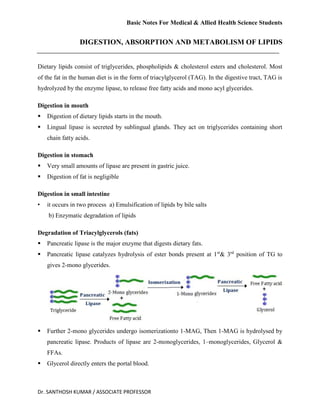
- 1. Basic Notes For Medical & Allied Health Science Students Dr. SANTHOSH KUMAR / ASSOCIATE PROFESSOR DIGESTION, ABSORPTION AND METABOLISM OF LIPIDS Dietary lipids consist of triglycerides, phospholipids & cholesterol esters and cholesterol. Most of the fat in the human diet is in the form of triacylglycerol (TAG). In the digestive tract, TAG is hydrolyzed by the enzyme lipase, to release free fatty acids and mono acyl glycerides. Digestion in mouth Digestion of dietary lipids starts in the mouth. Lingual lipase is secreted by sublingual glands. They act on triglycerides containing short chain fatty acids. Digestion in stomach Very small amounts of lipase are present in gastric juice. Digestion of fat is negligible Digestion in small intestine • it occurs in two process a) Emulsification of lipids by bile salts b) Enzymatic degradation of lipids Degradation of Triacylglycerols (fats) Pancreatic lipase is the major enzyme that digests dietary fats. Pancreatic lipase catalyzes hydrolysis of ester bonds present at 1st & 3rd position of TG to gives 2-mono glycerides. Further 2-mono glycerides undergo isomerizationto 1-MAG, Then 1-MAG is hydrolysed by pancreatic lipase. Products of lipase are 2-monoglycerides, 1–monoglycerides, Glycerol & FFAs. Glycerol directly enters the portal blood.
- 2. Basic Notes For Medical & Allied Health Science Students Dr. SANTHOSH KUMAR / ASSOCIATE PROFESSOR Degradation of cholesterol esters: Cholesteryl esterases hydrolyses of ester bonds present at 3rd Carbon of cholesteryl esters to gives cholesterol. Degradation of Phospholipids Pancreatic juice is rich in Phospholipase A2 isresponsible for the hydrolysis of phospholipids, which cleaves the fatty acid at the 2nd position of phospholipids. The products of phospholipase are FFAs & Lysophospholipids. The major digestible products (2-mono acylglycerol, FFA, cholesterol & lyso- phospholipids) now ready for absorption in the intestinal lumen by passive process. ABSORPTION OF LIPIDS Products combine with bile salts to form micelles. Micelles are absorbed into the intestinal mucosal cells. Micelles serve as the major vehicles for the transport of lipids from the intestinal lumen to intestinal mucosa. In intestinal mucosal cells, TGs are resynthesized from absorbed FFA & mono acyl glycerol. The resynthesized TGs cannot pass in to portal blood, but it enters the lymphatic vessels as a chylomicrons. The lymphatic duct then discharges the chylomicrons into bloodstream through thoracic duct & left sub clavian vein, reached to heart, then to peripheral tissues & finally to liver. Chylomicrons are hydrolyzed in adipose tissues by lipoprotein lipase to yield FFA & glycerol. In adipose tissues, the FFAs are re esterified with glycerol-3-P to synthesize TGs, which are stored as fuel reserve and provides the energy to skeletal muscle, cardiac muscle & liver.
- 3. Basic Notes For Medical & Allied Health Science Students Dr. SANTHOSH KUMAR / ASSOCIATE PROFESSOR METABOLISM OF TRIGLYCEROL LIPOGENESIS • Synthesis of TGs from fatty acyl CoA (from acetyl CoA) glycerol-3-P in adipose tissues. • Glycerol 3-P when it combined with two molecules of fatty acyl CoA to form lysophosphatidic acid first then phosphatidic acid by the enzymes acyl transferases. • Phosphatidic acid looses phosphate group and binds with third molecule of acyl CoA to form Triacylglycerides by acyl transferase. LIPOLYSIS: • Adipose tissue is a specialized connective tissue designed for synthesis, storage, hydrolysis of TGs. • Most of the body TGs stored as lipid droplets in cytoplasm of adipocyte whenever need of metabolic energy, the TGs undergo lipolysis. • Lipase catalyzes hydrolysis of ester bonds present at 1st , 3rd than 2nd position of TG to gives Di, mono glycerides & finally glycerol & FFAs. UTILIZATION OF FATTY ACIDS FOR PRODUCTION OF ENERGY Fatty acids in the circulation are taken up by cells and used for energy production, primarily in mitochondria, in a process integrated with energy generation from other sources. Utilization of fatty acids for energy production varies from tissue to tissues, depends on metabolic status, that is, fed or fasted. Oxidation of fatty acids: CoA esters of fatty acids are the substrates for oxidation. They are mainly o β-oxidation of fatty acid o α-Oxidation of fatty acid and ώ- oxidation of fatty acid Glycerol -3-Phoshate Lysophosphatidic acid Phosphatidic acid Di Acylglycerol Acyl Transferase FA- CoA CoA Acyl Transferase Phosphatase Acyl Transferase Triacylglycerol Reactions of Lipogenesis FA- CoA CoA FA- CoA CoA Pi
- 4. Basic Notes For Medical & Allied Health Science Students Dr. SANTHOSH KUMAR / ASSOCIATE PROFESSOR BETA (β) OXIDATION OF FATTY ACIDS Fatty acid oxidation occurs through the sequential removal of 2-carbon units by oxidation at the β-carbon position of the fatty acyl-CoA molecule. β - Oxidation of fatty acids takes place in the mitochondrial matrix. β - Oxidation of fatty acids involves in three stages I) Activation of Fatty Acids in cytosol II) Transport of fatty acyl CoA into mitochondria III) β – Oxidation reactions
- 5. Basic Notes For Medical & Allied Health Science Students Dr. SANTHOSH KUMAR / ASSOCIATE PROFESSOR I. Activation of Fatty Acidsin cytosol In the presence of ATP and coenzyme A, the enzyme acyl CoA synthetase catalyses the conversion of a free fatty acid to fatty acyl CoA (active form fatty acids). II. Transport of fatty acyl CoA into the mitochondrion Long-chain fatty acids cannot directly cross the inner mitochondrial membrane to mitochondrial matrix, so carnitine carrier system helps to transport the long chain fatty acids from cytosol to mitochondrial matrix. But Short-chain fatty acids are transported through the membrane freely. III. Steps of β-Oxidation: First reaction is the dehydrogenation. It is catalyzed by Acyl-CoA dehydrogenase to form ∆2 Trans enoyl-CoA from fatty acyl CoA. FAD converted to FADH2 Second reaction: water is added across the new double bond to make -β -hydroxy acyl-CoA from ∆2 trans-enoyl-CoA by enoyl-CoA hydratase. Third reaction: oxidation of the hydroxyl group at the β- position which forms a β - ketoacyl-CoA. This is the 2nd oxidation step catalyzed by β –OH-acyl-CoA dehydrogenase. NAD converted to NADH+H+ Final reaction: Thiolase catalyzed reaction, cleaves the β - ketoacyl-CoA using CoASH to produce acetyl-CoA and a fatty acyl CoA that has been shortened by two carbons from original FA-CoA. This sequence of four steps is repeated until the fatty acyl chain is completely degraded to acetyl CoA. OXIDATION OF PALMITOYL CoA: Beta oxidation of Palmitoyl CoA undergoes 7 cycles to yield 8 acetyl CoAs. I. In the β- oxidation - (7 cycles) Acyl-CoA dehydrogenase [1 FADH2] 1.5 ATPs β -OH-acyl-CoA Dehydrogenase [1NADH+H+ ] 2.5 ATP One cycle of β –oxidation produces 4 ATPs Total 7 β- oxidation cycles produces 7×4 = 28 ATPs Initial activation step use - 2 ATPs Total ATPs Produced 26 ATPs
- 6. Basic Notes For Medical & Allied Health Science Students Dr. SANTHOSH KUMAR / ASSOCIATE PROFESSOR II. from Acetyl CoA: One acetyl CoA can enter the TCA cycle & get completely oxidized to produce CO2 & H2O and produced 10 ATPs. Total 8 Acetyl CoA produced (8x 10 ATPs) = 80ATPs From one molecule of palmitoyl CoA completely oxidized to produce 106ATPs Beta oxidation 26 ATPs NET ATPs : 106 ATPs TCA cycle 80 ATPs METABOLISM OF KETONE BODIES Ketone bodies are Acetoacetic acid, β-OH butyric acid & acetone. Ketone bodies are formed from acetyl CoA resulting from β oxidation of fatty acids. KETOGENESIS: Ketogenesis is the process of formation of ketone bodies from acetyl CoA in the liver mitochondria. Two molecules of acetyl CoA react with each other to form acetoacetyl CoA. Acetyl CoA Acetoacetyl CoA HMG –CoA (β-Hydroxy –β-Methyl-Glutaryl CoA) Acetyl CoA Acetyl CoA CoA-SH Acetyl CoA Thiolase HMG-CoA Synthase HMG-CoA Lyase H2O CoASH + H+ Acetoacetate Acetone β- Hydroxybutyrate NADH+ H+ NAD+ H+ CO2 β- OH butyrate dehydrogenase Non- Enzymatic reaction Reactions of Ketogenesis
- 7. Basic Notes For Medical & Allied Health Science Students Dr. SANTHOSH KUMAR / ASSOCIATE PROFESSOR HMG CoA (β hydroxyl- β methyl glutaryl CoA) formed from the condensation of acetoacetyl CoA with acetyl CoA catalyzed by HMG CoA synthetase. HMG-CoA lyase enzyme catalyzes the cleavage of HMG-CoA to acetoacetate and acetyl CoA Acetoacetate produces β-hydroxybutyrate in a reaction catalyzed by β-hydroxybutyrate dehydrogenase in the present NADH. Both acetoacetate and β-hydroxybutyrate can be transported across the mitochondrial membrane then enter to the blood stream to be used as a fuel by other cells of the body. Small amounts of acetoacetate are spontaneously (non- enzymatically) decarboxylated to form acetone. While under normal conditions, acetone formation is negligible. Regulation: HMG CoA synthase is the regulatory enzyme of ketogenesis Ketogenesis induced by increased fatty acids in the blood. It is inhibited by high level of CoASH Inhibited by insulin and stimulated by glucagon KETOLYSIS / UTILYZATION OF KETONE BODIES Acetoacetyl CoA Acetyl CoA Acetyl CoA Acetoacetate β- Hydroxybutyrate NADH+H+ β- OH butyrate Dehydrogenase Acetoacetyl-CoA Thiolase CoA-SH NAD+ CoASH + ATP AMP + PPi Thiophorase Thiokinase Succinyl CoA Succinate
- 8. Basic Notes For Medical & Allied Health Science Students Dr. SANTHOSH KUMAR / ASSOCIATE PROFESSOR Ketolysis is the complete oxidation of ketone bodies in mitochondria of extrahepatic tissues, but not in the liver because of thiokinase and thiophorase enzymes deficiency. β-hydroxy butyrate is dehydrogenated to form acetoacetate, the reaction is catalyzed by β-hydroxy butyrate dehydrogenase. Activation of acetoacetate to acetoacetyl-CoA occurs by one of two pathways: a) One mechanism involves the activation of acetoacetate with ATP in presence of CoASH catalyzed by thiokinase. b) Other mechanism involves Succinyl CoAand the enzyme Thiophorase. Acetoacetyl CoA is split to acetyl CoA by thiolase and oxidized via citric acid cycle to CO2 and H2O CLINICAL SIGNIFICATION: Normal levels of ketone bodies in blood is less than 1 mg/dl and in urine 1mg/day KETOSIS: It is a condition, increase of the ketone bodies in the blood (Ketonemia), their appearance in the urine (ketonuria) and together with acetone odor in the breath is called ketosis. Causes: Uncontrolled diabetes mellitus, starvation & excessive alcohol consumption Unbalanced diet i.e. high fat, low carbohydrate diet. KETOACIDOSIS Ketoacidosis is a metabolic state associated with high conc. of ketone bodies and uncontrolled ketosis. In ketoacidosis, the body fails to adequately regulate ketone bodies production, so it leads to decreased the blood pH & acidosis In Diabetic ketoacidosis (DKA) It is mainly seen in patients with uncontrolled diabetes mellitus. Characterized by hyperglycemia causes severe osmotic dieresis leads to dehydration & loss of electrolytes.
- 9. Basic Notes For Medical & Allied Health Science Students Dr. SANTHOSH KUMAR / ASSOCIATE PROFESSOR Accumulation of acidic ketone bodies in blood leads to decreases pH, resulting in metabolic acidosis (ketoacidosis). Since it is seen due to insulin deficiency. Patients of DKA will show a deep gasping breathing pattern (referred to as "Kussmaul respiration") due to compensatory hyperventilation. Patient exhales acetone - Ketotic" (fruity) odor is present. Management: Administration of insulin Restoration of water & electrolyte balance Diagnosed by For ketone bodies - Urine dipstick test &Rothera’s test METABOLISM OF CHOLESTEROL The enzymes involved in cholesterol biosynthesis are present in cytosol The process of cholesterol synthesis: Squalene Squalene-2, 3-Oxide Squalene Mono oxygenase Lanosterol Cholesterol Isopentenyl Pyrophosphate (IPP) NADPH +H+ O2+NADPH+ H+ H2O+ NADP NADP +PPi 5 IPPs Squalene Synthase Acetyl CoA + Acetyl CoA Acetoacetyl CoA HMG-CoA Thiolase CoASH H2O HMG CoA Synthase Mevalonate HMG-CoA Reductase Isopentenyl Pyrophosphate (IPP) 2NADPH +2H+ 3ATP Acetyl CoA CoASH + H+ 2NADP+CoASH 3ADP+ Pi+CO2 3 Phosphorylations & one Decarboxylation reactions Cholesterol Synthesis
- 10. Basic Notes For Medical & Allied Health Science Students Dr. SANTHOSH KUMAR / ASSOCIATE PROFESSOR 1) Cholesterol is synthesized from cytosolic acetyl CoA. It starts by the condensation of three molecules of acetyl CoA to forming acetoacetyl CoA and finally HMG CoA by thiolase & HMG-CoA synthase. 2) HMG-CoA is converted to mevalonate (C6), catalyzed by HMG-CoA reductase. It consumes two NADPH. This reaction is the rate limiting step of cholesterol biosynthesis. 3) Mevalonate is then activated by three phosphorylated and decarboxylated reactions, catalyzed by kinase, ATP-dependent decarboxylase yields isopentenyl pyrophosphate (IPP). IPP is involved in isomerisation reaction to form dimethylallyl pyrophosphate (DMPP). 4) Four molecules of isopentenyl pyrophosphate condense with DMPP to generate squalene by squalene synthase (NADPH-dependent enzyme). 5) Squalene undergoes cyclization to yield lanosterol, catalyzed by squalene mono oxygenase. Then finally it forms cholesterol by a multi step process, resulting in the shortening of the carbon chain from 30C to 27C. Regulation of cholesterol synthesis: HMG CoA reductase, is a rate-limiting enzyme, It is the major control point for cholesterol biosynthesis, and is subject to different kinds of metabolic control like by covalent modifications, hormonal regulation, some drugs. COMPOUNDS DERIVED FROM CHOLESTEROL: Vitamin-D Bile Acids Bile Salts Steroid Hormones Derivatives of Cholesterol Vit -D3 (Cholecalciferol) formed from cholesterol.Vit-D3 enhancing intestinal absorption of Ca+ and helps in bone formation. Cholesterol involved in the synthesis of steroid hormones like glucocorticoids, mineralocorticoids & sex hormones (estrogen and progesterone), help control CHOLESTEROL
- 11. Basic Notes For Medical & Allied Health Science Students Dr. SANTHOSH KUMAR / ASSOCIATE PROFESSOR metabolisms, inflammation, immune functions, water and electrolyte balance, development of sexual characteristics. Cholesterol is required for the synthesis of bile salts that are important in lipid digestion &absorption. DISORDERS OF LIPID METABOLISM HYPERCHOLESTEROLEMIA: High levels of cholesterol in the blood. It is a combination of "hyperlipidemia" (elevated levels of lipids in the blood) & "hyperlipoproteinemia" (elevated levels of lipoproteins in the blood). Causes • Environmental factors - Dietary choices. • Genetic factors - due to a single gene defect such as in the case of familial hypercholesterolemia. Secondary causes: o High consumption alcohol, smoking o Obesity o Type 2 Diabetes mellitus o Nephrotic syndrome o Obstructive jaundice and o Hypothyroidism ATHEROSCLEROSIS: Thickening or hardening of arteries due to the accumulation of lipids & cholesterol in the inner arterial wall. • Hypercholesterolemia leads to formation of atheromatous plaques in the arteries (atherosclerosis) and progressive stenosis (narrowing) or even completes occlusion (blockage) of the involved arteries. • Finally to obstruct blood flow (low oxygen supply) in coronary artery, results in a myocardial infarction or heart attack. Primary causes: o Environmental factors: Dietary choices. Deposition of lipids (TG, Cholesterol& other lipids) in the inner arterial wall. ↓ Formation of plaque ↓ Endothelial damage ↓ Narrowing of the arterial lumen
- 12. Basic Notes For Medical & Allied Health Science Students Dr. SANTHOSH KUMAR / ASSOCIATE PROFESSOR o Genetic factors: additive effects of multiple genes & single gene defect (case of familial hypercholesterolemia). Secondary causes: o Obesity, high consumption alcohol & smoking, Diabetes mellitus, nephrotic syndrome, obstructive jaundice, hypothyroidism & Cushing’s syndrome o It can reduced by Consumption of PUFA with low cholesterol diet Dietary fibers Avoiding high carbohydrate diet & Taking statin drugs (lovastatin, simvastatin & atorvastatin) Treatment: o Eat low fat diet & exercise o Moderate consumption of alcohol, o Drugs therapy e.g., statin drugs. o Natural antioxidants such as Vit- E, C or β-carotene (protecting LDL against oxidation) OBESITY Anyone who is more than 20% above the standard weight for the same age, sex, and race, generally is considered to be overweight. Causes: Physiological: Overeating than caloric requirement, use of oral contraceptives for prolonged period & post menopausal women, genetic influences. Metabolically: Diabetes mellitus & Hyperlipidemia states Hormonal: Hypothyroidism, Hypogonadism, Hypopituitarism & cushing’s syndrome Obesity leads to hypertension, cardio vascular diseases, stroke, diabetes & atherosclerosis Metabolic changes in Obesity: o Serum triglyceride & Cholesterol levels are raised. o Mobilization of free fatty acid is reduced. o Promotes lipogenesis and inhibits lipolysis o BMR is normal. LIPOPROTEINEMIAS
- 13. Basic Notes For Medical & Allied Health Science Students Dr. SANTHOSH KUMAR / ASSOCIATE PROFESSOR Lipoproteinemias are high conc. of plasma lipoproteins. o Primary: genetic disorder o Secondary: underlying disease (thyroid, liver, renal diseases) HYPO LIPOPROTEINEMIAS (Decreased lipoproteins) Lipoproteinemias Defect Decreased levels A beta lipoproteinemias Absence of LDL Low plasma cholesterol, chylomicrons because of low VLDL & TG Familial α-lipoprotein deficiency (Tangier’s disease) Deficiency of HDL Plasma HDL completely absent & also Apo A-I & II HYPER LIPOPROTEINEMIAS: FREDERICK SON CLASSIFICATION OF LIPOPROTEINEMIAS Lipoproteinemias Defect Increased levels Type-I: Familial lipoproteinemias Lipoprotein lipase enzyme & Apo C-II TG, chylomicrons, LDL, VLDL & HDL Type –II: Familial hyper cholesterolaemia LDL receptors LDL, Cholesterol &TG Type –III: Familial Dys-Beta lipoproteinemias (broad beta disease) Defect in remnant metabolism LDL, VLDL, cholesterol, TG, Apo-E & Apo-B Type-IV: Familiar Hyper triglyceridaemia: VLDL, endogenous TG. Decreased HDL, LDL. Type-V : Combined hyper Lipidaemias Increased chylomicrons & VLDL & TG.Decreased con.c of HDL & LDL HYPOCHOLESTEROLEMIA: Decreased cholesterol below the normal levels in plasma Causes: o Hyper thyroidism, o Liver necrosis, o Malabsorption syndrome,
- 14. Basic Notes For Medical & Allied Health Science Students Dr. SANTHOSH KUMAR / ASSOCIATE PROFESSOR o Pernicious anemia & Haemolytic jaundice CHAPTER-17 DIGESTION AND ABSORPTION OF PROTEINS Digestion and absorption takes place in GI tract at different levels with proteolytic enzymes aided by gastro intestinal hormones Digestion in stomach Hydrochloric acid secreted by parietal cells of stomach denatures proteins and makes it susceptible to hydrolysis by protease enzymes. Pepsin: It is secreted as inactive zymogen –pepsinogens by chief cells of stomach Pepsinogens is converted to pepsin by HCl HCl Pepsinogens Pepsin Later, pepsin itself causes activation of pepsinogens to pepsinogens to pepsin (auto catalysis). Pepsin Pepsinogens Pepsin It is an endopeptidase with an optimum pH of 1 of 2. Its end products are proteases and peptones. It hydrolysis peptide bonds of phenyl alanine, tyrosine, tryptophan, glutamate Digestion in duodenum Proteolytic enzymes of pancreatic juice carry on further digestion Pancreatic enzymes secreted trypsinogen, chymotrypsinogen, procarboxypeptidase, proelastes and collagenase. These are zymogens released from pancrease mediated by the secretion of cholecystokinin and secretin ( two polypeptide hormones of digestive tract) Trypsin It is secreted as inactive trypsinogen. The activation is brought about by an enzyme enterokinase of intestinal juice. It is an endopeptidase, hydrolyses peptide bonds formed by basic amino acids arginine, histidine and lysine
- 15. Basic Notes For Medical & Allied Health Science Students Dr. SANTHOSH KUMAR / ASSOCIATE PROFESSOR It acts on proteins, proteoses, peptones converting them to polypeptides. Optimum pH is 8 to 9. Enterokinase Trypsinogen Trypsin Chymotrypsin It is secreted as inactive chymotrypsinogen. It is an endopeptidase specific for peptide bonds of aromatic amino acids (Phenyl alanine, tyrosine and tryptophan). Optimum pH is 7 to 8 Trypsin Chymotrypsinogen Chymotrypsin Proelastase: It is an endopeptidase. Hydrolyses peptide bonds next to small amino acid residues such as glycine, alanine and serine Trypsin Proelastase Elastase Carboxypeptidase: It is an exopeptidase. It hydrolyses peptide bonds adjacent to carboxyl terminal, liberating amino acids Trypsin Procarboxypeptidase Carboxypeptidase Proteins ↓ Proteoses ↓ Peptones ↓ Amino acids
- 16. Basic Notes For Medical & Allied Health Science Students Dr. SANTHOSH KUMAR / ASSOCIATE PROFESSOR In small intestine: Amino peptidase Present in intestinal juice, it is an exopeptidase. It hydrolyses peptide bonds adjacent to amino terminal, liberating amino acids. Dipeptidase: Present in intestinal juice It hydrolyses dipeptides to liberate amino acids Dipeptide Amino acid + Amino acid End products of protein digestion is amino acids ABSORPTION Amino acids are absorbed in small intestine by active transport, which is sodium dependent system These are six different carrier proteins for different groups of amino acids. L-amino acids are preferentially absorbed compared to D- amino acids. Amino acids are carried to liver and further metabolized Gamma glutamyl cycle (Meisters cycle) It takes place in kidney tubules and brain. Amino acids enter into the cell with help of glutathione. Meisters cycle Digestion of proteins
- 17. Basic Notes For Medical & Allied Health Science Students Dr. SANTHOSH KUMAR / ASSOCIATE PROFESSOR Tripeptide glutathione (GSH) reacts with the neutral amino acids to form gamma glutamyl amino acid .This is catalyzed by gamma glutamyl transferase The glutamyl amino acid is then cleaved to give the free amino acid inside the membrane. The net result is the transfer of an amino acid across the membrane. The transport of one molecule of amino acid and regeneration of GSH requires 3 molecules of ATP The transport of one amino acid with regeneration of glutathione required three molecules of ATP OVERVIEW OF DIGESTION OF PROTEINS S/ No Proteolytic Enzyme Zymogen Form Activation Substrate Products Optimum pH Specifications 01 Pepsin Stomach Gastric Juice Pepsinoge n By Gastric HCL Protein Proteoses Peptone Poly peptides 1-2 Low pH It is a endopeptidase specifically hydrolysis on interior peptide bond where the- NH2 group is coming from aromatic amino acid. Like phenyl alanine or tyrosine. 02 Trypsin Intestine Pancreatic Juice Trypsinog en Entero kinase (In intestine) Protein Proteoses Peptone Poly peptides 7-8 It is endopeptidase specifically hydrolyzing the interior peptide bond where the -NH2 group is coming from basic amino acid like arginine, histidine, and lysine etc 03 Chymotry psin Pancreatic Juice Chymotrip sinogen Trypsin pancreas Protein Proteoses Peptone Poly peptides 7-8 It is endopeptidase specifically hydrolyzing the interior peptide bond where the – COOH group coming from aromatic amino acid, like phenyl alanine or tyrosine. 04 Carboxy peptidase Pancreas Pancreatic Juice Procarbox y peptidase Trypsin Pancreas Polypepti des Tri peptides & Di peptides 7-8 It is an exopeptidase acts from carboxylic end of polypeptide cleaving one or 2 or 3 amino acids at a time producing tripeptides, dipeptides & amino acids. 05 Amino peptidase Intestinal mucosal - - Polypepti des Tri Peptides & Di 7-8 It is an exopeptidase acts from amino end of polypeptide cleaving 2 or 3 amino acids at a time producing tripeptides,
- 18. Basic Notes For Medical & Allied Health Science Students Dr. SANTHOSH KUMAR / ASSOCIATE PROFESSOR cells peptides dipeptides. 06 Tri peptidase Intestinal juice - - Tri peptides Di peptides & Amino acids 7-8 It is an exopeptidase acts from amino end of polypeptide cleaving one or 2 or 3 amino acids at a time producing tri- peptides, dipeptides & free a a. 07 Di peptidase Intestinal juice - - Di peptides Amino acids 7-8 It is an exopeptidase acts from amino end of polypeptide cleaving one or 2 or 3 a.a s at a time producing tripeptides, dipeptides & free a.a’s Note: Di & Tripeptidases also help in completion of protein digestion, final products are free amino acids. CHAPTER-18 GENERAL REACTIONS OF AMINO ACID The amount of free amino acids distributed throughout the body is called amino acid pool. Plasma level for most amino acids varies widely throughout the day. It ranges between 4 –8 mg/dl. It tends to increase in the fed state and tends to decrease in the post absorptive state.
- 19. Basic Notes For Medical & Allied Health Science Students Dr. SANTHOSH KUMAR / ASSOCIATE PROFESSOR All the catabolic pathway of amino acids involves decarboxylation, transamination, and deamination reactions to form α-keto acid. During the catabolism of amino acids, the α-keto acids that remain after removal of carboxylic group as carbon dioxide and amino group as ammonia from amino acids are called the carbon skeleton. Further carbon skeleton either enters carbohydrate (glucogenic) or lipid metabolisms (ketogenic) Ammonia is converted into urea in the liver. a) Decarboxylation reactions: Decarboxylation means removal of CO2 from amino acid with formation of corresponding amines. It is catalyzed by decarboxylase enzyme. Examples of decarboxylation reaction include: o Decarboxylation of histidine to form histamine. o Decarboxylation of tyrosine to form Tyramine o Glutamate to form gamma amino glutamic acid (GABA) b) Transamination reactions: Transamination means transfer of amino group from α-amino acid to α-keto acid with formation of a new α-amino acid and a new α-keto acid. It occurs in liver. Biosynthesis of nonessential amino acids Dietary protein Breakdown of tissue proteins Amino acids pool Sources of amino acid Fate of amino acids Formation of Structural protein (eg: tissue proteins) Biosynthesis of peptide hormones, haemoglobin, myoglobin & enzymes) Synthesis of biological important peptides (eg: Glutathione) Biosynthesis of NPN substances (eg: Urea, uric acid, creatine, creatinine)
- 20. Basic Notes For Medical & Allied Health Science Students Dr. SANTHOSH KUMAR / ASSOCIATE PROFESSOR All amino acids can be transaminated except lysine, threonine, proline and hydroxy proline. All transamination reactions are reversible. α- Amino acid New α- Keto acid + Transaminase / Amino transferase & PLP + α- Keto acid New α- Amino acid It is catalyzed by aminotransferases (transaminases). It needs pyridoxal phosphate as a coenzyme. Pyridoxal phosphate accepts the amino group from amino acid to form pyridoxamine phosphate, which in turn gives the amino group to α-keto acid. Examples of transaminases: Serum glutamate oxaloacetate transaminase (SGOT) /Aspartate amino transferase (AST) is specific for the cardiac lesions & also acute liver diseases, Hemolytic anemia. Serum glutamate pyruvate transaminase (SGPT) / Alanine amino transferase (ALT) is specific for liver diseases and also increases in acute hepatitis, hepatic Jaundice DEAMINATION REACTIONS: Deamination means the removal of amino group from α-amino acid in the form of ammonia with formation of α-keto acid in liver and kidney. Deamination occurs in two ways: A. Oxidative deamination. B. Non oxidative deamination. A. Oxidative deamination: It is catalyzed by enzymes L & D-amino acid oxidase and Glutamate dehydrogenase. Removal of ammonia from the amino acids with the link of oxidation process L- Alanine Pyruvate Alanine amino transferase (ALT) PLP + α- ketoglutarate + L-Glutamate L- Aspartate Oxaloacetate Aspartate amino transferase (AST) PLP + α- ketoglutarate L-Glutamate +
- 21. Basic Notes For Medical & Allied Health Science Students Dr. SANTHOSH KUMAR / ASSOCIATE PROFESSOR B. Non-oxidative deamination: Removal of ammonia from the amino acids without link of oxidation process Dehydratase enzyme deaminates amino acids containing hydroxyl group & it needs pyridoxal phosphate as coenzyme. METABOLISM OF AMMONIA: The transport of ammonia mostly occurs in the form of glutamine or alanine and not as free ammonia. Alanine is important for NH3 transport from muscle to liver by glucose – alanine cycle (in starvation). The ammonia is absorbed from the intestine into the portal venous blood. Liver promptly removes the ammonia from the portal blood. DISPOSAL OF AMMONIA: Ammonia has been disposed as Urea From amino acids via Transamination & deamination Degradation of biogenic amines AMMONIA Formation and Metabolic fate of ammonia in the body Converted to urea (urea cycle) Synthesis of non essential amino acids Formation of Purines & Pyrimidines Maintains the acid base balance via NH4 + ions From the amino group of purines and Pyrimidines acids By the action of intestinal bacteria (urease) on urea acids Synthesis of Glutamine Formation of amino sugars Serine + H2O Pyruvate + NH4 + Serine dehydratase Threonine dehydratase Threonine α- ketoglutarate + NH4 + Glutamic acid + H2O α- Ketoglutarate + NH4 + Glutamate dehydratase NADH+H+ NAD+
- 22. Basic Notes For Medical & Allied Health Science Students Dr. SANTHOSH KUMAR / ASSOCIATE PROFESSOR UREA Urea is the end product of protein catabolism. It is synthesized in liver & transported to kidneys for excretion in urine. The nitrogen of amino acids converted to ammonia first, it is toxic to the body. Then immediately it converted to urea by detoxification process (non toxic product). About 80-90% of N2 in the urine is in the form of urea. Structure of urea: Urea molecule has two —NH2 groups joined by a carbonyl (C=O) functional group. In this two —NH2 groups derived from one from ammonia & the other from aspartate. Carbonyl (C=O) group is supplied by CO2 UREA CYCLE (FORMATION OF UREA) The urea cycle takes place partly in the cytosol and partly in the mitochondria. Carbamyl phosphate synthetase 1 [CPS1]: This mitochondrial enzyme converts the ammonia into carbamyl phosphate, which is an unstable high energy compound. It requires two molecules of AMP + PPi to drive its synthesis. Ornithine transcarbamylase: The next reaction also takes place in the liver, mitochondrial matrix space, where ornithine is converted into citrulline. The remainder of the urea cycle takes place in the cytosol. Arginino-succinate synthetase: Once in the cytosol, citrulline Urea formation (Urea cycle)
- 23. Basic Notes For Medical & Allied Health Science Students Dr. SANTHOSH KUMAR / ASSOCIATE PROFESSOR condenses with aspartate and the reaction is driven by ATP. In this way aspartate contributes the second nitrogen atom to urea, the first having come from ammonia. [Production of arginino-succinate is an energetically expensive process, since the ATP is split to AMP and pyrophosphate. The pyrophosphate is then cleaved to inorganic phosphate using pyrophosphatase, so the overall reaction costs two equivalents of high energy phosphate per mole]. Arginino-succinate lyase: Elimination of fumarate from arginino-succinate then yields arginine. [Fumarate is formed as a by-product. Fumarate is not transported by mitochondria, so this requires the presence of cytosolic fumarase to form malate]. Arginase: Cleavage of arginine by arginase to produce urea regenerates ornithine, which is then available for another round of the cycle. Regulation of urea cycle: Carbamyl phosphate synthetase-I is rate limiting step. CPS-1 is strongly activated by N-acetyl glutamate (NAG), which controls the overall rate of urea production More intake of protein rich meal it leads to increasing NAG in liver. Then leads to increased urea synthesis. Inherited disorders of urea cycle: Inherited disorders Clinical features Hyper ammonemia type-I Carbamyl phosphate synthetase -1 (CRP-1) enzyme defect. Ammonia toxicity is occurs. Ornithinemia (or) Hyper ammonemia type-II Ornithine transcarbamylase enzyme defect Increased levels of glutamine, NH3& ornithine seen in blood. Citrullinemia Arginino succinate synthetase enzyme defect. Mental retardation occurs in this case. Increased levels of NH3& citrulline are seen in blood. Argininosuccinic acidurias It is inherited disorder in fatal (before 2year of age). Arginino succinase enzyme defect.
- 24. Basic Notes For Medical & Allied Health Science Students Dr. SANTHOSH KUMAR / ASSOCIATE PROFESSOR Mental retardation. Increased levels of Arginino-succinate are seen in blood & urine. Hyper argininemia Arginase enzyme defect. Hyper ammonemia is occurs. Increased excretion of lysine, cystine, ornithine & arginine in urine. CLINICAL ASPECT OF AMMONIA: The concentration of ammonia in blood from 40 to 70 μ gm/100ml, AMMONIA TOXICITY: A marginal elevation of NH3 in blood is found to be toxic to the body. Hyper ammonemia: 1) Acquired hyper ammonemia is usually the result of cirrhosis of the liver with the development of a collateral circulation, which shunts the portal blood around the organ, there by severely reducing the synthesis of urea. 2) Inherited hyper ammonemia results from genetic defects in the urea cycle enzymes. Ammonia intoxication is characterized by: o A peculiar flapping tremor. o Slurring of speech. o Blurring of vision. o And in severe cases coma and death due to increased NH3 concentration in blood and brain.
- 25. Basic Notes For Medical & Allied Health Science Students Dr. SANTHOSH KUMAR / ASSOCIATE PROFESSOR METABOLISM OF NON PROTEIN NITROGENOUS (NPN) SUBSTANCES Definition: The nitrogen containing substances other than protein are called non-protein nitrogenous substances. The important NPN substances are o Urea o Uric acid and o Creatinine Urea formations see in above page no: Clinical significance of urea: Normal levels: Blood urea conc is 15 to 45 mg/dl Hyper Uremia : Increased urea levels in blood more than 45mg/dl Hyper uremia mainly occurs in three conditions Pre Renal Conditions Renal Conditions Post Renal Conditions Dehydration, shock, severe burn, major surgeries & Hemorrhage Acute & chronic glomerulonephritis and Hydronephrosis (obstruction of the free flow of urine from the kidney) Enlargement of prostate, Stones in urinary tract and Tumors of bladder
- 26. Basic Notes For Medical & Allied Health Science Students Dr. SANTHOSH KUMAR / ASSOCIATE PROFESSOR Hypo Uremia: o Decreased urea levels seen in Severe liver diseases like liver hepatitis, Decreased protein intake And Protein malnutrition II) URIC ACID Uric acid is an end product of purine nucleotide catabolism Uric acid is a heterocyclic compound of carbon, nitrogen, oxygen, and hydrogen with the formula C5H4N4O3. Biomedical Importance of Uric acid It is a strong reducing agent. It plays as a potent antioxidant Act as a free radical scavenging molecule acting together with thiol groups of proteins. Removes both hydroxyl & peroxyl radicals. Formation of uric acid: Catabolism of the purine nucleotides leads to the production of uric acid. The enzyme xanthine oxidase makes uric acid from xanthine and hypoxanthine, which in turn are produced from purines. CLINICAL SIGNIFICANCE: Normal levels of Uric acids:
- 27. Basic Notes For Medical & Allied Health Science Students Dr. SANTHOSH KUMAR / ASSOCIATE PROFESSOR o In serum/ plasma: 3.0 - 7.0 mg/dl o In urine: 0.5 to 0.7gm/day HYPER URICAEMIA: - Increased uric acid levels in serum & decreased excretion by the kidney - Ethanol, increases the turnover of ATP & uric acid production A) GOUT: - It is a very painful arthritic condition. It may affect men between the ages of 40 to 50 years Gout is associated with either – Increased formation of uric acid - Decreased renal excretion Needle shaped urate crystals that get deposited in & around the joints of the extremities and cause inflammation (painful reddish swelling) called gouty tophi (clusters of urate crystals). Gout, apart from joints, may affect, kidneys and gall bladder in long standing untreated cases, associated with diabetes and obesity Classification of gout: Primary gout Secondary gout An inborn error of metabolism due to overproduction of uric acid. Due to the defective enzymes of purine biosynthesis Symptoms like painful arthritis & renal failure It results from a variety of diseases Increased production of uric acid leads to increased destruction or turnover of cells (Leukemia, Polycythemia & psoriasis) Hyper catabolic states (starvation, trauma) Treatment o Reduce dietary purine intake & restrict alcohol o Taking uricosuric drugs (Increase renal excretion & decrease the reabsorption of uric acid ) o Allopurinol is used mainly for Gout Allopurinol is an analog of hypoxanthine Competitive inhibitor of xanthine oxidase Decreasing the formation of uric acid Allopurinol
- 28. Basic Notes For Medical & Allied Health Science Students Dr. SANTHOSH KUMAR / ASSOCIATE PROFESSOR B) LESCH-NYHAN SYNDROME It is X- linked inherited disorder, That affects only males Mainly caused by a complete deficiency of an enzyme HGPRT (Hypoxanthine guanine phosphoribosyl transferase) It leads to decreased rate of salvage pathway, resulting in accumulation of PRPP (phosphoribosyl pyrophosphate) PRPP stimulates purine biosynthesis leads to increases production of uric acid Symptoms: o Self mutilation (destructive biting of fingers & lips) o Hyperuricemia, o Primary Gout, o Urinary tract stones and nephrolithiasis, o Neurological symptoms like mental retardation Allopurinol is used in treatment but does not alleviate the neurologic symptoms III) CREATINE / CREATININE: o Creatine is a nitrogenous organic acid that occurs naturally in vertebrates. o Helps to supply energy to all cells in the body, primarily muscle. Formation of Creatine and Creatine Phosphate The primary locations of creatine synthesis in the body mainly liver and kidneys. Three amino acids L-arginine, glycine, and L-methionine are involved in creatine formation.
- 29. Basic Notes For Medical & Allied Health Science Students Dr. SANTHOSH KUMAR / ASSOCIATE PROFESSOR The glycine and arginine react to produce two compounds ornithine and guanidinoacetate, catalyzed by a mitochondrial enzyme transamidinase in kidneys. This reaction is first rate- limiting step of creatine biosynthesis. S-adenosylmethionine (SAM) to donate the methyl group from methionine to guanidinoacetate thus producing creatine by guanidinoacetate N-methyl transferase in liver. Creatine reversible process catalyzed by creatine kinase, with the help of ATP to form phosphocreatine or creatine phosphate, then stored in skeletal muscles (95%), liver & kidneys (5%). Phospho creatine is a key component in energy metabolism in muscles. The creatine kinase is a key protein that is able to regenerate ATP for muscle exercise. FORMATION OF CREATININE The metabolism of creatinine is a spontaneous, non-enzymatic process. Creatinine is formed from dehydrate creatine and from dephosphorylated creatine phosphate, Creatinine is endogenous non-protein nitrogenous product formed in muscle from the high-energy compound creatine phosphate. The amount of creatinine produced is fairly constant & is primarily a function of muscle mass. Creatinine is removed from plasma by glomerular filtration & then excreted in urine. Methyl Transferase (In Liver) Guanidoacetate CREATINE Creatine Phosphate S-adenosylmethionine (SAM) ATP Glycine Transamidinase (In kidney) Ornithine Formation of Creatine and Creatinine Arginine S-adenosylhomocysteine (SAH) Creatine Kinase (In skeletal muscle) ADP CREATININE H2O Pi
- 30. Basic Notes For Medical & Allied Health Science Students Dr. SANTHOSH KUMAR / ASSOCIATE PROFESSOR Clinical significance: Elevated levels of creatinine in serum are associated with various renal diseases. Elevated levels of serum creatinine & is the half mark of chronic renal failure & may be observed in extensive muscle destruction also. Normal levels: Serum creatinine – 0.6 to 1.5 mg/100ml Daily excretion of creatinine in urine is 1 to 2 gms/day. Increased levels Decreased levels - Muscle dystrophy - Impaired renal functions - Reduced renal blood flow - Congenital heart failure. - Diuretics, such as coffee and tea, cause more frequent urination, thus potently decreasing creatinine levels. - A decrease in muscle mass will also cause a lower creatinine, as will pregnancy. - Vegetarians have been shown to have lower creatinine levels. Difference between creatine and creatinine Creatine Creatinine It is a muscle protein, present as creatine phosphate It is an anhydrous form of creatine It formed from glycine, arginine and methionine It formed in muscle from creatine phosphate It helps to supply energy to all muscle cells in the body. In the early stage of renal disease, creatinine clearance test is sensitive index of impaired renal function CHAPTER-19 METABOLISM OF INDIVIDUAL AMINO ACIDS METABOLISM OF GLYCINE Glycine is a simple, neutral, nutritionally non essential, glycogenic, optically inactive but metabolically very active amino acid. Formation:
- 31. Basic Notes For Medical & Allied Health Science Students Dr. SANTHOSH KUMAR / ASSOCIATE PROFESSOR Glycine synthesized from serine catalyzed by Serine OH methyl transferase SERINE GLYCINE THF Methylene THF Catabolism: Serine and glycine both enters anabolic and catabolic process. Serine undergoes non oxidative deamination by dehydratase with PLP to pyruvate and NH3, so glycine is glucogenic in nature. Glycine forms glyoxylate to oxalate on transamination reaction. Glycine + Pyruvate Glyoxylate +Alanine Oxalate also formed on deamination of Glycine. Glycine undergoes oxidative deamination to form NH3, CO2 and one carbon unit methylene -THFA, further ammonia converted to urea. Metabolic role of glycine: It is essential for protein synthesis. Heme is mainly synthesized by glycine and succinyl CoA. Glycine + Succinyl CoA γ ALA Heme It required for the formation of tissue protein like collagen (every 3rd amino acid was glycine) Glycine reacts with γ-glutamyl cysteine to form glutathione It helps in protects membranes of RBC from oxidation Play on important role in detoxifying H2O2 and lipids, peroxides and other harmful by products). Benzoic acid detoxified by glycine to form hippuric acid by conjugation in liver Hippuric acid (Detoxification) GLYCINE Amino acetone Glycocholic acid (Bile acid) Purines (C4, C5 & N7) Glyoxylate Glucose (gluconeogenesis) N5, N10 methylene FH4 Tissue protein synthesis Synthesis of Glutathione Formation of Creatine Heme synthesis Formation of serine
- 32. Basic Notes For Medical & Allied Health Science Students Dr. SANTHOSH KUMAR / ASSOCIATE PROFESSOR Glycine involved in the transport of amino acids through Meister cycle. Formation of creatine: glycine along with arginine and methionine to form creatine, than creatine stored as a form of high energy phosphate -creatine phosphate in muscle. Whole glycine molecule is incorporated into purine (C4, C5 and N7 of purine). Glycine involved in bile acids synthesis (Glycocholic acid) from cholesterol to help in emulsification process in digestion & absorption of lipids. Under starvation, glycine it gives glucose to the body. Glycine is present in brain stem and spinal cord; it opens chloride channels and acts as neurotransmitter. Glycine metabolic disorders: Primary Hyperoxaluria It is an autosomal recessive trait. Increased transamination of glycine forms more glyoxylate to form oxalates. Increased excretion of oxalates leads to oxaluria. Normal urine 50mg/day, but in this condition it leads to 600mg/day o More oxalates with calcium, deposits as crystals to results in stones in urinary tract, kidneys etc., o Recurrent urinary tract infections in infancy o Death in early life from renal failure / hypertension Treatment: Increases oxalate excretion by increased water intake, washed & minimized Oxalate free diet (restricting tea, cocoa, beetroot, spinach, sesame seeds and leafy vegetables). Glycinuria: It is inborn error of glycine metabolism. Characterized by glycine excreted in urine, due to defect in renal reabsorption of glycine. METABOLISM OF AROMATIC AMINO ACIDS: Aromatic amino acids are phenylalanine, tyrosine and tryptophan Phenylalanine (benzene group), tyrosine (indole group) are essential amino acids & non polar amino acids.
- 33. Basic Notes For Medical & Allied Health Science Students Dr. SANTHOSH KUMAR / ASSOCIATE PROFESSOR Tyrosine is a non essential amino acid, polar amino acid and contains phenol group. Conversion of Phenylalanine to Tyrosine • Phenylalanine is hydroxylated at para-position by phenylalanine hydroxylase to produce tyrosine (p-OH phenylalanine). • This is an irreversible reaction and requires a specific coenzyme biopterin. • The enzyme phenylalanine hydroxylase is present in the liver Formation of Tyrosine Catabolism of tyrosine: Reactions: Tyrosine on transamination to forms para hydroxy phenyl pyruvate by tyrosine transaminase enzyme. On oxidation p-OH phenyl pyruvate to forms homogentisic acid Aromatic ring of homogentisic acid opens by oxidase to converts 4-maleyl acetoacetate 4-maleylacetoacetate isomerizes to form 4-Fumaryl acetoacetate 4-Fumarylacetoacetate hydrolyses to Fumarate to inters into TCA cycle & Acetoacetate enters to fat metabolism, so phenylalanine, tyrosine is glucogenic & ketogenic amino acid
- 34. Basic Notes For Medical & Allied Health Science Students Dr. SANTHOSH KUMAR / ASSOCIATE PROFESSOR Catabolism of Tyrosine (How Tyrosine is a Glucogenic and Ketogenic amino acid showed on above reaction) FATE OF TYROSINE: Dopamine and norepinephrine serves as neurotransmitters in the brain and autonomic nervous system. Norepinephrine and epinephrine stimulate the degradation of triglycerides & glycogen. They cause an increase in the blood pressure.
- 35. Basic Notes For Medical & Allied Health Science Students Dr. SANTHOSH KUMAR / ASSOCIATE PROFESSOR Tyrosine is the precursor for melanin (pigment of skin, hair and eye) occurs in melanosomes present in melanocytes. T3 hormone synthesized from the tyrosine residues of the protein - thyroglobulin and activated by iodine. Derivatives derived from Tyrosine Iodination of tyrosine ring occurs to produce mono-and diiodotyrosine from which triiodothyronine (T3) and thyroxine (T4) are synthesized. The protein thyroglobulin undergoes proteolytic breakdown to release the free hormones, T3 and T4. DISORDERS OF PHENYLALANINE &TYROSINE Phenylketonuria Phenylketonuria (PKU) is the most common metabolic disorder in amino acid metabolism. The incidence of PKU is 1 in 10,000 births. It is due to the deficiency of the hepatic enzyme, phenylalanine hydroxylase. Causes: The accumulation of phenylalanine in tissues and blood, and results in its increased excretion in urine. Excessive production of phenyl-pyruvate, phenyl-acetate, phenyl-lactate and phenyl- glutamine excreted in urine in high concentration. Effects on CNS system -Mental and growth retardation, failure to walk or talk & seizures and tremor Effect on pigmentation - Inhibits tyrosinase and impairs melanin formation. It result is hypo pigmentation that causes light skin colour, fair hair, etc. TYROSINE Dopamine DOPA Norepinephrine Thyroid Hormones (T3 & T4) Epinephrine Melanin (Color pigment)
- 36. Basic Notes For Medical & Allied Health Science Students Dr. SANTHOSH KUMAR / ASSOCIATE PROFESSOR Phenyl acetate presents in this patient’s urine and gives the urine mousy odor. This is usually assayed by Guthrie test, which is a bacterial (Bacillus subtilis) bioassay for phenylalanine and phenylpyruvate in urine can be detected by ferric chloride test (a green colour is obtained). Tyrosinemia type II: Due to a defect in the enzyme Tyrosine transaminase. Accumulation and excretion of tyrosine and its metabolites – (p-OH phenyl pyruvate, p- OH phenyl lactate, p-OH phenyl acetate, N-acetyl tyrosine and tyramine). Neonatal tyrosinemia The absence of the enzyme p-hydroxyphenyl pyruvate dioxygenase causes neonatal tyrosinemia. This is mostly a temporary condition and usually responds to ascorbic acid. Alkaptonuria It is a autosomal recessive disorder, due to the defective enzyme homogentisate oxidase in tyrosine metabolism. Homogentisate accumulates in tissues and blood, and is excreted into urine. Homogentisate, on standing, gets oxidized to the corresponding quinones, which polymerize to give black or brown coloured alkaptones. For this reason, the urine of alkaptonuric patients resembles DARK BROWN in colour. In diagnosis, change in colour of the urine on standing to brown or dark .The urine gives a positive test with ferric chloride and silver nitrate. Tyrosinosis or tyrosinemia type - I This is due to the deficiency of the enzymes fumaryl acetoacetate hydroxylase and /or maleyl -acetoacetate isomerase. Causes: o Liver failure, Rickets, o Renal tubular dysfunction and polyneuropathy, diarrhea, Vomiting and o ‘Cabbage-like’ odor. ALBINISM Albinism is an inborn error, due to the lack of synthesis of the pigment melanin.
- 37. Basic Notes For Medical & Allied Health Science Students Dr. SANTHOSH KUMAR / ASSOCIATE PROFESSOR Biochemical basis: Deficiency or lack of the enzyme is tyrosinase. Decrease in melanosomes of melanocytes. Impairment in melanin polymerization Lack or protein matrix in melanosomes. Limitation of substrate (tyrosine) availability. Lack of melanin in albinos makes them sensitive to sunlight. Increased susceptibility to skin cancer (carcinoma) is observed. Photophobia (intolerance to light) is associated with lack of pigment in the eyes. METABOLISM OF BRANCHED CHAIN AMINO ACIDS Valine, leucine and isoleucine are the branched chain and essential amino acids. Valine is glycogenic amino acid, leucine is ketogenic amino acid and isoleucine was glycogenic & ketogenic amino acid. Reaction: All the three amino acids undergo same sequence of reactions initially and later diverge to form different end products.
- 38. Basic Notes For Medical & Allied Health Science Students Dr. SANTHOSH KUMAR / ASSOCIATE PROFESSOR Common reaction: Transamination – valine, leucine and isoleucine undergo transamination to form their respective keto acids Decarboxylation – alpha keto acid dehydrogenase complex converts alpha keto acids to the corresponding α,β unsaturated acyl CoA thioesters. Dehydrogenation – acyl CoA dehydrogenase catalyses to form corresponding acyl CoA. Separate steps: Valine finally to form propionyl CoA Leucine is catabolized to acetyl CoA & acetoacetate. Isoleucine is degraded to form Propionyl CoA & acetyl CoA. Based on the end products obtained, the branched chain amino acids are either glycogenic & ketogenic. Disorders: Maple syrup urine disease: It is a rare genetic defect due to the defect of enzyme branched chain -keto acid dehydrogenase. The urine of the affected individuals smells like maple syrup or burnt sugar.
- 39. Basic Notes For Medical & Allied Health Science Students Dr. SANTHOSH KUMAR / ASSOCIATE PROFESSOR Causes: Blockade in the conversion of -keto acids to the respective acyl CoA thioesters. The plasma and urine conc of BCAAs and their keto acids are highly elevated. Complications and symptoms Accumulation of branched chain amino acids causes impairment in transport and function of other amino acids. Protein biosynthesis is reduced. Branched chain amino acids competitively inhibit glutamate dehydrogenase. It results in acidosis, lethargy, convulsions, mental retardation, coma and finally death within one year after birth. Diagnosis- FeCl3 test, DNPH test , Rothera's test Treatment: Low diet of branched chain amino acid