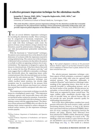
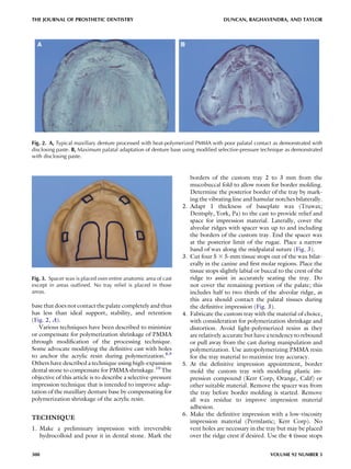
This article describes a selective-pressure impression technique for edentulous maxilla that aims to improve palatal adaptation of dentures. The technique involves placing spacer wax over the alveolar ridges and palate, leaving parts of the palate uncovered. This creates a deeper vault on the definitive cast, compensating for shrinkage of the denture base material during processing. The resulting denture has improved contact with palatal tissues compared to standard techniques.