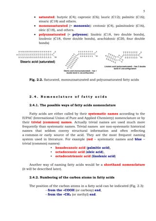
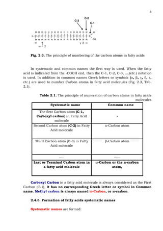
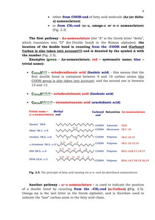
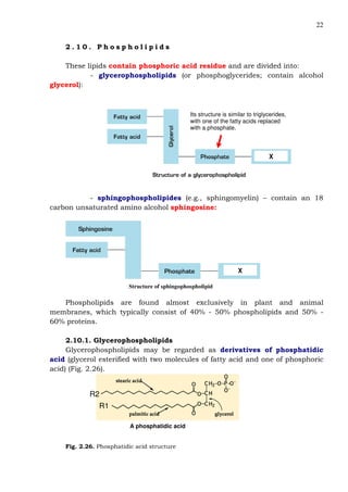
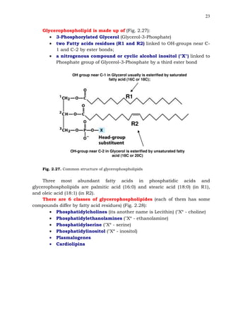
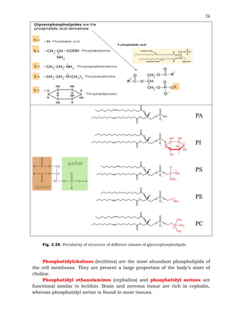
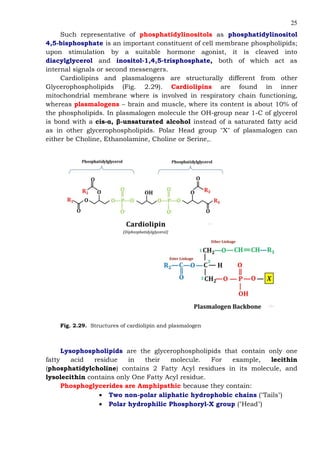
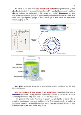
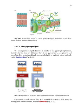
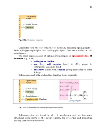
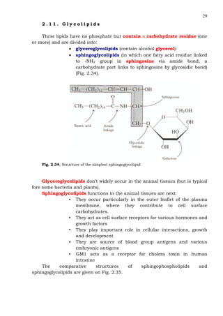
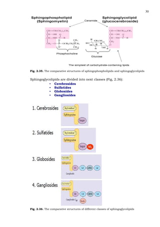
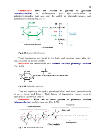
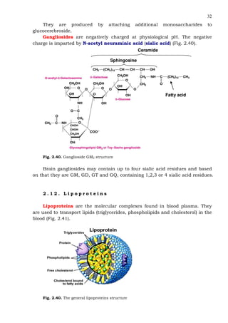
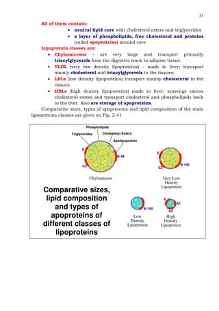
Taras Shevchenko National University of Kyiv
The Institute of Biology and Medicine
METHODICAL POINTING
from the course
“Biological and bioorganic chemistry”
(part 2. Fatty acids and lipids)
for the studens of a 1 course
with English of educating
Compiler – the candidate of biological sciences,
the associate professor
Synelnyk Tatyana Borysivna
Readers:
It is ratified to printing by meeting of scientific advice of The
Institute of Biology and Medicine (“____”________________ 2018,
protocol №____)
Kyiv-2018
2
CONTENT
2.1. General information ……………………………………..
2.2. Lipids classification……………………………………..
2.3. Fatty acids as main lipids components ………….
2.4. Nomenclature of fatty acids ………………………….
2.4.1. The possible ways of fatty acids
nomenclature …………………………………………………
2.4.2. Numbering of the carbon atoms in fatty
acids……………………………………..………………………
2.4.3. Formation of fatty acids systematic
names ……………………………………..……………………
2.4.4. Shorthand nomenclature ……………………..
2.5. Some properties of fatty acids ……………………..
2.6. The neutral fats ………………………………………….
2.6.1. The triacylglycerols classification,
nomenclature, localization in body and
functions……………………………………………………….
2.6.2. The physical and chemical properties of
triacylglycerols ……………………………………………
2.7. Waxes…………………………………………………………..
2.8. Steroids ……………………………………………………….
2.9. Complex lipids: classification……………………..
2.10. Phospholipids ……………………..……………………..
2.10.1. Glycerophospholipids …………………………
2.10.2. Sphingophospholipids …………………………
2.11. Glycolipids…………………………………………………
2.12. Lipoproteins…………………………………………………
2.13. Eicosanoids…………………………………………………
2.14. Isoprenoids…………………………………………………
2.15. Test questions ……………………………………………
3
3
4
5
5
5
6
7
10
11
12
12
17
18
21
22
22
27
29
32
34
35
37
3
2 . 1 . G e n e r a l i n f o r m a t i o n
The lipids are biological substances with various structures that are
insoluble in water and soluble in non-polar organic solvents such as
chloroform, benzene and ether. They are hydrophobic in nature because of the
high amount of hydrocarbons in their structure.
Lipids functions are next:
• structural components of cell membranes;
• regulation of cell membranes permeability;
• participation in the creation of intercellular contacts;
• participation in the nerve impulse transmittion;
• energy storage;
• since fat is а bad conductor of heat, it provides excellent insulation.
• protection of the internal organs.
• precursors of a number of biologically active substances.
2 . 2 . L i p i d s c l a s s i f i c a t i o n
Lipids are divided into simple and complex lipids as well as lipids
derivatives.
Simple lipids are classified into:
• neutral fats, or acylglycerols – fats and oils that are esters of Fatty
Acids and Glycerol
• waxes - are esters of fatty acids and higher molecular weight
monohydric alcohols
• steroids - contain the cyclopentanperhydrofenanthrene nucleus
Complex lipids - contain additional components:
• phospholipids containing phosphoric acid residue:
- glycerophospholipides
- sphingophospholipides;
• glycolipids containing hexoses, sulfated hexoses, hexosamines,
sialic acids:
- glyceroglycolipids (not in mammalians but in plants)
- glycosphingolipids (cerebrosides, globosides, gangliosides,
sulphatides)
• lipoproteins
Lipid Derivatives:
• fatty acids
• derivatives of polyunsaturated arachidonic acid – eicosanoisds
(prostaglandins, thromboxanes and leukotrienes).
• derivatives of cholesterol - steroid hormones, bile acids, vitamin
D3;
4
• izoprenoids (fat soluble vitamins - vitamin A, vitamin K, vitamin E,
terpenes)
Lipids are also divided into saponifiable and non saponifiable.
Saponification is the base hydrolysis of ester bonds.
Saponifiable lipids are lipids that have an ester functional group and so
can be hydrolyzed under basic conditions. The hydrolyzed products always
include fatty acids (or salts of fatty acids), and in some cases glycerol and other
molecular components contained in the lipid. The examples of saponifiable
lipids are:
• neutral fats (acylglycerols),
• phospholipids,
• glycolipids
• waxes
Non-saponifiable lipids are those lipids that cannot be hydrolyzed to fatty
acids, for example:
• terpenes (have several five-carbon building blocks called
isoprene units - for ex., such fat-soluble vitamins as Vit. A,
E, K) (Fig. 2.1)
• steroids
Fig. 2.1. Structure of isoprene - five-carbon building blocks of terpenes
and some other compounds
2 . 3 . F a t t y a c i d s a s m a i n l i p i d s c o m p o n e n t s
They:
• are derived from saponifiable lipids
• usually have non-branching chain and even number of carbon
atoms
• the length of the aliphatic chain is usually from between 10 and 20
carbons
• saturated (alkanes) and unsaturated (alkenes)
• in most unsaturated fatty acids, the cis isomer predominates; the
trans isomer is rare.
There are three groups of fatty acids (Fig. 1.2):
5
• saturated: butyric (C4); capronic (C6); lauric (C12); palmitic (C16);
stearic (C18) and others.
• monounsaturated (= monoenic): crotonic (C4), palmitooleic (C16),
oleic (C18), and others.
• polyunsaturated (= polyenes): linoleic (C18, two double bonds),
linolenic (C18, three double bonds), arachidonic (C20, four double
bonds)
Stearic acid (saturated)
Oleinic acid (monounsaturated) – has 1
double bond in cis-configuration
Linoleic acid (polyunsaturated) – has 2 double
bond in cis-configuration
Fig. 2.2. Saturated, monounsaturated and polyunsaturated fatty acids
2 . 4 . N o m e n c l a t u r e o f f a t t y a c i d s
2.4.1. The possible ways of fatty acids nomenclature
Fatty acids are either called by their systematic names according to the
IUPAC (International Union of Pure and Applied Chemistry) nomenclature or by
their trivial (common) names. Actually trivial names are used much more
frequently than systematic names. Trivial names are non-systematic historical
names that seldom convey structural information and often reflecting
a common or early source of the acid. They are the most frequent naming
system used in literature. For example (red – systematic names and blue –
trivial (common) names):
• hexadecanoic acid (palmitic acid),
• octadecenoic acid (oleic acid),
• octadecatrienoic acid (linolenic acid)
Another way of naming fatty acids would be a shorthand nomenclature
(it will be described later).
2.4.2. Numbering of the carbon atoms in fatty acids
The position of the carbon atoms in a fatty acid can be indicated (Fig. 2.3):
- from the –COOH (or carboxy) end,
- from the –CH3 (or methyl) end.
6
C-2
C-1
C-3
Fig. 2.3. The principle of numbering of the carbon atoms in fatty acids
In systematic and common names the first way is used. When the fatty
acid is indicated from the -COOH end, then the C-1, C-2, C-3, ….(etc.) notation
is used. In addition in common names Greek letters or symbols (α, β, ɣ, δ, ε,
etc.) are used to number Carbon atoms in fatty acid molecules (Fig. 2.3, Tab.
2.1).
Table 2.1. The principle of numeration of carbon atoms in fatty acids
molecules
Systematic name Common name
The first Carbon atom (C-1,
Carboxyl carbon) in Fatty Acid
molecule
-
Second Carbon atom (C-2) in Fatty
Acid molecule
α-Carbon atom
Third Carbon atom (C-3) in Fatty
Acid molecule
β-Carbon atom
….. ……
Last or Terminal Carbon atom in
a fatty acid molecule
ω-Carbon or the n-carbon
atom,
Carboxyl Carbon in a fatty acid molecule is always considered as the First
Carbon (C-1), it has no corresponding Greek letter or symbol in Common
name. Methyl carbon is always named ω-Carbon, or n-carbon.
2.4.3. Formation of fatty acids systematic names
Systematic names are formed:
7
• from alkane name with the same number of carbon atoms
by replacing the ending -e with suffix -oic acid (for
saturated fatty acids);
• from alkene name with the same number of carbon atoms
by replacing the ending -e with suffix -enoic acid (for
monounsaturated fatty acids).
Examples of systematic and trivial names of saturated fatty acids:
16:0 – hexadecanoic acid (Palmitic acid - from Latin Palm tree) - from
Hexadecane;
18:0 – octadecanoic acid (Stearic acid) - from octadecane
20:1 - eicosanoic acid (Arachidic acid - from Greek Arachne, spider) –
from eicosane
Examples of systematic and trivial names of monounsaturated fatty
acids:
16:1 – hexadecenenoic acid (palmitooleic acid) – from hexadecene
18:1 – octadecenoic acid (oleic acid) – from octadecene
Examples of systematic and trivial names of polyunsaturated fatty
acids:
С18:2 – Octadecadienoic acid (linoleic acid) – from octadecadiene
С18:3 – Octadecatrienoic acid (linolenic acid) – from octadecatriene
С20:4 – eicosatetraenoic acid (arachidonic acid) – from eicosatetraene
2.4.4. Shorthand nomenclature
In shorthand nomenclature fatty acids are named by their number of
carbon atoms, and their number of double bonds after a colon, e.g. 16:0 (Fig.
2.4).
Fig. 2.4. The principle of writing the name of a fatty acid via shorthand
nomenclature
For unsaturated fatty acids the shorthand nomenclature can also contain
the positioning of the double bonds – it can be pointed:
•16:0 (palmitic)
•18: 1 (oleic)
•20: 3 (linolenic)
The first number refers to the
total number of carbon atoms, and
the second one – to the number
of double bonds
8
• either from COOH-end of fatty acid molecule (∆x (or delta-
x) nomenclature)
• or from CH3-end (ω−x, omega-x or n−x nomenclature)
(Fig. 2.3)
The first pathway - ∆x-nomenclature (the “∆” is the Greek letter “delta”,
which translates into “D” (for Double bond) in the Roman alphabet): the
location of the double bond is counting from the -COOH end (Carboxyl
Carbon is also taken into account!!!) and is denoted by the symbol ∆ with
the number (Fig. 2.5).
Examples (green - ∆x-nomenclature; red – systematic name; blue –
trivial name):
• С18:2,∆9,12 – octadecadienoic acid (linoleic acid) - this means that the
first double bond is contained between 9 and 10 carbon atoms (the
COOH group is also taken into account), and the second one is between
12 and 13.
• С18:3,∆9,12,15 – octadecatrienoic acid (linolenic acid)
• С20:4,∆5,8,11,14 – eicosatetraenoic acid (arachidonic acid)
Trivial name + Methyl
ω−x-nomenclature end
Carboxyl Saturation ∆x-nomenclature
end
Fig. 2.5. The principle of fatty acid naming via ω−x- and ∆x-shorthand nomenclature
Another pathway - ω−x nomenclature - is used to indicate the position
of a double bond by counting from the –СН3-end (ω-Carbon) ((Fig. 2.5).
Omega (ω) is the last letter in the Greek alphabet, and is therefore used to
indicate the “last” carbon atom in the fatty acid chain.
9
Examples (green - ω−x -nomenclature; blue – trivial name):
• 18:3ω3 – linolenic acid - this means that the first double bond is
contained between 3 and 4 carbon atoms, if we count from the –CH3
group (this group is also taken into account)
• 18:1ω9 - oleic acid – it contains a double bond between C9 and
C10 counting from the ω-carbon atom (i.e., from the last Carbon
atom in the fatty acid molecule);
• 18:2ω6 - linoleic acid – it contains the first double bond between
C6 and C7 counting from the ω-carbon atom,
In Omega numbering system the position of the second double bond is not
indicated; it is because of double bonds in polyunsaturated fatty acids ARE
NOT in the Conjugated form such as:
– CH = CH – CH = CH –
Incorrect
They are separated by two single bonds, not by just one single bond:
– CH = CH – CH2 – CH = CH – CH2 – CH = CH –
Correct
Thus, in linoleic acid (C18:2ω6) the second double bond will be between
C9 and C10 from the ω-carbon.
According to ω−x–nomenclature, unsaturated fatty acids are divided into
groups, the most important of which are next:
• ω9 series – there are fatty acids in which the first double bond is
between C9 and C10 counting from the ω-carbon (for ex., Oleic
acid)
• ω6 series - these fatty acids have the first double bond between C6
and C7 counting from the ω-carbon (for ex., linoleic acid and
arachidonic acid)
• ω3 series - fatty acids in which the first double bond is between C3
and C4 counting from the ω-carbon (example - linolenic acid)
The unsaturated fatty acids that cannot be biosynthesized in tissues of
some animals including humans (because of luck the saturases – enzymes
needed for some double bonds formation) are called essential fatty acids. So
thus they must be obtained in the diet. The most of the essential fatty acids are
members of the ω6 and ω3 series – for example, linoleic and linolenic acids.
Some animals including humans can synthesize arachidonic acid from linoleic
acid obtained in the diet.
10
2 . 5 . S o m e p r o p e r t i e s o f f a t t y a c i d s
Fatty acids are amphipathic, because of the hydrophobic tail and
hydrophilic (–COOH) head. The longer the hydrocarbon chain of the fatty acid,
the higher its melting point, and the greater the number of double bonds in the
fatty acid - the lower its melting point. Therefore unsaturated fatty acids have
substantially lower melting points than saturated ones, and the greater the
degree of unsaturation, the lower the melting point (Fig. 2.6).
Unsaturated Fatty Acids
Saturated Fatty Acids
20:4
18:3
18:2
18:1
16:1
20:0
18:0
16:0
14:0
12:0
Carbon Atoms:
Double Bonds
Melting Point
(°C)
Common
Name
-49
-11
-5
16
1
77
70
63
58
44
Arachidonic acid
Linolenic acid
Linoleic acid
Oleic acid
Palmitoleic acid
Arachidic acid
Stearic acid
Palmitic acid
Myristic acid
Lauric acidHigher
mp
Lower
mp
Fig. 2.6. Some fatty acids and their melting points
This is due to hydrocarbon chains of saturated fatty acids can lie parallel
with strong dispersion forces between their chains (Fig. 2.7); they pack into
well-ordered, compact crystalline forms and melt above room temperature. In
contrast, because of the cis configuration of the double bonds in unsaturated
fatty acids, their hydrocarbon chains have a less ordered structure and
dispersion forces between them are weaker; these triglycerides have melting
points below room temperature
Fig. 2.7. The peculiarities of saturated and unsaturated fatty acids hydrocarbon chains
structure
11
Salts of fatty acids are called soaps and form micelles in aqueous (water is
solvent) solutions. These spherical structures allow the charged head-groups to
be in contact with water while at the same time removing the non-polar tails
from contact with water. More details about these formations will be given later.
2 . 6 . T h e n e u t r a l f a t s
Neutral fats are represenrated by glycerides, more correctly known
as acylglycerols. These compounds are esters formed from glycerol and fatty
acids. Glycerol, which is also called glycerin, is alcohol that has three hydroxyl
functional groups, which can be esterified with one, two, or three fatty acids to
form:
• monoacylglycerols,
• diacylglycerols
• triacylglycerols
These compounds are given on Fig. 2.8
Glycerol Monoacylglycerol Diacylglycerol Triacylglicerol
Fig. 2.8. The structures of glycerol, monoacylglycerol, diacylglycerol and triacylglycerol
The fatty acids can react with the hydroxyl groups to form esters. For
triglycerides, all three hydroxyls of the glycerol have a fatty acid residue
attached to it (Fig. 2.9).
12
CH2
CH
CH2
O
O
O
glycerol
residue
C
O
CH2CH2CH2CH2CH2CH2CH2CH2CH2CH2CH2CH2CH2CH2CH3
C
O
CH2CH2CH2CH2CH2CH2CH2CH2CH2CH2CH2CH2CH2CH2CH3
C
O
CH2CH2CH2CH2CH2CH2CH2CH2CH2CH2CH2CH2CH2CH2CH3
fatty acid
residues
Fig. 2.9. Structure of triacylglycerol and scheme of its formation
Neutral fats are neutral because they are uncharged and do not contain
acidic or basic groups. They are nonpolar and hydrophobic.
2.6.1. The triacylglycerols classification, nomenclature, localization in
body and functions
The triacylglycerols can be divided into simple and mixed ones.
The simple triacylglycerols contain identical fatty acid residues. For
example:
• triolein (or tri-oleoyl-glycerol) contains three molecules of
oleic acid residues esterified to a molecule of glycerol,
• tristearin (or tri-stearoyl-glycerol) contains three stearic
acid residues esterified to a molecule of glycerol
Mixed Triacylglycerols contain two or three different types of fatty acid
residues esterified to a molecule of glycerol. They are named according to the
placement of the fatty acid residues on the glycerol molecule:
- 1-palmitoleoyl-2-linoleoyl-3-stearoyl-glycerol;
- 1,3-dipalmitoleoylstearoyl-glycerol.
Body Triacylglycerols may be:
• the structural component of fat cells – adipocytes in the
adipose tissue – these triacylglycerols are the form of
stored energy
• the structural component of other cells – for example,
hepatocytes, tubular cells of kidney – in obesity
• the components of lipoproteins
2.6.2. The physical and chemical properties of triacylglycerols
Physical properties of triacylglycerols depend on the fatty acid components.
Their melting point increases as the number of carbons in its hydrocarbon
chains increases and as the number of double bonds decreases. Triglycerides
13
rich in unsaturated fatty acids are generally liquid at room and form oils;
triglycerides from plants tend to have a higher proportion of unsaturated fatty
acids. Triglycerides rich in saturated fatty acids are generally solids at room
temperature and form fats. Triglycerides from animals tend to have a higher
proportion of saturated fatty acids.
Reactions that involve triglycerides include:
• Hydrogenation
• Oxidation
• Base-catalyzed hydrolysis (saponification)
Hydrogenation reaction is typical for triacylglycerols rich on
unsaturated fatty acids that can be hydrogenated to produce saturated fats.
In hydrogenation, double bonds in unsaturated fatty acids react with H2 in the
presence of a Ni or Pt catalyst:
ssaattuurraatteedduunnssaattuurraatteedd
C C
H
H
H
H
H2 C
H
H
H
CH
H
H
+
platinum
catalyst
If triacylglycerol in its composition contains several residues of
unsaturated fatty acids, the hydrogenation may be complete or partial. In
complete hydrogenation all unsaturated fatty acid residues of this
triacylglycerol are subject to reduction, whereas in partial hydrogenation only
some unsaturated fatty acid-components of triacylglycerol are converted into
corresponding saturated fatty acids (Fig. 2.10)
Total
(complete)
hydrogenation
Liquid Solid
14
Partial
(incomplete)
hydrogenation
Liquid Solid
Fig. 2.10. The schemes of complete and partial hydrogenation
In fats unsaturated fatty acids normally found as a cis-isomers (Fig. 2.11).
Their trans-isomers called trans-fatty acids occur in trans fats.
Fig. 2.11. Cis- and trans-fatty acids
Trans-fatty acids are formed from unsaturated fatty acids as a side
product of their partial hydrogenation (Fig. 2.12).
Fig. 2.12. The scheme of trans-fatty acids forming at partial hydrogenation
15
Trans fat are found in margarine, deep-fried foods like fried chicken and
French-fried potatoes, snack chips, imitation cheese, ect (Fig. 2.13).
C
O
O
C
O
O
C
O
O
H
H
H
H
CH2
CH
CH2
saturated fat
trans unsaturated fat
cis unsaturated fat
FFaatt ((TTrriiaaccyyllggllyycceerriiddee))
Fig. 2.13. The trans fat structure and example of trans fat courses
Trans fat tends to raise cholesterol levels in the blood, although not as
much as saturated fat.
Rancidity of fat is the physico-chemical changes in the natural properties
of fat leading to the development of unpleasant odor or taste or abnormal color
because of atmospheric oxygen, light, bacterial or fungal enzymes or heat
action. There are two main causes of fat rancidity (Fig. 2.14):
- fat oxidation (peroxides, aldehydes, ketones and dicarboxylic fatty
acids are formed that smell bad)
- fat hydrolysis (with free fatty acids and glycerol formation, and free
fatty acids have unpleasant odor)
Fig. 2.14. The rancidity pathways
16
Saturated fats are more resistant to rancidity then unsaturated. The
products of rancidity are toxic and can occur food poisoning and cancer.
Moreover, rancidity destroys fat-soluble vitamins and polyunsaturated
essentially fat acids.
Oxidation of triglycerides and rancidity of fat. In fat oxidation
peroxides, aldehydes, ketones and dicarboxylic fatty acids are formed that
smell bad (Fig. 2.15).
These compounds are smell bad
Triglycerides oxidation
Linoleic acid oxidation
Fig. 2.15. The scheme of rancidity of fat caused by oxidation
Saponification (base-catalyzed hydrolysis) of fat. Saponification is the
base-catalyzed hydrolysis of the ester bonds in a triglyceride. The products of
this hydrolysis are carboxylic fatty acids and an alcohol glycerol. Because the
reaction is base-catalyzed, the fatty acids then react with base forming salts of
fatty acids – soaps (Fig. 2.16)
Fig. 2.16. The scheme of fat saponification
17
Soaps form micelle in the water solutes – in these structures hydrophobic
chains of fatty acids are directed inside of micelle, whereas carboxylic group
together with ions (Na+ or K+) are directed to water (Fig. 2.17).
Fig. 2.17. The scheme of micelle structure
2 . 7 . W a x e s
Waxes are esters of long chain fatty acids and long chain alcohol (Fig.
2.18).
1144--3366 ccaarrbboonnss 1166--3300 ccaarrbboonnss
Fig. 2.18. The wax structure
Waxes are very hydrophobic and are used by plants and animals for
protective, water-proof coatings. Some examples of waxes are given on Fig. 2.19
and on Tab. 2.2.
18
Fig. 2.19. The beeswax structure
Table 2.2. Composition of some waxes
2.8. Steroids
Steroids are a type of lipid that is not derived from a fatty acid. The
structure of steroids is based on the tetracyclic (four-ring) system –
cyclopentanoperhydrophenanthrene (steroid) nucleus:
•
Cyclopentanoperhydrophenanthrene
(steroid) nucleus
Three of the rings are six-membered, while the fourth is five-membered.
Cholesterol is the steroid that used as the starting point for the synthesis
of other steroids (Fig. 2.20). Besides being used to synthesize the other steroids,
cholesterol is dissolved in membranes to keep them fluid.
19
H3 C
HO
H3 C
H H
H
Fig. 2.20. Cholesterol structure
The most of cholesterol is bonded through ester links to fatty acids
(esterified cholesterol), but some is found in a free form (free cholesterol).
Bile acids are one of the main cholesterol derivatives (Fig. 2.21). They
are synthesized in the liver, stored in the gallbladder, and secreted into the
intestine where their function is emulsify dietary fats and aid in their
absorption and digestion.
HO
H H
H
H
OH
OH COOH
Cholic acid
Fig. 2.21. Structure of trihydroxy bile acid – cholic acid
Male and female sex hormones - androgens and estrogens, respectively –
are also cholesterol derivatives (Fig. 2.22) as well as corticosteroids (Fig. 2.23).
Fig. 2.22. Structure of the main sex hormones
20
Corticosteroids are a class of steroid hormones – derivatives of
cholesterol, that are produced in the adrenal cortex of vertebrates and include
both glucocorticoids and mineralocorticoids. Glucocorticoids (for example,
cortisol) are involved in glucose metabolism, whereas mineralocorticoids
regulate blood pressure and volume by stimulating the kidneys to absorb Na+.
Aldosterone
C=O
CH2 OH
O
H
H3 C H
H
CHO
OH
Fig. 2.22. Structures of the corticosteroids
Vitamin D (cholecalciferol) in its active form – 1,25-dioxycholecalciferol -
play a role in the regulation of calcium and phosphorus metabolism (Fig. 2.23).
Fig. 2.23. Structures of Vitamin D3
Cardiac Glycosides increase the force of cardiac muscle contraction via
acting on the cellular Na+/K+ ATPase pump. The general structure of a cardiac
glycoside consists of a steroid ring attached to a sugar and an R group (Fig.
2.24).
Fig. 2.24. The general structures of cardiac glycoside
Examples of cardic glycosides are convallotoxin (from Convallaria majalis),
ouabain and strophanthin (from Strophanthus kombe), digoxin and digitoxin
(from Digitalis purpurea) and oleandrin (Nerium oleander) (Fig. 2.25).
21
Digitoxin
From digitalis purpurea
Fig. 2.24. The structure of digitoxin
They are used as treatments for congestive heart failure and cardiac
arrhythmias; however, their relative toxicity prevents them from being widely
used.
2 . 9 . C o m p l e x l i p i d s : c l a s s i f i c a t i o n
They are the major constituents of cell membranes but are found also in
circulating fluids. They can be classified into two main groups (Fig. 2.25):
• Lipids with a phosphate residue called phospholipids
• Lipids containing a carbohydrate residue (one or more) called
glycolipids
Both groups of lipids can be divided into two subgroups dependent on
alcohol that is contained in their structures – glycerol (such lipids are called
glycerophospholipids or glyceroglycolipids, respectively) or sphingosine
(sphingophospholipids or sphingoglycolipids).
There are also lipoproteins – structures that includes different types of
lipids plus protein part called apoproteins.
Fig. 2.25. Complex lipids classification
22
2 . 1 0 . P h o s p h o l i p i d s
These lipids contain phosphoric acid residue and are divided into:
- glycerophospholipids (or phosphoglycerides; contain alcohol
glycerol):
X
Its structure is similar to triglycerides,
with one of the fatty acids replaced
with a phosphate.
- sphingophospholipides (e.g., sphingomyelin) – contain an 18
carbon unsaturated amino alcohol sphingosine:
Structure of sphingophospholipid
X
Phospholipids are found almost exclusively in plant and animal
membranes, which typically consist of 40% - 50% phospholipids and 50% -
60% proteins.
2.10.1. Glycerophospholipids
Glycerophospholipids may be regarded as derivatives of phosphatidic
acid (glycerol esterified with two molecules of fatty acid and one of phosphoric
acid) (Fig. 2.26).
R1
R2
A phosphatidic acid
Fig. 2.26. Phosphatidic acid structure
23
Glycerophospholipid is made up of (Fig. 2.27):
• 3-Phosphorylated Glycerol (Glycerol-3-Phosphate)
• two Fatty acids residues (R1 and R2) linked to OH-groups near C-
1 and C-2 by ester bonds;
• a nitrogenous compound or cyclic alcohol inositol ("X") linked to
Phosphate group of Glycerol-3-Phosphate by a third ester bond
OH group near C-1 in Glycerol usually is esterified by saturated
fatty acid (16C or 18C);
OH-group near C-2 in Glycerol is esterified by unsaturated fatty
acid (18C or 20C)
R1
R2
Fig. 2.27. Common structure of glycerophospholipids
Three most abundant fatty acids in phosphatidic acids and
glycerophospholipids are palmitic acid (16:0) and stearic acid (18:0) (in R1),
and oleic acid (18:1) (in R2).
There are 6 classes of glycerophospholipides (each of them has some
compounds differ by fatty acid residues) (Fig. 2.28):
• Phosphatidylcholines (its another name is Lecithin) ("X“ - choline)
• Phosphatidylethanolamines ("X“ - ethanolamine)
• Phosphatidylserine ("X“ - serine)
• Phosphatidylinositol ("X“ - inositol)
• Plasmalogenes
• Cardiolipins
24
X =
X =
X =
X =
X =
X
Fig. 2.28. Peculiarity of structure of different classes of glycerophospholipids
Phosphatidylcholines (lecithins) are the most abundant phospholipids of
the cell membrane. They are present a large proportion of the body's store of
choline.
Phosphatidyl ethanolamines (cephalins) and phosphatidyl serines are
functional similar to lecithin. Brain and nervous tissue are rich in cephalin,
whereas phosphatidyl serine is found in most tissues.
25
Such representative of phosphatidylinositols as phosphatidylinositol
4,5-bisphosphate is an important constituent of cell membrane phospholipids;
upon stimulation by a suitable hormone agonist, it is cleaved into
diacylglycerol and inositol-1,4,5-trisphosphate, both of which act as
internal signals or second messengers.
Cardiolipins and plasmalogens are structurally different from other
Glycerophospholipids (Fig. 2.29). Cardiolipins are found in inner
mitochondrial membrane where is involved in respiratory chain functioning,
whereas plasmalogens – brain and muscle, where its content is about 10% of
the phospholipids. In plasmalogen molecule the OH-group near 1-C of glycerol
is bond with a cis-α, β-unsaturated alcohol instead of a saturated fatty acid
as in other glycerophospholipids. Polar Head group "X" of plasmalogen can
either be Choline, Ethanolamine, Choline or Serine,.
Fig. 2.29. Structures of cardiolipin and plasmalogen
Lysophospholipids are the glycerophospholipids that contain only one
fatty acid residue in their molecule. For example, lecithin
(phosphatidylcholine) contains 2 Fatty Acyl residues in its molecule, and
lysolecithin contains only One Fatty Acyl residue.
Phosphoglycerides are Amphipathic because they contain:
• Two non-polar aliphatic hydrophobic chains ("Tails")
• Polar hydrophilic Phosphoryl-X group ("Head")
26
So when these molecules are mixed with water they spontaneously form
micelles (spherical structures that are formed by one lipid monolayer) for lipid
bilayers (planar or spherical – the latter are called liposomes); in this
structures hydrophobic groups of glycerophospholipids are contacted each with
other, and hydrophilic groups - with water (it is the basis of membrane
structure) (Fig. 2.30).
Fig. 2.30. Amphipatic properties of glycerophospholipids; monolayer, micelle, lipid
bilayer and liposome
On the surface of the water / air separation, phospholipids form a
monolayer in which the hydrophilic heads are directed to the aqueous phase
and the hydrophobic fatty acid tails - to the air (Fig. 2.30).
In general, the main function of glycerophospholipids is their role in
biological membranes structure and functions: fluid-mosaic model of biological
membrane showing the lipid bilayer and membrane proteins on the inner and
outer surfaces of the membrane (Fig. 2.31).
27
Fig. 2.31. Phospholipid bilayer as a main part of biological membranes (a) and fluid-
mosaic model of biological membrane (b)
2.10.2. Sphingophospholipids
The sphingophospholipids function is similar to the glycerophospholipids,
but structurally they are different: there is no glycerol core, and glycerol and
one of the fatty acids found in glycerophospholipids is replaced with a molecule
called Sphingosine (Fig. 2.32).
Fig. 2.32. Comparative structures of glycerophospholipids and sphingophospholipids
Compound formed when a fatty acid molecule is linked to -NH2 group in
sphingosine via amide bond is called ceramide (Fig. 3.33).
28
Fig. 2.32. Ceramide structure
Ceramides form the core structure of naturally occurring sphingolipids –
both sphingophospholipids and sphingoglycolipids that are founded in cell
membranes
The main representative of sphingophospholipids is sphingomyeline. It
contains (Fig. 2.33):
• sphingosine residue
• one fatty acid residue linked to -NH2 group in
sphingosine via amide bond
• phosphate linked with choline (phosphocholine) via ester
linkage
Sphingosine and fatty acid residue together forms ceramide.
Choline
Fig. 2.33. Common structure of sphingophospholipids
Sphingomyelins are found in all cell membranes and are important
structural components of the myelin sheath, the protective and insulating
coating that surrounds nerves.
29
2 . 1 1 . G l y c o l i p i d s
These lipids have no phosphate but contain a carbohydrate residue (one
or more) and are divided into:
• glyceroglycolipids (contain alcohol glycerol):
• sphingoglycolipids (in which one fatty acid residue linked
to -NH2 group in sphingosine via amide bond; a
carbohydrate part links to sphingosine by glycosidic bond)
(Fig. 2.34).
Fig. 2.34. Structure of the simplest sphingoglycolipid
Glyceroglycolipids don’t widely occur in the animal tissues (but is typical
fore some bacteria and plants).
Sphingoglycolipids functions in the animal tissues are next:
• They occur particularly in the outer leaflet of the plasma
membrane, where they contribute to cell surface
carbohydrates.
• They act as cell surface receptors for various hormones and
growth factors
• They play important role in cellular interactions, growth
and development
• They are source of blood group antigens and various
embryonic antigens
• GM1 acts as a receptor for cholera toxin in human
intestine
The comparative structures of sphingophospholipids and
sphingoglycolipids are given on Fig. 2.35.
30
Fig. 2.35. The comparative structures of sphingophospholipids and sphingoglycolipids
Sphingoglycolipids are divided into next classes (Fig, 2.36):
• Cerebrosides
• Sulfatides
• Globosides
• Gangliosides
Fig. 2.36. The comparative structures of different classes of sphingoglycolipids
31
Cerebrosides have one residue of glucose or galactose
(monosaccharide) as carbohydrate part (glucocerebrosides or
galactocerebrosides that also may be called as glucosylceramides and
galactosylceramides) (Fig. 2.37).
Fig. 2.37. Cerebroside structure
These compounds are found in the brain and nervous tissue with high
concentration in myelin sheath.
Sulfatides are cerebrosides that contain sulfated galactosyl residues
(Fig. 2.38).
Fig. 2.38. Sulfatides structure
They are negatively charged at physiological pH and found predominantly
in nerve tissue and kidney. Their failure of degradation causes them to
accumulate in nervous tissues.
Globosides – have two or more glucose or galactose residues
(oligosaccharide) in their structure (Fig. 2.39).
Fatty acid
Fig. 2.39. Globoside structure
32
They are produced by attaching additional monosaccharides to
glucocerebroside.
Gangliosides are negatively charged at physiological pH. The negative
charge is imparted by N-acetyl neuraminic acid (sialic acid) (Fig. 2.40).
Ceramide
Fatty acid
Sphingosine
Fig. 2.40. Ganglioside GM2 structure
Brain gangliosides may contain up to four sialic acid residues and based
on that they are GM, GD, GT and GQ, containing 1,2,3 or 4 sialic acid residues.
2 . 1 2 . L i p o p r o t e i n s
Lipoproteins are the molecular complexes found in blood plasma. They
are used to transport lipids (triglycerides, phospholipids and cholesterol) in the
blood (Fig. 2.41).
Fig. 2.40. The general lipoproteins structure
33
All of them contain:
• neutral lipid core with cholesterol esters and triglycerides
• a layer of phospholipids, free cholesterol and proteins
(called apoproteins) around core
Lipoprotein classes are:
• Chylomicrons – are very large and transport primarily
triacylglycerols from the digestive track to adipose tissue.
• VLDL (very low density lipoproteins) – made in liver; transport
mainly cholesterol and triacylglycerols to the tissues;
• LDLs (low density lipoproteins) transport mainly cholesterol to the
tissues.
• HDLs (high density lipoproteins) made in liver; scavenge excess
cholesterol esters and transport cholesterol and phospholipids back
to the liver. Also are storage of apoproteins.
Comparative sizes, types of apoproteins and lipid composition of the main
lipoproteins classes are given on Fig. 2.41
Comparative sizes,
lipid composition
and types of
apoproteins of
different classes of
lipoproteins
34
Fig. 2.41. Comparative sizes, types of apoproteins and lipid composition of the main
lipoproteins classes
Cholesterol of HDLs is a “good cholesterol”, and cholesterol of VLDLs and
LDLs is a “bad cholesterol” – its high levels may cause atherosclerosis
development
2 . 1 3 . E i c o s a n o i d s
Eicosanoids are derived from arachidonic acid:
Their examples are (Fig. 2.42):
• Leukotrienes (LTs)
• Prostaglandins (PGs),
• Prostacyclins (PGIs) and
• Thromboxanes (TXs)
35
Fig. 2.42. Examples of eicosanoids
2 . 1 4 . I s o p r e n o i d s
Isoprenoids contain a repeating five-carbon unit know as isoprene:
They are synthesized from isopentenyl pyrophosphate. One of the lagest
groups of isoprenoids are terpenes – an enormous group of molecules that are
found largely in the essential oils of plants. Terpenes are divided into:
• Monoterpenes: 2 isoprene units - geraniol (in germaniums)
• Sesquiterpenes: 3 isoprene units - farnesene (part of citronells oil)
• Diterpenes: 4 isoprene units - phytol (a plant alcohol)
• Tetraterpenes: 8 isoprene units - carotenoids (orange pigment in
plants)
Their structures are given on Fig. 2.43.
Fig. 2.43. Examples of terpenes
36
There are also mixed terpenoids – biomolecules which have isoprenoid
(isoprenyl) components. Their examples include some fat soluble vitamins
(vitamin E, vitamin K, vitamin A) and ubiquinone. Moreover, some proteins
(for example, G proteins which is an important component of cell transduction)
can be isoprenylated (attached to isopreniod groups) – it causes .their
attachment to plasmatic membrane.
Vitamin K - the name of this vitamin comes from the German word
Koagulation, signifying its important role in the blood-clotting process:
Vitamin E (α-tocopherol) functions as an antioxidant:
Vitamin A occurs only in the animal world. It is found in the plant world
in the form of a provitamin in a group of pigments called carotenes:
37
2 . 1 5 . T e s t q u e s t i o n s
1. Which of these lipids built from fatty acids and glycerol?
A. waxes
B. neutral fats
C. glycerophospholipids
D. glycerosphingolipids
E. steroids
2. Which of these lipids belong to complex lipids?
A. waxes
B. neutral fats
C. glycerophospholipids
D. derivatives of cholesterol
E. steroids
3. Bile acids and vitamin D3 are derivatives of:
A. Arachidonic acid
B. Cholesterol
C. Waxes
D. Neutral fat
E. Sphingolipids
4. Vitamin A, vitamin K, vitamin E as well as terpenes belong to:
A. derivatives of cholesterol
B. fatty acids derivatives
C. eicosanoisds
D. izoprenoids
E. waxes
5. The examples of non-saponifiable lipid are:
A. neutral fats,
B. phospholipids,
C. steroids
D. glycolipids
E. waxes
6. Which of these fatty acids has such shorthand nomenclature -
С20:4,∆5,8,11,14:
A. palmitic
B. oleic
C. linolenic
D. linoleic acid
E. arachidonic acid
38
7. Triolein and 1-palmitoleoyl-2-linoleoyl-3-stearoyl-glycerol are:
A. neutral fats,
B. phospholipids,
C. steroids
D. glycolipids
E. waxes
8. Adipocytes are the cells of the adipose tissue. Their function is to
deposite:
A. waxes
B. neutral fats
C. glycerophospholipids
D. derivatives of cholesterol
E. steroids
9. The physico-chemical changes in the natural properties of fat leading
to the development of unpleasant odor or taste or abnormal color because
of atmospheric oxygen, light, bacterial or fungal enzymes or heat action
are called:
A. Saponification
B. Partial (incomplete) hydrogenation
C. Complete hydrogenation
D. Rancidity
E. Esterification
10. Phosphatidylethanolamines and phosphatidylcholines belong to:
A. waxes
B. neutral fats
C. glycerophospholipids
D. derivatives of cholesterol
E. sphingophospholipids

3 6 lipids

  • 1.
    Taras Shevchenko NationalUniversity of Kyiv The Institute of Biology and Medicine METHODICAL POINTING from the course “Biological and bioorganic chemistry” (part 2. Fatty acids and lipids) for the studens of a 1 course with English of educating Compiler – the candidate of biological sciences, the associate professor Synelnyk Tatyana Borysivna Readers: It is ratified to printing by meeting of scientific advice of The Institute of Biology and Medicine (“____”________________ 2018, protocol №____) Kyiv-2018
  • 2.
    2 CONTENT 2.1. General information…………………………………….. 2.2. Lipids classification…………………………………….. 2.3. Fatty acids as main lipids components …………. 2.4. Nomenclature of fatty acids …………………………. 2.4.1. The possible ways of fatty acids nomenclature ………………………………………………… 2.4.2. Numbering of the carbon atoms in fatty acids……………………………………..……………………… 2.4.3. Formation of fatty acids systematic names ……………………………………..…………………… 2.4.4. Shorthand nomenclature …………………….. 2.5. Some properties of fatty acids …………………….. 2.6. The neutral fats …………………………………………. 2.6.1. The triacylglycerols classification, nomenclature, localization in body and functions………………………………………………………. 2.6.2. The physical and chemical properties of triacylglycerols …………………………………………… 2.7. Waxes………………………………………………………….. 2.8. Steroids ………………………………………………………. 2.9. Complex lipids: classification…………………….. 2.10. Phospholipids ……………………..…………………….. 2.10.1. Glycerophospholipids ………………………… 2.10.2. Sphingophospholipids ………………………… 2.11. Glycolipids………………………………………………… 2.12. Lipoproteins………………………………………………… 2.13. Eicosanoids………………………………………………… 2.14. Isoprenoids………………………………………………… 2.15. Test questions …………………………………………… 3 3 4 5 5 5 6 7 10 11 12 12 17 18 21 22 22 27 29 32 34 35 37
  • 3.
    3 2 . 1. G e n e r a l i n f o r m a t i o n The lipids are biological substances with various structures that are insoluble in water and soluble in non-polar organic solvents such as chloroform, benzene and ether. They are hydrophobic in nature because of the high amount of hydrocarbons in their structure. Lipids functions are next: • structural components of cell membranes; • regulation of cell membranes permeability; • participation in the creation of intercellular contacts; • participation in the nerve impulse transmittion; • energy storage; • since fat is а bad conductor of heat, it provides excellent insulation. • protection of the internal organs. • precursors of a number of biologically active substances. 2 . 2 . L i p i d s c l a s s i f i c a t i o n Lipids are divided into simple and complex lipids as well as lipids derivatives. Simple lipids are classified into: • neutral fats, or acylglycerols – fats and oils that are esters of Fatty Acids and Glycerol • waxes - are esters of fatty acids and higher molecular weight monohydric alcohols • steroids - contain the cyclopentanperhydrofenanthrene nucleus Complex lipids - contain additional components: • phospholipids containing phosphoric acid residue: - glycerophospholipides - sphingophospholipides; • glycolipids containing hexoses, sulfated hexoses, hexosamines, sialic acids: - glyceroglycolipids (not in mammalians but in plants) - glycosphingolipids (cerebrosides, globosides, gangliosides, sulphatides) • lipoproteins Lipid Derivatives: • fatty acids • derivatives of polyunsaturated arachidonic acid – eicosanoisds (prostaglandins, thromboxanes and leukotrienes). • derivatives of cholesterol - steroid hormones, bile acids, vitamin D3;
  • 4.
    4 • izoprenoids (fatsoluble vitamins - vitamin A, vitamin K, vitamin E, terpenes) Lipids are also divided into saponifiable and non saponifiable. Saponification is the base hydrolysis of ester bonds. Saponifiable lipids are lipids that have an ester functional group and so can be hydrolyzed under basic conditions. The hydrolyzed products always include fatty acids (or salts of fatty acids), and in some cases glycerol and other molecular components contained in the lipid. The examples of saponifiable lipids are: • neutral fats (acylglycerols), • phospholipids, • glycolipids • waxes Non-saponifiable lipids are those lipids that cannot be hydrolyzed to fatty acids, for example: • terpenes (have several five-carbon building blocks called isoprene units - for ex., such fat-soluble vitamins as Vit. A, E, K) (Fig. 2.1) • steroids Fig. 2.1. Structure of isoprene - five-carbon building blocks of terpenes and some other compounds 2 . 3 . F a t t y a c i d s a s m a i n l i p i d s c o m p o n e n t s They: • are derived from saponifiable lipids • usually have non-branching chain and even number of carbon atoms • the length of the aliphatic chain is usually from between 10 and 20 carbons • saturated (alkanes) and unsaturated (alkenes) • in most unsaturated fatty acids, the cis isomer predominates; the trans isomer is rare. There are three groups of fatty acids (Fig. 1.2):
  • 5.
    5 • saturated: butyric(C4); capronic (C6); lauric (C12); palmitic (C16); stearic (C18) and others. • monounsaturated (= monoenic): crotonic (C4), palmitooleic (C16), oleic (C18), and others. • polyunsaturated (= polyenes): linoleic (C18, two double bonds), linolenic (C18, three double bonds), arachidonic (C20, four double bonds) Stearic acid (saturated) Oleinic acid (monounsaturated) – has 1 double bond in cis-configuration Linoleic acid (polyunsaturated) – has 2 double bond in cis-configuration Fig. 2.2. Saturated, monounsaturated and polyunsaturated fatty acids 2 . 4 . N o m e n c l a t u r e o f f a t t y a c i d s 2.4.1. The possible ways of fatty acids nomenclature Fatty acids are either called by their systematic names according to the IUPAC (International Union of Pure and Applied Chemistry) nomenclature or by their trivial (common) names. Actually trivial names are used much more frequently than systematic names. Trivial names are non-systematic historical names that seldom convey structural information and often reflecting a common or early source of the acid. They are the most frequent naming system used in literature. For example (red – systematic names and blue – trivial (common) names): • hexadecanoic acid (palmitic acid), • octadecenoic acid (oleic acid), • octadecatrienoic acid (linolenic acid) Another way of naming fatty acids would be a shorthand nomenclature (it will be described later). 2.4.2. Numbering of the carbon atoms in fatty acids The position of the carbon atoms in a fatty acid can be indicated (Fig. 2.3): - from the –COOH (or carboxy) end, - from the –CH3 (or methyl) end.
  • 6.
    6 C-2 C-1 C-3 Fig. 2.3. Theprinciple of numbering of the carbon atoms in fatty acids In systematic and common names the first way is used. When the fatty acid is indicated from the -COOH end, then the C-1, C-2, C-3, ….(etc.) notation is used. In addition in common names Greek letters or symbols (α, β, ɣ, δ, ε, etc.) are used to number Carbon atoms in fatty acid molecules (Fig. 2.3, Tab. 2.1). Table 2.1. The principle of numeration of carbon atoms in fatty acids molecules Systematic name Common name The first Carbon atom (C-1, Carboxyl carbon) in Fatty Acid molecule - Second Carbon atom (C-2) in Fatty Acid molecule α-Carbon atom Third Carbon atom (C-3) in Fatty Acid molecule β-Carbon atom ….. …… Last or Terminal Carbon atom in a fatty acid molecule ω-Carbon or the n-carbon atom, Carboxyl Carbon in a fatty acid molecule is always considered as the First Carbon (C-1), it has no corresponding Greek letter or symbol in Common name. Methyl carbon is always named ω-Carbon, or n-carbon. 2.4.3. Formation of fatty acids systematic names Systematic names are formed:
  • 7.
    7 • from alkanename with the same number of carbon atoms by replacing the ending -e with suffix -oic acid (for saturated fatty acids); • from alkene name with the same number of carbon atoms by replacing the ending -e with suffix -enoic acid (for monounsaturated fatty acids). Examples of systematic and trivial names of saturated fatty acids: 16:0 – hexadecanoic acid (Palmitic acid - from Latin Palm tree) - from Hexadecane; 18:0 – octadecanoic acid (Stearic acid) - from octadecane 20:1 - eicosanoic acid (Arachidic acid - from Greek Arachne, spider) – from eicosane Examples of systematic and trivial names of monounsaturated fatty acids: 16:1 – hexadecenenoic acid (palmitooleic acid) – from hexadecene 18:1 – octadecenoic acid (oleic acid) – from octadecene Examples of systematic and trivial names of polyunsaturated fatty acids: С18:2 – Octadecadienoic acid (linoleic acid) – from octadecadiene С18:3 – Octadecatrienoic acid (linolenic acid) – from octadecatriene С20:4 – eicosatetraenoic acid (arachidonic acid) – from eicosatetraene 2.4.4. Shorthand nomenclature In shorthand nomenclature fatty acids are named by their number of carbon atoms, and their number of double bonds after a colon, e.g. 16:0 (Fig. 2.4). Fig. 2.4. The principle of writing the name of a fatty acid via shorthand nomenclature For unsaturated fatty acids the shorthand nomenclature can also contain the positioning of the double bonds – it can be pointed: •16:0 (palmitic) •18: 1 (oleic) •20: 3 (linolenic) The first number refers to the total number of carbon atoms, and the second one – to the number of double bonds
  • 8.
    8 • either fromCOOH-end of fatty acid molecule (∆x (or delta- x) nomenclature) • or from CH3-end (ω−x, omega-x or n−x nomenclature) (Fig. 2.3) The first pathway - ∆x-nomenclature (the “∆” is the Greek letter “delta”, which translates into “D” (for Double bond) in the Roman alphabet): the location of the double bond is counting from the -COOH end (Carboxyl Carbon is also taken into account!!!) and is denoted by the symbol ∆ with the number (Fig. 2.5). Examples (green - ∆x-nomenclature; red – systematic name; blue – trivial name): • С18:2,∆9,12 – octadecadienoic acid (linoleic acid) - this means that the first double bond is contained between 9 and 10 carbon atoms (the COOH group is also taken into account), and the second one is between 12 and 13. • С18:3,∆9,12,15 – octadecatrienoic acid (linolenic acid) • С20:4,∆5,8,11,14 – eicosatetraenoic acid (arachidonic acid) Trivial name + Methyl ω−x-nomenclature end Carboxyl Saturation ∆x-nomenclature end Fig. 2.5. The principle of fatty acid naming via ω−x- and ∆x-shorthand nomenclature Another pathway - ω−x nomenclature - is used to indicate the position of a double bond by counting from the –СН3-end (ω-Carbon) ((Fig. 2.5). Omega (ω) is the last letter in the Greek alphabet, and is therefore used to indicate the “last” carbon atom in the fatty acid chain.
  • 9.
    9 Examples (green -ω−x -nomenclature; blue – trivial name): • 18:3ω3 – linolenic acid - this means that the first double bond is contained between 3 and 4 carbon atoms, if we count from the –CH3 group (this group is also taken into account) • 18:1ω9 - oleic acid – it contains a double bond between C9 and C10 counting from the ω-carbon atom (i.e., from the last Carbon atom in the fatty acid molecule); • 18:2ω6 - linoleic acid – it contains the first double bond between C6 and C7 counting from the ω-carbon atom, In Omega numbering system the position of the second double bond is not indicated; it is because of double bonds in polyunsaturated fatty acids ARE NOT in the Conjugated form such as: – CH = CH – CH = CH – Incorrect They are separated by two single bonds, not by just one single bond: – CH = CH – CH2 – CH = CH – CH2 – CH = CH – Correct Thus, in linoleic acid (C18:2ω6) the second double bond will be between C9 and C10 from the ω-carbon. According to ω−x–nomenclature, unsaturated fatty acids are divided into groups, the most important of which are next: • ω9 series – there are fatty acids in which the first double bond is between C9 and C10 counting from the ω-carbon (for ex., Oleic acid) • ω6 series - these fatty acids have the first double bond between C6 and C7 counting from the ω-carbon (for ex., linoleic acid and arachidonic acid) • ω3 series - fatty acids in which the first double bond is between C3 and C4 counting from the ω-carbon (example - linolenic acid) The unsaturated fatty acids that cannot be biosynthesized in tissues of some animals including humans (because of luck the saturases – enzymes needed for some double bonds formation) are called essential fatty acids. So thus they must be obtained in the diet. The most of the essential fatty acids are members of the ω6 and ω3 series – for example, linoleic and linolenic acids. Some animals including humans can synthesize arachidonic acid from linoleic acid obtained in the diet.
  • 10.
    10 2 . 5. S o m e p r o p e r t i e s o f f a t t y a c i d s Fatty acids are amphipathic, because of the hydrophobic tail and hydrophilic (–COOH) head. The longer the hydrocarbon chain of the fatty acid, the higher its melting point, and the greater the number of double bonds in the fatty acid - the lower its melting point. Therefore unsaturated fatty acids have substantially lower melting points than saturated ones, and the greater the degree of unsaturation, the lower the melting point (Fig. 2.6). Unsaturated Fatty Acids Saturated Fatty Acids 20:4 18:3 18:2 18:1 16:1 20:0 18:0 16:0 14:0 12:0 Carbon Atoms: Double Bonds Melting Point (°C) Common Name -49 -11 -5 16 1 77 70 63 58 44 Arachidonic acid Linolenic acid Linoleic acid Oleic acid Palmitoleic acid Arachidic acid Stearic acid Palmitic acid Myristic acid Lauric acidHigher mp Lower mp Fig. 2.6. Some fatty acids and their melting points This is due to hydrocarbon chains of saturated fatty acids can lie parallel with strong dispersion forces between their chains (Fig. 2.7); they pack into well-ordered, compact crystalline forms and melt above room temperature. In contrast, because of the cis configuration of the double bonds in unsaturated fatty acids, their hydrocarbon chains have a less ordered structure and dispersion forces between them are weaker; these triglycerides have melting points below room temperature Fig. 2.7. The peculiarities of saturated and unsaturated fatty acids hydrocarbon chains structure
  • 11.
    11 Salts of fattyacids are called soaps and form micelles in aqueous (water is solvent) solutions. These spherical structures allow the charged head-groups to be in contact with water while at the same time removing the non-polar tails from contact with water. More details about these formations will be given later. 2 . 6 . T h e n e u t r a l f a t s Neutral fats are represenrated by glycerides, more correctly known as acylglycerols. These compounds are esters formed from glycerol and fatty acids. Glycerol, which is also called glycerin, is alcohol that has three hydroxyl functional groups, which can be esterified with one, two, or three fatty acids to form: • monoacylglycerols, • diacylglycerols • triacylglycerols These compounds are given on Fig. 2.8 Glycerol Monoacylglycerol Diacylglycerol Triacylglicerol Fig. 2.8. The structures of glycerol, monoacylglycerol, diacylglycerol and triacylglycerol The fatty acids can react with the hydroxyl groups to form esters. For triglycerides, all three hydroxyls of the glycerol have a fatty acid residue attached to it (Fig. 2.9).
  • 12.
    12 CH2 CH CH2 O O O glycerol residue C O CH2CH2CH2CH2CH2CH2CH2CH2CH2CH2CH2CH2CH2CH2CH3 C O CH2CH2CH2CH2CH2CH2CH2CH2CH2CH2CH2CH2CH2CH2CH3 C O CH2CH2CH2CH2CH2CH2CH2CH2CH2CH2CH2CH2CH2CH2CH3 fatty acid residues Fig. 2.9.Structure of triacylglycerol and scheme of its formation Neutral fats are neutral because they are uncharged and do not contain acidic or basic groups. They are nonpolar and hydrophobic. 2.6.1. The triacylglycerols classification, nomenclature, localization in body and functions The triacylglycerols can be divided into simple and mixed ones. The simple triacylglycerols contain identical fatty acid residues. For example: • triolein (or tri-oleoyl-glycerol) contains three molecules of oleic acid residues esterified to a molecule of glycerol, • tristearin (or tri-stearoyl-glycerol) contains three stearic acid residues esterified to a molecule of glycerol Mixed Triacylglycerols contain two or three different types of fatty acid residues esterified to a molecule of glycerol. They are named according to the placement of the fatty acid residues on the glycerol molecule: - 1-palmitoleoyl-2-linoleoyl-3-stearoyl-glycerol; - 1,3-dipalmitoleoylstearoyl-glycerol. Body Triacylglycerols may be: • the structural component of fat cells – adipocytes in the adipose tissue – these triacylglycerols are the form of stored energy • the structural component of other cells – for example, hepatocytes, tubular cells of kidney – in obesity • the components of lipoproteins 2.6.2. The physical and chemical properties of triacylglycerols Physical properties of triacylglycerols depend on the fatty acid components. Their melting point increases as the number of carbons in its hydrocarbon chains increases and as the number of double bonds decreases. Triglycerides
  • 13.
    13 rich in unsaturatedfatty acids are generally liquid at room and form oils; triglycerides from plants tend to have a higher proportion of unsaturated fatty acids. Triglycerides rich in saturated fatty acids are generally solids at room temperature and form fats. Triglycerides from animals tend to have a higher proportion of saturated fatty acids. Reactions that involve triglycerides include: • Hydrogenation • Oxidation • Base-catalyzed hydrolysis (saponification) Hydrogenation reaction is typical for triacylglycerols rich on unsaturated fatty acids that can be hydrogenated to produce saturated fats. In hydrogenation, double bonds in unsaturated fatty acids react with H2 in the presence of a Ni or Pt catalyst: ssaattuurraatteedduunnssaattuurraatteedd C C H H H H H2 C H H H CH H H + platinum catalyst If triacylglycerol in its composition contains several residues of unsaturated fatty acids, the hydrogenation may be complete or partial. In complete hydrogenation all unsaturated fatty acid residues of this triacylglycerol are subject to reduction, whereas in partial hydrogenation only some unsaturated fatty acid-components of triacylglycerol are converted into corresponding saturated fatty acids (Fig. 2.10) Total (complete) hydrogenation Liquid Solid
  • 14.
    14 Partial (incomplete) hydrogenation Liquid Solid Fig. 2.10.The schemes of complete and partial hydrogenation In fats unsaturated fatty acids normally found as a cis-isomers (Fig. 2.11). Their trans-isomers called trans-fatty acids occur in trans fats. Fig. 2.11. Cis- and trans-fatty acids Trans-fatty acids are formed from unsaturated fatty acids as a side product of their partial hydrogenation (Fig. 2.12). Fig. 2.12. The scheme of trans-fatty acids forming at partial hydrogenation
  • 15.
    15 Trans fat arefound in margarine, deep-fried foods like fried chicken and French-fried potatoes, snack chips, imitation cheese, ect (Fig. 2.13). C O O C O O C O O H H H H CH2 CH CH2 saturated fat trans unsaturated fat cis unsaturated fat FFaatt ((TTrriiaaccyyllggllyycceerriiddee)) Fig. 2.13. The trans fat structure and example of trans fat courses Trans fat tends to raise cholesterol levels in the blood, although not as much as saturated fat. Rancidity of fat is the physico-chemical changes in the natural properties of fat leading to the development of unpleasant odor or taste or abnormal color because of atmospheric oxygen, light, bacterial or fungal enzymes or heat action. There are two main causes of fat rancidity (Fig. 2.14): - fat oxidation (peroxides, aldehydes, ketones and dicarboxylic fatty acids are formed that smell bad) - fat hydrolysis (with free fatty acids and glycerol formation, and free fatty acids have unpleasant odor) Fig. 2.14. The rancidity pathways
  • 16.
    16 Saturated fats aremore resistant to rancidity then unsaturated. The products of rancidity are toxic and can occur food poisoning and cancer. Moreover, rancidity destroys fat-soluble vitamins and polyunsaturated essentially fat acids. Oxidation of triglycerides and rancidity of fat. In fat oxidation peroxides, aldehydes, ketones and dicarboxylic fatty acids are formed that smell bad (Fig. 2.15). These compounds are smell bad Triglycerides oxidation Linoleic acid oxidation Fig. 2.15. The scheme of rancidity of fat caused by oxidation Saponification (base-catalyzed hydrolysis) of fat. Saponification is the base-catalyzed hydrolysis of the ester bonds in a triglyceride. The products of this hydrolysis are carboxylic fatty acids and an alcohol glycerol. Because the reaction is base-catalyzed, the fatty acids then react with base forming salts of fatty acids – soaps (Fig. 2.16) Fig. 2.16. The scheme of fat saponification
  • 17.
    17 Soaps form micellein the water solutes – in these structures hydrophobic chains of fatty acids are directed inside of micelle, whereas carboxylic group together with ions (Na+ or K+) are directed to water (Fig. 2.17). Fig. 2.17. The scheme of micelle structure 2 . 7 . W a x e s Waxes are esters of long chain fatty acids and long chain alcohol (Fig. 2.18). 1144--3366 ccaarrbboonnss 1166--3300 ccaarrbboonnss Fig. 2.18. The wax structure Waxes are very hydrophobic and are used by plants and animals for protective, water-proof coatings. Some examples of waxes are given on Fig. 2.19 and on Tab. 2.2.
  • 18.
    18 Fig. 2.19. Thebeeswax structure Table 2.2. Composition of some waxes 2.8. Steroids Steroids are a type of lipid that is not derived from a fatty acid. The structure of steroids is based on the tetracyclic (four-ring) system – cyclopentanoperhydrophenanthrene (steroid) nucleus: • Cyclopentanoperhydrophenanthrene (steroid) nucleus Three of the rings are six-membered, while the fourth is five-membered. Cholesterol is the steroid that used as the starting point for the synthesis of other steroids (Fig. 2.20). Besides being used to synthesize the other steroids, cholesterol is dissolved in membranes to keep them fluid.
  • 19.
    19 H3 C HO H3 C HH H Fig. 2.20. Cholesterol structure The most of cholesterol is bonded through ester links to fatty acids (esterified cholesterol), but some is found in a free form (free cholesterol). Bile acids are one of the main cholesterol derivatives (Fig. 2.21). They are synthesized in the liver, stored in the gallbladder, and secreted into the intestine where their function is emulsify dietary fats and aid in their absorption and digestion. HO H H H H OH OH COOH Cholic acid Fig. 2.21. Structure of trihydroxy bile acid – cholic acid Male and female sex hormones - androgens and estrogens, respectively – are also cholesterol derivatives (Fig. 2.22) as well as corticosteroids (Fig. 2.23). Fig. 2.22. Structure of the main sex hormones
  • 20.
    20 Corticosteroids are aclass of steroid hormones – derivatives of cholesterol, that are produced in the adrenal cortex of vertebrates and include both glucocorticoids and mineralocorticoids. Glucocorticoids (for example, cortisol) are involved in glucose metabolism, whereas mineralocorticoids regulate blood pressure and volume by stimulating the kidneys to absorb Na+. Aldosterone C=O CH2 OH O H H3 C H H CHO OH Fig. 2.22. Structures of the corticosteroids Vitamin D (cholecalciferol) in its active form – 1,25-dioxycholecalciferol - play a role in the regulation of calcium and phosphorus metabolism (Fig. 2.23). Fig. 2.23. Structures of Vitamin D3 Cardiac Glycosides increase the force of cardiac muscle contraction via acting on the cellular Na+/K+ ATPase pump. The general structure of a cardiac glycoside consists of a steroid ring attached to a sugar and an R group (Fig. 2.24). Fig. 2.24. The general structures of cardiac glycoside Examples of cardic glycosides are convallotoxin (from Convallaria majalis), ouabain and strophanthin (from Strophanthus kombe), digoxin and digitoxin (from Digitalis purpurea) and oleandrin (Nerium oleander) (Fig. 2.25).
  • 21.
    21 Digitoxin From digitalis purpurea Fig.2.24. The structure of digitoxin They are used as treatments for congestive heart failure and cardiac arrhythmias; however, their relative toxicity prevents them from being widely used. 2 . 9 . C o m p l e x l i p i d s : c l a s s i f i c a t i o n They are the major constituents of cell membranes but are found also in circulating fluids. They can be classified into two main groups (Fig. 2.25): • Lipids with a phosphate residue called phospholipids • Lipids containing a carbohydrate residue (one or more) called glycolipids Both groups of lipids can be divided into two subgroups dependent on alcohol that is contained in their structures – glycerol (such lipids are called glycerophospholipids or glyceroglycolipids, respectively) or sphingosine (sphingophospholipids or sphingoglycolipids). There are also lipoproteins – structures that includes different types of lipids plus protein part called apoproteins. Fig. 2.25. Complex lipids classification
  • 22.
    22 2 . 10 . P h o s p h o l i p i d s These lipids contain phosphoric acid residue and are divided into: - glycerophospholipids (or phosphoglycerides; contain alcohol glycerol): X Its structure is similar to triglycerides, with one of the fatty acids replaced with a phosphate. - sphingophospholipides (e.g., sphingomyelin) – contain an 18 carbon unsaturated amino alcohol sphingosine: Structure of sphingophospholipid X Phospholipids are found almost exclusively in plant and animal membranes, which typically consist of 40% - 50% phospholipids and 50% - 60% proteins. 2.10.1. Glycerophospholipids Glycerophospholipids may be regarded as derivatives of phosphatidic acid (glycerol esterified with two molecules of fatty acid and one of phosphoric acid) (Fig. 2.26). R1 R2 A phosphatidic acid Fig. 2.26. Phosphatidic acid structure
  • 23.
    23 Glycerophospholipid is madeup of (Fig. 2.27): • 3-Phosphorylated Glycerol (Glycerol-3-Phosphate) • two Fatty acids residues (R1 and R2) linked to OH-groups near C- 1 and C-2 by ester bonds; • a nitrogenous compound or cyclic alcohol inositol ("X") linked to Phosphate group of Glycerol-3-Phosphate by a third ester bond OH group near C-1 in Glycerol usually is esterified by saturated fatty acid (16C or 18C); OH-group near C-2 in Glycerol is esterified by unsaturated fatty acid (18C or 20C) R1 R2 Fig. 2.27. Common structure of glycerophospholipids Three most abundant fatty acids in phosphatidic acids and glycerophospholipids are palmitic acid (16:0) and stearic acid (18:0) (in R1), and oleic acid (18:1) (in R2). There are 6 classes of glycerophospholipides (each of them has some compounds differ by fatty acid residues) (Fig. 2.28): • Phosphatidylcholines (its another name is Lecithin) ("X“ - choline) • Phosphatidylethanolamines ("X“ - ethanolamine) • Phosphatidylserine ("X“ - serine) • Phosphatidylinositol ("X“ - inositol) • Plasmalogenes • Cardiolipins
  • 24.
    24 X = X = X= X = X = X Fig. 2.28. Peculiarity of structure of different classes of glycerophospholipids Phosphatidylcholines (lecithins) are the most abundant phospholipids of the cell membrane. They are present a large proportion of the body's store of choline. Phosphatidyl ethanolamines (cephalins) and phosphatidyl serines are functional similar to lecithin. Brain and nervous tissue are rich in cephalin, whereas phosphatidyl serine is found in most tissues.
  • 25.
    25 Such representative ofphosphatidylinositols as phosphatidylinositol 4,5-bisphosphate is an important constituent of cell membrane phospholipids; upon stimulation by a suitable hormone agonist, it is cleaved into diacylglycerol and inositol-1,4,5-trisphosphate, both of which act as internal signals or second messengers. Cardiolipins and plasmalogens are structurally different from other Glycerophospholipids (Fig. 2.29). Cardiolipins are found in inner mitochondrial membrane where is involved in respiratory chain functioning, whereas plasmalogens – brain and muscle, where its content is about 10% of the phospholipids. In plasmalogen molecule the OH-group near 1-C of glycerol is bond with a cis-α, β-unsaturated alcohol instead of a saturated fatty acid as in other glycerophospholipids. Polar Head group "X" of plasmalogen can either be Choline, Ethanolamine, Choline or Serine,. Fig. 2.29. Structures of cardiolipin and plasmalogen Lysophospholipids are the glycerophospholipids that contain only one fatty acid residue in their molecule. For example, lecithin (phosphatidylcholine) contains 2 Fatty Acyl residues in its molecule, and lysolecithin contains only One Fatty Acyl residue. Phosphoglycerides are Amphipathic because they contain: • Two non-polar aliphatic hydrophobic chains ("Tails") • Polar hydrophilic Phosphoryl-X group ("Head")
  • 26.
    26 So when thesemolecules are mixed with water they spontaneously form micelles (spherical structures that are formed by one lipid monolayer) for lipid bilayers (planar or spherical – the latter are called liposomes); in this structures hydrophobic groups of glycerophospholipids are contacted each with other, and hydrophilic groups - with water (it is the basis of membrane structure) (Fig. 2.30). Fig. 2.30. Amphipatic properties of glycerophospholipids; monolayer, micelle, lipid bilayer and liposome On the surface of the water / air separation, phospholipids form a monolayer in which the hydrophilic heads are directed to the aqueous phase and the hydrophobic fatty acid tails - to the air (Fig. 2.30). In general, the main function of glycerophospholipids is their role in biological membranes structure and functions: fluid-mosaic model of biological membrane showing the lipid bilayer and membrane proteins on the inner and outer surfaces of the membrane (Fig. 2.31).
  • 27.
    27 Fig. 2.31. Phospholipidbilayer as a main part of biological membranes (a) and fluid- mosaic model of biological membrane (b) 2.10.2. Sphingophospholipids The sphingophospholipids function is similar to the glycerophospholipids, but structurally they are different: there is no glycerol core, and glycerol and one of the fatty acids found in glycerophospholipids is replaced with a molecule called Sphingosine (Fig. 2.32). Fig. 2.32. Comparative structures of glycerophospholipids and sphingophospholipids Compound formed when a fatty acid molecule is linked to -NH2 group in sphingosine via amide bond is called ceramide (Fig. 3.33).
  • 28.
    28 Fig. 2.32. Ceramidestructure Ceramides form the core structure of naturally occurring sphingolipids – both sphingophospholipids and sphingoglycolipids that are founded in cell membranes The main representative of sphingophospholipids is sphingomyeline. It contains (Fig. 2.33): • sphingosine residue • one fatty acid residue linked to -NH2 group in sphingosine via amide bond • phosphate linked with choline (phosphocholine) via ester linkage Sphingosine and fatty acid residue together forms ceramide. Choline Fig. 2.33. Common structure of sphingophospholipids Sphingomyelins are found in all cell membranes and are important structural components of the myelin sheath, the protective and insulating coating that surrounds nerves.
  • 29.
    29 2 . 11 . G l y c o l i p i d s These lipids have no phosphate but contain a carbohydrate residue (one or more) and are divided into: • glyceroglycolipids (contain alcohol glycerol): • sphingoglycolipids (in which one fatty acid residue linked to -NH2 group in sphingosine via amide bond; a carbohydrate part links to sphingosine by glycosidic bond) (Fig. 2.34). Fig. 2.34. Structure of the simplest sphingoglycolipid Glyceroglycolipids don’t widely occur in the animal tissues (but is typical fore some bacteria and plants). Sphingoglycolipids functions in the animal tissues are next: • They occur particularly in the outer leaflet of the plasma membrane, where they contribute to cell surface carbohydrates. • They act as cell surface receptors for various hormones and growth factors • They play important role in cellular interactions, growth and development • They are source of blood group antigens and various embryonic antigens • GM1 acts as a receptor for cholera toxin in human intestine The comparative structures of sphingophospholipids and sphingoglycolipids are given on Fig. 2.35.
  • 30.
    30 Fig. 2.35. Thecomparative structures of sphingophospholipids and sphingoglycolipids Sphingoglycolipids are divided into next classes (Fig, 2.36): • Cerebrosides • Sulfatides • Globosides • Gangliosides Fig. 2.36. The comparative structures of different classes of sphingoglycolipids
  • 31.
    31 Cerebrosides have oneresidue of glucose or galactose (monosaccharide) as carbohydrate part (glucocerebrosides or galactocerebrosides that also may be called as glucosylceramides and galactosylceramides) (Fig. 2.37). Fig. 2.37. Cerebroside structure These compounds are found in the brain and nervous tissue with high concentration in myelin sheath. Sulfatides are cerebrosides that contain sulfated galactosyl residues (Fig. 2.38). Fig. 2.38. Sulfatides structure They are negatively charged at physiological pH and found predominantly in nerve tissue and kidney. Their failure of degradation causes them to accumulate in nervous tissues. Globosides – have two or more glucose or galactose residues (oligosaccharide) in their structure (Fig. 2.39). Fatty acid Fig. 2.39. Globoside structure
  • 32.
    32 They are producedby attaching additional monosaccharides to glucocerebroside. Gangliosides are negatively charged at physiological pH. The negative charge is imparted by N-acetyl neuraminic acid (sialic acid) (Fig. 2.40). Ceramide Fatty acid Sphingosine Fig. 2.40. Ganglioside GM2 structure Brain gangliosides may contain up to four sialic acid residues and based on that they are GM, GD, GT and GQ, containing 1,2,3 or 4 sialic acid residues. 2 . 1 2 . L i p o p r o t e i n s Lipoproteins are the molecular complexes found in blood plasma. They are used to transport lipids (triglycerides, phospholipids and cholesterol) in the blood (Fig. 2.41). Fig. 2.40. The general lipoproteins structure
  • 33.
    33 All of themcontain: • neutral lipid core with cholesterol esters and triglycerides • a layer of phospholipids, free cholesterol and proteins (called apoproteins) around core Lipoprotein classes are: • Chylomicrons – are very large and transport primarily triacylglycerols from the digestive track to adipose tissue. • VLDL (very low density lipoproteins) – made in liver; transport mainly cholesterol and triacylglycerols to the tissues; • LDLs (low density lipoproteins) transport mainly cholesterol to the tissues. • HDLs (high density lipoproteins) made in liver; scavenge excess cholesterol esters and transport cholesterol and phospholipids back to the liver. Also are storage of apoproteins. Comparative sizes, types of apoproteins and lipid composition of the main lipoproteins classes are given on Fig. 2.41 Comparative sizes, lipid composition and types of apoproteins of different classes of lipoproteins
  • 34.
    34 Fig. 2.41. Comparativesizes, types of apoproteins and lipid composition of the main lipoproteins classes Cholesterol of HDLs is a “good cholesterol”, and cholesterol of VLDLs and LDLs is a “bad cholesterol” – its high levels may cause atherosclerosis development 2 . 1 3 . E i c o s a n o i d s Eicosanoids are derived from arachidonic acid: Their examples are (Fig. 2.42): • Leukotrienes (LTs) • Prostaglandins (PGs), • Prostacyclins (PGIs) and • Thromboxanes (TXs)
  • 35.
    35 Fig. 2.42. Examplesof eicosanoids 2 . 1 4 . I s o p r e n o i d s Isoprenoids contain a repeating five-carbon unit know as isoprene: They are synthesized from isopentenyl pyrophosphate. One of the lagest groups of isoprenoids are terpenes – an enormous group of molecules that are found largely in the essential oils of plants. Terpenes are divided into: • Monoterpenes: 2 isoprene units - geraniol (in germaniums) • Sesquiterpenes: 3 isoprene units - farnesene (part of citronells oil) • Diterpenes: 4 isoprene units - phytol (a plant alcohol) • Tetraterpenes: 8 isoprene units - carotenoids (orange pigment in plants) Their structures are given on Fig. 2.43. Fig. 2.43. Examples of terpenes
  • 36.
    36 There are alsomixed terpenoids – biomolecules which have isoprenoid (isoprenyl) components. Their examples include some fat soluble vitamins (vitamin E, vitamin K, vitamin A) and ubiquinone. Moreover, some proteins (for example, G proteins which is an important component of cell transduction) can be isoprenylated (attached to isopreniod groups) – it causes .their attachment to plasmatic membrane. Vitamin K - the name of this vitamin comes from the German word Koagulation, signifying its important role in the blood-clotting process: Vitamin E (α-tocopherol) functions as an antioxidant: Vitamin A occurs only in the animal world. It is found in the plant world in the form of a provitamin in a group of pigments called carotenes:
  • 37.
    37 2 . 15 . T e s t q u e s t i o n s 1. Which of these lipids built from fatty acids and glycerol? A. waxes B. neutral fats C. glycerophospholipids D. glycerosphingolipids E. steroids 2. Which of these lipids belong to complex lipids? A. waxes B. neutral fats C. glycerophospholipids D. derivatives of cholesterol E. steroids 3. Bile acids and vitamin D3 are derivatives of: A. Arachidonic acid B. Cholesterol C. Waxes D. Neutral fat E. Sphingolipids 4. Vitamin A, vitamin K, vitamin E as well as terpenes belong to: A. derivatives of cholesterol B. fatty acids derivatives C. eicosanoisds D. izoprenoids E. waxes 5. The examples of non-saponifiable lipid are: A. neutral fats, B. phospholipids, C. steroids D. glycolipids E. waxes 6. Which of these fatty acids has such shorthand nomenclature - С20:4,∆5,8,11,14: A. palmitic B. oleic C. linolenic D. linoleic acid E. arachidonic acid
  • 38.
    38 7. Triolein and1-palmitoleoyl-2-linoleoyl-3-stearoyl-glycerol are: A. neutral fats, B. phospholipids, C. steroids D. glycolipids E. waxes 8. Adipocytes are the cells of the adipose tissue. Their function is to deposite: A. waxes B. neutral fats C. glycerophospholipids D. derivatives of cholesterol E. steroids 9. The physico-chemical changes in the natural properties of fat leading to the development of unpleasant odor or taste or abnormal color because of atmospheric oxygen, light, bacterial or fungal enzymes or heat action are called: A. Saponification B. Partial (incomplete) hydrogenation C. Complete hydrogenation D. Rancidity E. Esterification 10. Phosphatidylethanolamines and phosphatidylcholines belong to: A. waxes B. neutral fats C. glycerophospholipids D. derivatives of cholesterol E. sphingophospholipids