Download to read offline
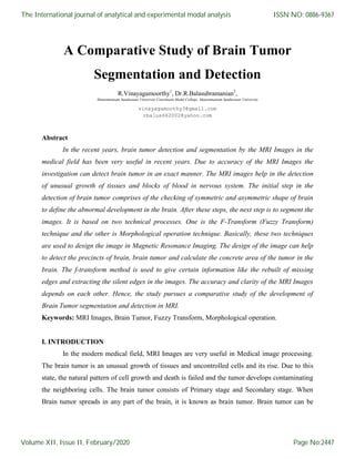
![The next step is the Pre-processing Stage. It consists of Noise removal methods. This
method can be done by using various spatial filters or linear or nonlinear filters. Other artifacts
like text are removed by some sort of morphological operations.
Fig. 1 Basic Block Diagram of Brain Tumor Detection and Segmentation Process
In the pre-processing stages, RGB to grey conversion and reshaping also takes place. It
includes of median filter for noise removal methods. In general, the possibilities of noise
interaction in modern MRI scan are very low. It is because of the thermal Effect.
Image Smoothing is the method of simplifying an image while at the time of preserving
important information in the image. The chief aim is to reduce noise or useless details without
introducing high level of distortion and to simplify subsequent analysis in the procedure.
Image Registration is the next process of bringing two or more images into proper
alignment. In medical imaging, image registration allows for the concurrent use of images taken
with different modalities such as MRI and CT scanning methods at dissimilar times or with
dissimilar patient positions. For example, images are acquired preoperative, as well as during
intra-operative surgery. Due to time constraints, the real-time intra-operative images have lower
resolution than the pre-operative images. Moreover, deformations that occur at the time of
surgery makes difficult to compare with the high resolution pre-operative image to the low
resolution intra-operative surgery of the patient. Hence, Image registration attempts to help the
surgeon relate the two sets of images [8].
The International journal of analytical and experimental modal analysis
Volume XII, Issue II, February/2020
ISSN NO: 0886-9367
Page No:2449](https://image.slidesharecdn.com/285-ijaema-february-3487-230731010917-94c07d87/85/285-IJAEMA-FEBRUARY-3487-pdf-3-320.jpg)
![Image Segmentation is the key stage to identify the image properly because it affects the
accuracy of the subsequent diagnostic steps. On the other hand, proper segmentation is intricate
because of the different verities in lesion shapes, sizes, and colors along with different types of
skin and textures. Moreover, some lesions have asymmetrical boundaries and some have smooth
transition between the lesion and the skin. Several algorithms have been proposed to solve the
problem. They are classified as thresholding, edge-based or region-based methods and of
supervised and unsupervised techniques. The processes are termed as Threshold segmentation,
Water shed segmentation, Gradient Vector Flow, K-mean Clustering and Fuzzy C-means
Clustering.
After proper segmentation, morphological processing has been applied to remove the
unwanted parts. This process consists of image opening, image closing, dilation, and erosion
operations.
At the end of these segmentation and detection process, decision has been taken weather
that MRI image consists of any tumor or not and the normal or the abnormal state of weather has
been checked.
III. REVIEW OF LITERATURE
The 2016 World Health Organization says on the classification of tumor of central
nervous system is a conceptual one as well as pertain overview of predecessor. WHO classifies
CNS tumor by molecular parameters for its diagnosis structure. Further than 2016 CNS WHO
presence the new diffuse glomas and other tumor and defines the new feature like both histology
as well as molecule [1].
There are different types of brain tumors. They are Glioma, Papillary, Glioneuronal
tumor etc. The histological variants are capable of different edge distribution, location,
symptoms and the behaviours or clinical [2].
Fuzzy clustering is method which has been widely used in biomedical field to detect the
image. Effective fuzzy clustering algorithm is used in abnormal MRI brain image segmentation.
By using clustering in brain tumor segmentation we can diagnose accurately the region of cancer
[3].
Now-a-days, brain tumor is one of the major hazardous diseases. Its detection should be
fast and accurate and can be detected by automated tumor detection techniques. One of the
automated tumor detection techniques is the use of MRI images. It defines the tumor growth
The International journal of analytical and experimental modal analysis
Volume XII, Issue II, February/2020
ISSN NO: 0886-9367
Page No:2450](https://image.slidesharecdn.com/285-ijaema-february-3487-230731010917-94c07d87/85/285-IJAEMA-FEBRUARY-3487-pdf-4-320.jpg)
![region and the edges detection. As compare to other techniques with this it gives more accurate
as well as clear and advantages of automated tumor detection techniques is used for removal of
tumor if needed [4].
The neural networks are a new technology has been discovered. The neural networks are
an “HOT” research area, like a cardiology, radiology, oncology etc. To solve highly complex
problem three is combination of neurons into layers permits for artificial neural network. In an
medical applications the neural network are like ANNs etc. and the medical application the
neural network are used to map an input into a desired output [5].
It is a new technique of detection of brain tumor and for very good result and accuracy.
The watershed method is combined with edge detection operation. The color brain MRI images
can be obtained by this algorithm. In this the RGB image is converts into on HSV color image so
that the image is separated in 3 regions which are known as hue, saturation and intensity. The
canny edge detector is applied is applied to an output image for rebuilt process of edge occurs in
this .at last combining the three images and the final resultant brain tumor segmented image is
obtained. This algorithm is applied on 20 brain MRI images for excellent result [6].
In an MRI image the highly irregular boundaries of tumor tissues is seen. For a
segmentation of medical image, the deformable modes and region base methods are used. The
main problems are there in MRI images like undefined location of tumor are unseen boundaries
or data loss at boundaries and a silent edge not extended. By using this algorithm the silent edge
is extended and found boundary of tumor location or area and once the boundary or location of
tumor is seen clearly. Then removal of tumor can be take place [7].
Mariam Saii, Zaid Kraitem (2017) in their research “Automatic Brain Tumor Detection in
MRI Using Image Processing Techniques” offers a fully automatic method for tumor
segmentation on Magnetic Resonance Images MRI. In this method, at first in the preprocessing
level, anisotropic diffusion filter is applied to the image by 8-connected neighborhood for
removing noise from it. [9] In the second step, Support Vector Machine SVM Classifier had
been used for tumor detection accurately. After creating appropriate mask image, based on its
symmetry in axial and coronary MRI, the tumor had been detected and segmented (Dice
coefficient > 0.90) in a few seconds. In short, the method applied on several MRI images with
different types had been regardless of the degree of complexity.
The International journal of analytical and experimental modal analysis
Volume XII, Issue II, February/2020
ISSN NO: 0886-9367
Page No:2451](https://image.slidesharecdn.com/285-ijaema-february-3487-230731010917-94c07d87/85/285-IJAEMA-FEBRUARY-3487-pdf-5-320.jpg)
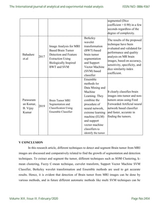
![Bahadure et.al (2017) in their article “Image Analysis for MRI Based Brain Tumor
Detection and Feature Extraction Using Biologically Inspired BWT and SVM” had studied on
the segmentation, detection, and extraction of infected tumor area from MRI images. It is the
primary concern and a tedious task performed by radiologists and other clinical experts. The
accuracy depends on the experience of the experts. So in the modern times, the use of computer
aided technology in medical field becomes very necessary to overcome such limitations. The
researchers had studied to improve the performance and condense the complexity involves in the
MRI image segmentation process. They have investigated Berkeley wavelet transformation
(BWT) based brain tumor segmentation in the study. In addition, to improve the accuracy and
quality rate of the support vector machine (SVM) based classifier; relevant features are extracted
from each segmented tissue. [10] The results of the proposed technique have been evaluated and
validated for performance and quality analysis on MR brain images, based on accuracy,
sensitivity, specificity, and dice similarity index coefficient. The experimental results achieved
96.51% accuracy, 94.2% specificity, and 97.72% sensitivity, demonstrating the effectiveness of
the proposed technique for identifying normal and abnormal tissues from brain MR images. [10]
Parasuraman Kumar, B. Vijay Kumar (2019) in their article “Brain Tumor MRI
Segmentation and Classification Using Ensemble Classifier” had discussed on Brain tumor and
abnormal formation within the brain. They stressed it is becoming a major cause for death in
many cases. The detection of these cells has been a difficult problem and MRI gives the solution
to it. It is very essential to compare brain tumor from the MRI treatment. It is very difficult to
have vision about the abnormal structures of human brain using simple imaging techniques.
According to them, Ensemble methods are the most influential development in Data Mining and
Machine Learning in early decades. They combine the procedure of neural network, extreme
learning machine (ELM) and support vector machine classifiers to identify the tumours
accurately. This system consists of manifold phases such as Preprocessing, segmentation, feature
extraction, and classification. This phase clearly classifies brain images into tumor and non-
tumors areas using Feed Forwarded Artificial neural network based classifier. Hence, these
experiments have proved robust to initialization, faster and accurate finding of the tumors. [11]
IV COMPARISON AND GROWTH OF YESTER RESEARCHES
Table-1 Techniques and Results in Brain Tumor Segmentation and Detection
Author Year Paper Name Technique Result
The International journal of analytical and experimental modal analysis
Volume XII, Issue II, February/2020
ISSN NO: 0886-9367
Page No:2452](https://image.slidesharecdn.com/285-ijaema-february-3487-230731010917-94c07d87/85/285-IJAEMA-FEBRUARY-3487-pdf-6-320.jpg)
![adopted to achieve more accuracy and more efficient results; and these developments can avoid
fatal causalities by accurate identification and target based proper diagnosis.
REFERENCES
[1] D.N.Louis, et al, “The 2007 WHO classification of tumor of central nervous system,” Act d
neuropathological, Vol 114, pp 97-109, 2007.
[2] P.Kleihues, et al. “The new WHO classification of brain tumor, brain pathology”, Vol 3, pp.
255-268, 1993.
[3] D.J.Hemanth, et al, “Effective fuzzy clustering algorithm for abnormal MR brain image
segmentation,” Advance Computing Conference 2009, pp-609-614.
[4] S.Chrutha and M.J.Jayashree, “An efficient brain tumor detection by integrating modified
texture based region growing and cellular automata edge detection,” Control
Instrumentation, Communication and Computational Technology (ICCICCT), 2014,
pp.1193-1199.
[5] A. Abdullah, et al., “Implementation of an improved cellular neural network algorithm for
brain tumor detection," Biomedical Engineering (Isobel), 2012, pp. 611- 615.
[6] I. Maiti and M. Chakra borty, “A new method for brain tumor segmentation based on
watershed and edge detection algorithms in HSV color model,” Computing and
Communication Systems (NCCCS), 2012, pp. 1-5.
[7] R. Preetha and G. R. Suresh, "Performance Analysis of Fuzzy C Means Algorithm in
Automated Detection of Brain Tumor," Computing and Communication Technologies
(WCCCT), 2014, pp. 30-33.
[8] Bandana Sharma et al. “Review Paper on Brain Tumor Detection Using Pattern Recognition
Techniques” International Journal of Recent Research Aspects, ISSN: 2349-7688, Vol. 3,
Issue 2, June 2016, pp. 151-156
[9] Mariam Saii, Zaid Kraitem. “Automatic Brain Tumor Detection in MRI Using Image
Processing Techniques.” Biomedical Statistics and Informatics. Vol. 2, No. 2, 2017, pp. 73-
76. doi: 10.11648/j.bsi.20170202.16
[10] Nilesh Bhaskarrao Bahadure et.al. “Image Analysis for MRI Based Brain Tumor Detection
and Feature Extraction Using Biologically Inspired BWT and SVM”. International Journal
of Biomedical Imaging, Volume 2017, Article ID 9749108,
The International journal of analytical and experimental modal analysis
Volume XII, Issue II, February/2020
ISSN NO: 0886-9367
Page No:2455](https://image.slidesharecdn.com/285-ijaema-february-3487-230731010917-94c07d87/85/285-IJAEMA-FEBRUARY-3487-pdf-9-320.jpg)
![https://doi.org/10.1155/2017/9749108.
[11] Parasuraman Kumar, B. VijayKumar “Brain Tumor MRI Segmentation and Classification
Using Ensemble Classifier” International Journal of Recent Technology and Engineering
(IJRTE), ISSN: 2277-3878, Volume-8, Issue-14, June 2019.
The International journal of analytical and experimental modal analysis
Volume XII, Issue II, February/2020
ISSN NO: 0886-9367
Page No:2456](https://image.slidesharecdn.com/285-ijaema-february-3487-230731010917-94c07d87/85/285-IJAEMA-FEBRUARY-3487-pdf-10-320.jpg)

This document presents a comparative study of brain tumor segmentation and detection techniques in MRI images. It discusses several techniques used for brain tumor segmentation including fuzzy transform, morphological operations, thresholding, edge-based and region-based methods. The document also reviews various literature on brain tumor detection algorithms using techniques such as fuzzy clustering, neural networks, watershed segmentation, and support vector machines. Accuracy levels of different algorithms are presented ranging from dice coefficients above 0.90 to overall accuracy of 96.51%.