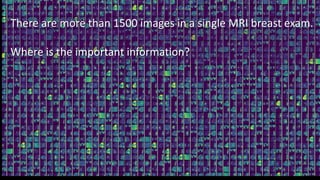
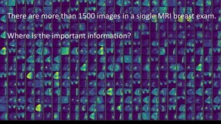
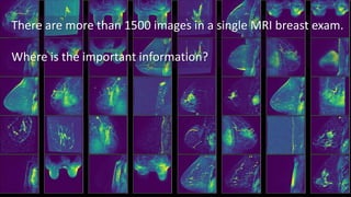
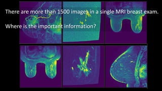
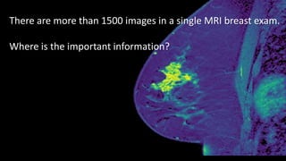
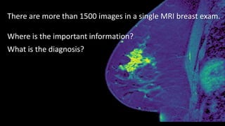
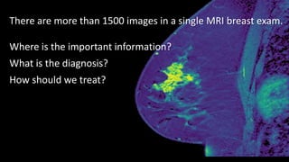
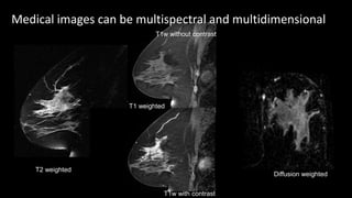
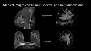
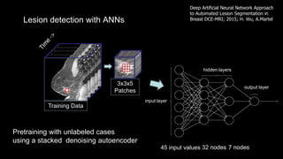
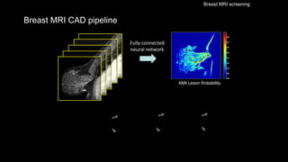
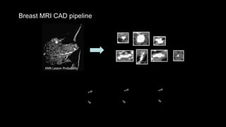
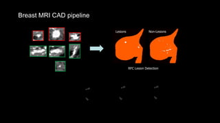
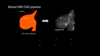
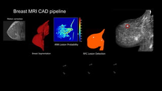
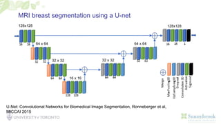
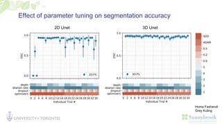
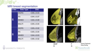
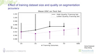
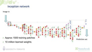
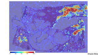
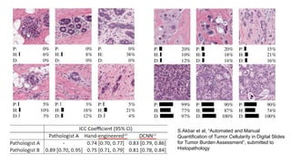
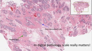
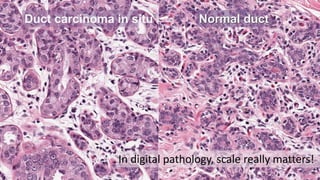
Anne Martel, PhD
Senior Scientist, Physical Sciences
Sunnybrook Research Institute
Professor, Department of Medical Biophysics
University of Toronto
Vector Institute Faculty Affiliate
Machine Learning for Medical Imaging
Toronto Machine Learning Summit, 2
A Web-Based Image
Management Solution for
Advanced Workflows
Co-Founder, shareholder and CSO of Pathcore Inc, Toronto
There are more than 1500 images in a single MRI breast exam.
Where is the important information?
There are more than 1500 images in a single MRI breast exam.
Where is the important information?
There are more than 1500 images in a single MRI breast exam.
Where is the important information?
There are more than 1500 images in a single MRI breast exam.
Where is the important information?
There are more than 1500 images in a single MRI breast exam.
Where is the important information?
There are more than 1500 images in a single MRI breast exam.
Where is the important information?
What is the diagnosis?
There are more than 1500 images in a single MRI breast exam.
Where is the important information?
What is the diagnosis?
How should we treat?
Medical images can be multispectral and multidimensional
T2 weighted
T1 weighted
Diffusion weighted
T1w with contrast
T1w without contrast
Medical images can be multispectral and multidimensional
Sagittal view
Axial view
Training Data
3x3x5
Patches
Lesion detection with ANNs
Deep Artificial Neural Network Approach
to Automated Lesion Segmentation in
Breast DCE-MRI; 2015; H. Wu, A.Martel
45 input values 32 nodes 7 nodes
Pretraining with unlabeled cases
using a stacked denoising autoencoder
hidden layers
output layer
input layer
Breast MRI CAD pipeline
ANN Lesion Probability
Fully connected
neural network
Breast MRI screening
Breast MRI CAD pipeline
ANN Lesion Probability
Breast MRI CAD pipeline
RFC Lesion Detection
Lesions Non-Lesions
False positive
detection removal
Breast MRI CAD pipeline
RFC Lesion Detection
Lesions
Sagittal MIP
Lesion
Classification
Breast Segmentation
Breast MRI CAD pipeline
Lesion
Classification
Motion correction
RFC Lesion Detection
ANN Lesion Probability
MRI breast segmentation using a U-net
128x128 128x128
64 x 64 64 x 64
32 x 32 32 x 32
16 x 16
U-Net: Convolutional Networks for Biomedical Image Segmentation, Ronneberger et al,
MICCAI 2015
Effect of parameter tuning on segmentation accuracy
2D Unet 3D Unet
Homa Fashendi
Grey Kuling
MRI breast segmentation
U-Net Data Type DSC
2D WOFS 0.96 ± 0.03
FS 0.94 ± 0.07
MIXED 0.94 ± 0.09
MULTI 0.95 ± 0.04
3D
WOFS 0.95 ± 0.02
FS 0.95 ± 0.04
MIXED 0.94 ± 0.03
MULTI 0.96 ± 0.02
WOF
S
F
S
Homa Fashendi
Grey Kuling
Effect of training dataset size and quality on segmentation
accuracy
Homa Fashendi
Grey Kuling
SPIE-AAPM-NCI BreastPathQ: Cancer Cellularity Challenge
http://spiechallenges.cloudapp.net/competitions/14
Assessing tumour burden
C
Automatic cellularity assessment from post-treated
breast surgical specimens.
Peikari et al, 2017, Cytometry A, 91:1078-1087
Inception network
Image in
Prediction out
 Approx 1500 training patches
 10 million learned weights
Shazia Akbar
S.Akbar et al, “Automated and Manual
Quantification of Tumor Cellularity in Digital Slides
for Tumor Burden Assessment”, submitted to
Histopathology
In digital pathology, scale really matters!
Normal duct
Duct carcinoma in situ
Duct carcinoma in situ Normal duct
In digital pathology, scale really matters!
Why is AI in medical imaging challenging?
 Performance has to be excellent
 Interpretability is essential for clinical acceptance
 Need to understand how the images are acquired and what they
represent
 Labelled training data sets are scare and expensive
• Mohammad
• Peikari
• Shazia Akbar
Nikhil
Seth • Azadeh
• Yazanpanah
• Rushin Shojaii
Cristina Gallego-Ortiz
• Andrei
• Mouraviev
• Yingli Lu
• Sharmila
• Balasingham
Hongbo Wu
• Sylvester Chiang
Radiology
• Belinda Curpen
Radiation oncology
• Eileen Rackovitch, MD
Anatomic Pathology
• Sharon Nofech-Mozes, MD
• Sherine Salama, M

Machine Learning for Medical Imaging

  • 1.
    Anne Martel, PhD SeniorScientist, Physical Sciences Sunnybrook Research Institute Professor, Department of Medical Biophysics University of Toronto Vector Institute Faculty Affiliate Machine Learning for Medical Imaging Toronto Machine Learning Summit, 2
  • 2.
    A Web-Based Image ManagementSolution for Advanced Workflows Co-Founder, shareholder and CSO of Pathcore Inc, Toronto
  • 3.
    There are morethan 1500 images in a single MRI breast exam. Where is the important information?
  • 4.
    There are morethan 1500 images in a single MRI breast exam. Where is the important information?
  • 5.
    There are morethan 1500 images in a single MRI breast exam. Where is the important information?
  • 6.
    There are morethan 1500 images in a single MRI breast exam. Where is the important information?
  • 7.
    There are morethan 1500 images in a single MRI breast exam. Where is the important information?
  • 8.
    There are morethan 1500 images in a single MRI breast exam. Where is the important information? What is the diagnosis?
  • 9.
    There are morethan 1500 images in a single MRI breast exam. Where is the important information? What is the diagnosis? How should we treat?
  • 10.
    Medical images canbe multispectral and multidimensional T2 weighted T1 weighted Diffusion weighted T1w with contrast T1w without contrast
  • 11.
    Medical images canbe multispectral and multidimensional Sagittal view Axial view
  • 12.
    Training Data 3x3x5 Patches Lesion detectionwith ANNs Deep Artificial Neural Network Approach to Automated Lesion Segmentation in Breast DCE-MRI; 2015; H. Wu, A.Martel 45 input values 32 nodes 7 nodes Pretraining with unlabeled cases using a stacked denoising autoencoder hidden layers output layer input layer
  • 13.
    Breast MRI CADpipeline ANN Lesion Probability Fully connected neural network Breast MRI screening
  • 14.
    Breast MRI CADpipeline ANN Lesion Probability
  • 15.
    Breast MRI CADpipeline RFC Lesion Detection Lesions Non-Lesions False positive detection removal
  • 16.
    Breast MRI CADpipeline RFC Lesion Detection Lesions Sagittal MIP Lesion Classification
  • 17.
    Breast Segmentation Breast MRICAD pipeline Lesion Classification Motion correction RFC Lesion Detection ANN Lesion Probability
  • 18.
    MRI breast segmentationusing a U-net 128x128 128x128 64 x 64 64 x 64 32 x 32 32 x 32 16 x 16 U-Net: Convolutional Networks for Biomedical Image Segmentation, Ronneberger et al, MICCAI 2015
  • 19.
    Effect of parametertuning on segmentation accuracy 2D Unet 3D Unet Homa Fashendi Grey Kuling
  • 20.
    MRI breast segmentation U-NetData Type DSC 2D WOFS 0.96 ± 0.03 FS 0.94 ± 0.07 MIXED 0.94 ± 0.09 MULTI 0.95 ± 0.04 3D WOFS 0.95 ± 0.02 FS 0.95 ± 0.04 MIXED 0.94 ± 0.03 MULTI 0.96 ± 0.02 WOF S F S Homa Fashendi Grey Kuling
  • 21.
    Effect of trainingdataset size and quality on segmentation accuracy Homa Fashendi Grey Kuling
  • 22.
    SPIE-AAPM-NCI BreastPathQ: CancerCellularity Challenge http://spiechallenges.cloudapp.net/competitions/14
  • 23.
    Assessing tumour burden C Automaticcellularity assessment from post-treated breast surgical specimens. Peikari et al, 2017, Cytometry A, 91:1078-1087
  • 24.
    Inception network Image in Predictionout  Approx 1500 training patches  10 million learned weights
  • 25.
  • 26.
    S.Akbar et al,“Automated and Manual Quantification of Tumor Cellularity in Digital Slides for Tumor Burden Assessment”, submitted to Histopathology
  • 27.
    In digital pathology,scale really matters! Normal duct Duct carcinoma in situ
  • 28.
    Duct carcinoma insitu Normal duct In digital pathology, scale really matters!
  • 29.
    Why is AIin medical imaging challenging?  Performance has to be excellent  Interpretability is essential for clinical acceptance  Need to understand how the images are acquired and what they represent  Labelled training data sets are scare and expensive
  • 30.
    • Mohammad • Peikari •Shazia Akbar Nikhil Seth • Azadeh • Yazanpanah • Rushin Shojaii Cristina Gallego-Ortiz • Andrei • Mouraviev • Yingli Lu • Sharmila • Balasingham Hongbo Wu • Sylvester Chiang Radiology • Belinda Curpen Radiation oncology • Eileen Rackovitch, MD Anatomic Pathology • Sharon Nofech-Mozes, MD • Sherine Salama, M

Editor's Notes

  • #2 1.2 million patient visits each year. 165,000 imaging exams in 2017-2018 11,000 staff Sunnybrook is home to Canada's largest trauma centre also 6th largest comprehensive cancer centre in North America > 200 scientists
  • #8 Locate areas of interest automatically, eg for back pain, automatically find specific vertebra. Helps with “hanging protocols”. This is the area where companies like Siemens are investing heavily. No need for FDA approval, good return on investment as it speeds up radiologists work flow, which makes vendors products more attractive. Measurements of volume etc also come under this category.
  • #9 This is possibly the most difficult area for AI as there are huge barriers. Need FDA approval, legal implications for a missed diagnosis. Difficult to gather enough data for rarer conditions. Eg in prostate cancer, may be hard to find a lot of grade 5 tumours, in breast confounding enhancement from normal tissue is a big problem. What about incidental findings?
  • #10 Potentially the greatest return on investment as, in most cases, radiologists don’t have a lot of methods that can predict response to therapy, largely because they don’t often get that feedback. Ability to grade tumours and predict response to therapy allows better stratification of patients into treatment groups for example.
  • #11 In medical imaging we may have multidimensional data. Multiple sequences, different time points.
  • #12 2D, 3D, 4D and even 5D are possible. Slice thickness is very important in deciding whether to use 2D or 3D approach,
  • #13 Work in the Martel lab to carry out breast MRI CAD
  • #17 #6203
  • #18 Detected 97% of biopsied lesions, 67/71 malignant lesions, 113/140 benign, cost of 29 false positives in normal breasts.
  • #19 U-nets are most common architecture for segmentation in medical imaging
  • #20 Submitted to Medical Physics, 2018, under review
  • #21 19 volumes, 3 readers in the test dataset. Won’t really have a good indication of which method works best until this is deployed in the field
  • #22 Submitted to Medical Physics, 2018, under review
  • #24 97% accuracy in differentiating between tumour and non tumour Main difficulty is with time – takes over an hour to process a slide Advantage is that is easy for pathologists to understand – follows their workflow
  • #26 CNNs are orders f magnitude faster to deploy
  • #27 93% accuracy for cancer/no cancer Heatmap showing cellularity scores (0-100%) overlaid on a digital slide based on recent work submitted to Modern Pathology The slide was taken from one of the test cases in the Tumor Burden dataset
  • #29 There are 41,000 tiles of size 256x256 in the single whole slide image. In other studies we have higher magnification so would be 160,000 patches!