2018 l3-chemical bondings
•
0 likes•21 views

This document provides an overview of chemical bonding concepts including ionic bonds, covalent bonds, electronegativity, and molecular shapes. Key points covered include: 1) Ionic bonds form between cations and anions via electrostatic attraction while covalent bonds form through the sharing of electron pairs. 2) Electronegativity determines the polarity of covalent bonds, with more electronegative atoms attracting bonding electrons. 3) VSEPR theory predicts molecular geometry based on electron pair-atom repulsion.

Recommended
More Related Content
What's hot
What's hot (19)
Similar to 2018 l3-chemical bondings
Similar to 2018 l3-chemical bondings (20)
Recently uploaded
Recently uploaded (20)
2018 l3-chemical bondings
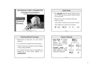
- 1. 1 Christopher G. Hamaker, Illinois State University, Normal IL © 2008, Prentice Hall Chapter 12 Chemical Bonding INTRODUCTORY CHEMISTRY Concepts & Connections Fifth Edition by Charles H. Corwin Chapter 12 2 • Recall that an atom has core and valence electrons. • Core electrons are found close to the nucleus. • Valence electrons are found in the most distant s and p energy subshells. • It is valence electrons that are responsible for holding two or more atoms together in a chemical bond. Chemical Bond Concept Chapter 12 3 • The octet rule states that atoms bond in such a way so that each atom acquires eight electrons in its outer shell. • There are two ways in which an atom may achieve an octet. (a) by transfer of electrons from one atom to another (b) by sharing one or more pairs of electrons Octet Rule Chapter 12 4 • Ionic bonds are formed from the complete transfer of electrons between atoms to form ionic compounds. • Covalent bonds are formed when two atoms share electrons to form molecular compounds. Types of Bonds
- 2. 2 Chapter 12 5 • An ionic bond is formed by the attraction between positively charged kations and negatively charged anions. • This “electrostatic attraction” is similar to the attraction between opposite poles on two magnets. Ionic Bonds Chapter 12 6 • The ionic bonds formed from the combination of anions and cations are very strong and result in the formation of a rigid, crystalline structure. The structure for NaCl, ordinary table salt, is shown here. Ionic Bonds Chapter 12 7 • Cations are formed when an atom loses valence electrons to become positively charged. • Most main group metals achieve a noble gas electron configuration by losing their valence electrons and are isoelectronic with a noble gas. • Magnesium (Group IIA/2) loses its two valence electrons to become Mg2+. • A magnesium ion has 10 electrons (12 – 2 = 10 e-) and is isoelectronic with neon. Formation of Cations Chapter 12 8 • We can use electron dot formulas to look at the formation of cations. • Each of the metals in Period 3 form cations by losing 1, 2, or 3 electrons, respectively. Each metal atom becomes isoelectronic with the preceding noble gas, neon. Formation of Cations
- 3. 3 Chapter 12 9 • Anions are formed when an atom gains electrons and becomes negatively charged. • Most non-metals achieve a noble gas electron configuration by gaining electrons to become isoelectronic with a noble gas. • Chlorine (Group VIIA/17) gains one valence electron and becomes Cl–. • A chloride ion has 18 electrons (17 + 1 = 18 e-) and is isoelectronic with argon. Formation of Anions Chapter 12 10 • We can also use electron dot formulas to look at the formation of anions. • The non-metals in Period 3 gain 1, 2, or 3 electrons, respectively, to form anions. Each non-metal ion is isoelectronic with the following noble gas, argon. Formation of Anions Chapter 12 11 • The radius of a cation is smaller than the radius of its starting atom. • The radius of an anion is larger than the radius of its starting atom. Ionic Radii Chapter 12 12 • Covalent bonds are formed when two non-metal atoms share electrons and the shared electrons in the covalent bond belong to both atoms. • When hydrogen chloride (HCl) is formed, the hydrogen atom shares its one valence electron with the chlorine, This gives the chlorine atom eight electrons in its valence shell, making it isoelectronic with argon. • The chlorine atom shares one of its valence electrons with the hydrogen, giving it two electrons in its valence shell and making it isoelectronic with helium. Covalent Bonds
- 4. 4 Chapter 12 13 Chapter 12 14 • When a covalent bond is formed, the valence shells of the two atoms overlap with each other. • In HCl, the hydrogen 1s energy sublevel overlaps with the chlorine 3p energy sublevel. The mixing of sublevels draws the atoms closer together. • The distance between the two atoms is smaller than the sum of their atomic radii and is the bond length. Bond Length Chapter 12 15 • Energy is released when two atoms form a covalent bond: H(g) + Cl(g) HCl(g) + heat • Conversely, energy is needed to break a covalent bond. • The energy required to break a covalent bond is referred to as the bond energy. • The amount of energy required to break a covalent bond is the same as the amount of energy released when the bond is formed: HCl(g) + heat H(g) + Cl(g) Bond Energy Kekuatan Ikatan Kovalen • Kekuatan ikatan diindikasikan dengan besarnya energi yang diperlukan untuk memutuskan ikatan tersebut. • Entalpi ikatan: energi yang diperlukan untuk memutuskan 1 mol ikatan. • Makin panjang ikatan kovalen, makin kecil energi ikatannya.
- 5. 5 Chapter 12 19 • In Section 6.8 we drew electron dot formulas for atoms. • The number of dots around each atom is equal to the number of valence electrons the atom has. • We will now draw electron dot formulas for molecules (also called Lewis structures). • A Lewis structure shows the bonds between atoms and helps us to visualize the arrangement of atoms in a molecule. Electron Dot Formulas of Molecules Chapter 12 20 1. Calculate the total number of valence electrons by adding all of the valence electrons for each atom in the molecule. 2. Divide the total valence electrons by 2 to find the number of electron pairs in the molecule. 3. Surround the central atom with 4 electron pairs. Use the remaining electron pairs to complete the octet around the other atoms. The only exception is hydrogen, which only needs two electrons. Guidelines for Electron Dot Formulas
- 6. 6 Chapter 12 21 4. Electron pairs that are shared by atoms are called bonding electrons. The other electrons complete octets and are called nonbonding electrons, or lone pairs. 5. If there are not enough electron pairs to provide each atom with an octet, move a nonbonding electron pair between two atoms that already share an electron pair. Guidelines for Electron Dot Formulas Chapter 12 22 1. First, count the total number of valence electrons: oxygen has 6 and each hydrogen has 1 for a total of 8 electrons [6 + 2(1) = 8 e-]. The number of electron pairs is 4 [8/2 = 4]. 2. Place 8 electrons around the central oxygen atom. 3. We can then place the two hydrogen atoms in any of the four electron pair positions. Notice there are 2 bonding and 2 nonbonding electron pairs. Electron Dot Formula for H2O Chapter 12 23 • To simplify, we represent bonding electron pairs with a single dash line called a single bond. • The resulting structure is referred to as the structural formula of the molecule. Electron Dot Formula for H2O Chapter 12 24 1. First, count the total number of valence electrons: each oxygen has 6 and sulfur has 6 for a total of 24 electrons [3(6) + 6 = 24 e-]. This gives us 12 electron pairs. 2. Place 4 electron pairs around the central sulfur atom and attach the three oxygens. We started with 12 electron pairs and have 8 left. 3. Place the remaining electron pairs around the oxygen atoms to complete each octet. 4. One of the oxygens does not have an octet, so move a nonbonding pair from the sulfur to provide 2 pairs between the sulfur and the oxygen. Electron Dot Formula for SO3
- 7. 7 Chapter 12 25 • The two shared electron pairs constitute a double bond. • The double bond can be placed between the sulfur and any of the 3 oxygen atoms and the structural formula can be shown as any of the structures below. This phenomenon is called resonance. Resonance Chapter 12 26 1. The total number of valence electrons is 5 – 4(1) – 1 = 8 e-. We must subtract one electron for the positive charge. We have 4 pairs of electrons. 2. Place 4 electron pairs around the central nitrogen atom and attach the four hydrogens. 3. Enclose the polyatomic ion in brackets and indicate the charge outside the brackets. Electron Dot Formula for NH4 + Chapter 12 27 1. The total number of valence electrons is 4 + 3(6) + 2 = 24 e-. We must add one electron for the negative charge. We have 12 pairs of electrons. 2. Place 4 electron pairs around the central carbon atom and attach the three oxygens. Use the remaining electron pairs to give the oxygen atoms their octets. 3. One oxygen does not have an octet. Make a double bond and enclose the ion in brackets. Electron Dot Formula for CO3 2- Chapter 12 28 • Covalent bonds result from the sharing of valence electrons. • Often, the two atoms do not share the electrons equally. One of the atoms holds onto the electrons more tightly than the other. • When one of the atoms holds the shared electrons more tightly, the bond is polarized. • A polar covalent bond is one in which the electrons are not shared equally. Polar Covalent Bonds
- 8. 8 Chapter 12 29 • Each element has an innate ability to attract valence electrons. • Electronegativity is the ability of an atom to attract electrons in a chemical bond. • Linus Pauling devised a method for measuring the electronegativity of each of the elements. • Fluorine is the most electronegative element. Electronegativity Chapter 12 30 • Electronegativity increases as you go left to right across a period. • Electronegativity increases as you go from bottom to top in a family. Electronegativity Chapter 12 31 • The electronegativity of H is 2.1; Cl is 3.0. • Since there is a difference in electronegativity between the two elements (3.0 – 2.1 = 0.9), the bond in H–Cl is polar. • Since Cl is more electronegative, the bonding electrons are attracted toward the Cl atom and away from the H atom. This will give the Cl atom a slightly negative charge and the H atom a slightly positive charge. Electronegativity Differences Chapter 12 32 • We use the Greek letter delta, d, to indicate a polar bond. • The negatively charged atom is indicated by the symbol d–, and the positively charged atom is indicated by the symbol d+. This is referred to as delta notation for polar bonds. d+ H–Cl d– Delta (δ) Notation for Polar Bonds
- 9. 9 Chapter 12 33 • The hydrogen halides HF, HCl, HBr, and HI all have polar covalent bonds. • The halides are all more electronegative than hydrogen and are designated with a d–. Delta Notation for Polar Bonds Chapter 12 34 • What if the two atoms in a covalent bond have the same or similar electronegativities? • The bond is not polarized and it is a nonpolar covalent bond. If the electronegativity difference is less than 0.5, it is usually considered a nonpolar bond. • The diatomic halogen molecules have nonpolar covalent bonds. Nonpolar Covalent Bonds Chapter 12 35 • A covalent bond resulting from one atom donating a lone pair of electrons to another atom is called a coordinate covalent bond. • A good example of a molecule with a coordinate covalent bond is ozone, O3. Coordinate Covalent Bonds Chapter 12 36 Hydrogen Bonds • The bond between H and O in water is very polar. • Therefore, the oxygen is partially negative, and the hydrogens are partially positive. • As a result, the hydrogen atom on one molecule is attracted to the oxygen atom on another. • This intermolecular interaction is referred to as a hydrogen bond.
- 10. 10 Chapter 12 37 • Electron pairs surrounding an atom repel each other. This is referred to as Valence Shell Electron Pair Repulsion (VSEPR) theory. • The electron pair geometry indicates the arrangement of bonding and nonbonding electron pairs around the central atom. • The molecular shape gives the arrangement of atoms around the central atom as a result of electron repulsion. Shapes of Molecules Chapter 12 38 • Methane, CH4, has four pairs of bonding electrons around the central carbon atom. • The four bonding pairs (and, therefore, atoms) are repelled to the four corners of a tetrahedron. The electron pair geometry is tetrahedral. • The molecular shape is also tetrahedral. Tetrahedral Molecules Chapter 12 39 • In ammonia, NH3, the central nitrogen atom is surrounded by three bonding pairs and one nonbonding pair. • The electron pair geometry is tetrahedral and the molecular shape is trigonal pyramidal. Trigonal Pyramidal Molecules Chapter 12 40 • In water, H2O, the central O atom is surrounded by two nonbonding pairs and two bonding pairs. • The electron pair geometry is tetrahedral and the molecular shape is bent. Bent Molecules
- 11. 11 Chapter 12 41 • In carbon dioxide, CO2, the central C atom is bonded to each oxygen by two electron pairs (a double bond). • According to VSEPR, the electron pairs will repel each other, and they will be at opposite sides of the C atom. • The electron pair geometry and the molecular shape are both linear. Linear Molecules Chapter 12 42 Summary of VSEPR Theory Chapter 12 43 Nonpolar Molecules with Polar Bonds • CCl4 has polar bonds, but the overall molecule is nonpolar • Using VSEPR theory, the four chlorine atoms are at the four corners of a tetrahedron • The chlorines are each δ–, while the carbon is δ+. • The net effect of the polar bonds is zero, so the molecule is nonpolar. Chapter 12 44 Diamond vs. Graphite • Why is diamond colorless and hard, while graphite is black and soft if both are pure carbon? • Diamond has a 3-dimensional structure, while graphite has a 2-dimensional structure. • The layers in graphite are able to slide past each other easily. graphite diamond
- 12. 12 Chapter 12 45 • Chemical bonds hold atoms together in molecules. • Atoms bond in such a way as to have eight electrons in their valence shell: the octet rule. • There are 2 types of bonds: ionic and covalent. • Ionic bonds are formed between a cation and an anion. • Covalent bonds are formed from the sharing of electrons. Chapter Summary Chapter 12 46 • Electron dot formulas help us to visualize the arrangements of atoms in a molecule. • Electrons are shared unequally in polar covalent bonds. • Electronegativity is a measure of the ability of an atom to attract electrons in a chemical bond. • Electronegativity increases from left to right and from bottom to top on the periodic table. Chapter Summary, continued Chapter 12 47 • VSEPR theory can be used to predict the shapes of molecules. • The electron pair geometry gives the arrangement of bonding and nonbonding pairs around a central atom. • The molecular shape gives the arrangement of atoms in a molecule. Chapter Summary, continued