Birinci Trimester Ultrasonografi:Önem Henüz birkaç santimetre olan embriyoda binlerce anatomik yapı mevcuttur.İleride gelişecek anomaliler bu embriyoner dönemde ortaya çıkmaya başlamakta ancak bir çoğu daha sonraki dönemlerde bulgu vermektedir
3.
Hemen hepsi geçicihaberciler olan bazı belirteçler,gebelik haftası ilerleyince kaybolabilmekte,eşlik eden anomaliler yavaş yavaş rutin ultrasonografi ile görülebilir hale gelmektedir.Gelişim sürecinde embriyo ile ekstraembriyoner yapılar arasında var olan dengeli oranlar,normallik ile anormallik arasındaki sınırı çizmemize olanak tanımaktadır.
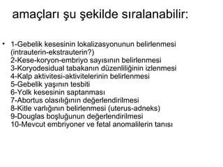
Klasik kitaplarda yeralan bu amaçlara artık bir yenisi eklenmiştir:Embriyoner ve fetal yapıların anatomik bütünlüğünün incelenmesi ve ileride görülebilecek olası anomalilerin ve kromozom defektlerinin önceden saptanabilmesi
7.
11-14 haftalarında anensefali,ensefalosel,hidrosefali,holoprozensefali,Meckel-Grubersendromu,iniensefali,spina bifida,major kalp anomalileri,omfalosel, gastroşisiz,diyafragmatik herni,renal agenezi,infantil polikistik böbrek,hidronefroz,megasistis,iskelet sistemi anomalileri,kaudal regresyon sendromu gibi fetal defekt,anomali ve sendromların tanısı konulabilmektedir[11,12].
8.
öncelikle gebeliğin erkendönemlerinden itibaren görülen normal bulgular ele alınacak,daha sonra spesifik anormallikler açıklanmaya çalışılacaktır.
9.
Normal Erken GebelikUltrasonografi Bulguları 1-Desidua 2-Gebelik kesesi: 3-Çift desidual kese belirtisi: 4-Yolk kesesi: 4- Çift kabarcık belirtisi: 5-Embriyo ve kalp atımı: 6-CRL: 7-Amniyon ve amniyotik kese
10.
Normal Erken GebelikUltrasonografi Bulguları 1-Desidua Beklenen adet dönemine yakın günlerde (24-28 günler) daha ödemli, kalın desidualı (> 18 mm),ekojen bir endometriyum tanı koydurmamakla birlikte gebeliği düşündürür.Bu durumda hassas gebelik testlerinin değeri daha yüksektir[13].Yeh ve arkadaşlarına göre, ultrasonografide ilk belirti (23.gün) implantasyon tarafında desidual kalınlaşma ile birlikte fokal ekojenik bir zonun görülmesidir
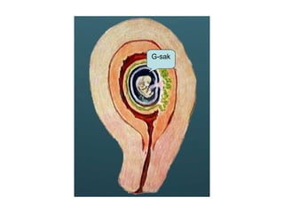
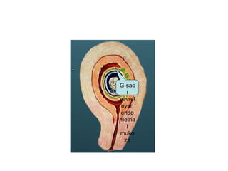
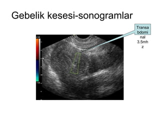
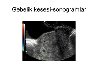
2-Gebelik kesesi: Aslında gebelik kesesi terimi anatomik bir terim değildir,ancak ultrasonografi literatüründe yaygın olarak kullanılmaktadır Aslının "koryonik kese" olması gerekir.Kalınlaşmış desidua içinde yer alan parlak çevreli hipoekojen kompleks kese,gebeliğin ilk objektif belirtisidir.
13.
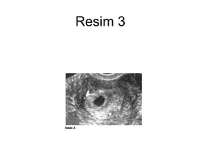
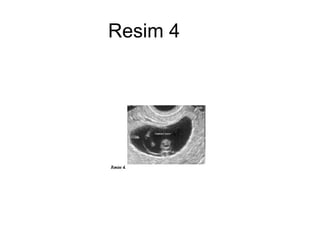
Bu sıvı dolukese yapısı aslında,embriyoner disk, amniyon,sekonder Yolk kesesi ve ekstraembriyoner sölomu barındıran koryonik bir boşluktur (Resim-3 ve 4).Ekojen halka görüntüsünün nedeni desidua içine doğru büyüyen villuslardır.
IVF tecrübesine göre4hafta 2gün iken (ortalama: 29-32.günler) kese 2-3 mm çapında görülebilir.Genelde fundus posterioruna asimetrik olarak yerleşir ve diğer yapılar görülene kadar günde 1-2 mm büyür. Görülebilmesi için transvaginal muayene ve yüksek büyütme kullanılması gereklidir.
Transvaginal ultrasonografide kese8 mm'den büyük ve Yolk kesesi yoksa veya kese 20 mm'den büyük ve embriyo yoksa o gebelik bozulmuştur.Eğer halka yapısı yok,köşeli,irregüler,berrak,hidropik bir kese görünümü mevcutsa buna “Psödogestasyonel kese” denir,ektopik veya oluşmamış gebelikte görülür [13,15].
2-Çift desidual kesebelirtisi: Bu bulgu intrauterin gebeliğin bir belirtisidir.Son adetin ilk gününden sonraki 39-42.günler arasında,Yolk kesesinin ilk görüldüğü günlerde,desidua vera içinde desidua kapsularis ve koryon leave ile sınırlanmış iki ayrı kesenin görülmesi ile karakterizedir.Desidua bazalis eksantrik ekojen bir kalınlaşma olarak görülebilir [16].
25.
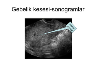
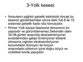
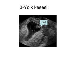
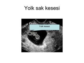
3-Yolk kesesi: İntrauterin sağlıklı gebelik belirtisidir.Ancak bu kesenin görülmesinden sonra dahi %8.8 ile 15 arasında gebelik kaybı söz konusudur. Primer Yolk kesesi blastosist döneminin bir yapısıdır ve görüntülenemez.Sekonder olanı 34-38.günler arasında ekstraembriyoner endodermden gelişir.Embriyoner kökenli olmasına rağmen ekstraembriyoner konumdadır.Amniyon ile koryon arasındadır,sölomun içine doğru büyür ve umbilikal korda yapışıktır.
26.
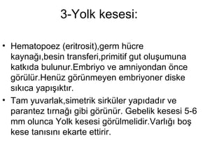
3-Yolk kesesi: Hematopoez (eritrosit),germ hücre kaynağı,besin transferi,primitif gut oluşumuna katkıda bulunur.Embriyo ve amniyondan önce görülür.Henüz görünmeyen embriyoner diske sıkıca yapışıktır. Tam yuvarlak,simetrik sirküler yapıdadır ve parantez tırnağı gibi görünür. Gebelik kesesi 5-6 mm olunca Yolk kesesi görülmelidir.Varlığı boş kese tanısını ekarte ettirir.
27.
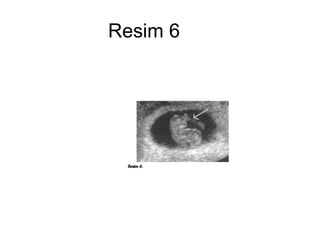
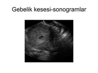
3-Yolk kesesi: Gebelikkesesi 8 mm iken,halen yolk kesesi görülmüyorsa o gebelik bozulmuştur.Gebelik kesesi 1 mm büyüdükçe Yolk kesesi 0.1 mm büyür;15 mm'den sonra her 1 mm için Yolk kesesi 0.3 mm büyür Amniyon arttıkça bu kese kenara itilir.Önceleri koryonik kesenin büyük bölümünü doldururken,kalp atımı görülmeden hemen önce koryonik kesenin haciminin 1/3'üne düşer.Yolk kesesi görüldükten 3-7 gün sonra kalp atımları görülmelidir.Boyutları 4-6 mm arasında olup, sabittir.Çapı 2 mm'den az,7 mm'den fazla veya kalsifiye ise prognoz kötüdür [13,17](Resim-5).
4- Çift kabarcıkbelirtisi: Postmenstruel 39. gün (5.5 hafta) civarında geçici bir bulgudur. Üç ile yedi gün sonra kalp atımları görülür.Embriyonun görülmesi ile bu belirti kaybolur.İkiz ile karıştırılmamalıdır[18].
32.
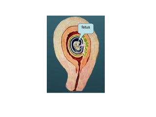
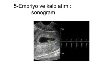
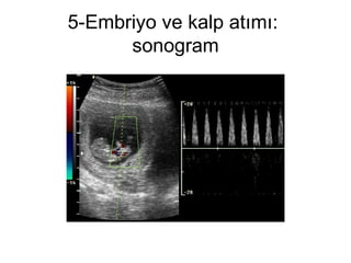
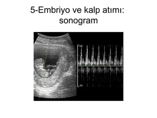
5-Embriyo ve kalpatımı: Postmenstruel 39-42.günlerde (5.5-6 hafta),Yolk kesesinden 3-7 gün sonra,dikkatli bakıldığnda görülür.Embriyo 2-3 mm olarak belirir. Gebeliğin 6.5 haftasından önce 80-100 vuru/dak, 9.haftasında 160-190 vuru/dak iken,daha sonra normal ritmi olan 120-160 vuru/dak'ya düşer. Kalp aktivitesinin ilk görünümünden sonra kayıp riski %10'dur. Gebeliğin 8.haftasından sonra bu risk %2-4'e düşer[13].Embriyo 4-5 mm'ye ulaşana kadar kalp aktivitesi görülmeyebilir
33.
5-Embriyo ve kalpatımı: hatta transabdominal incelemelerde 9 mm'de bile negatif olabilir[19].Kalp atım hızı 85'in altında ise gebelik prognozunun kötü olduğu bildirilmiştir [20].
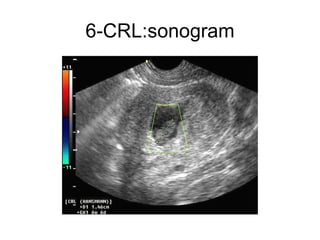
6-CRL: Embriyonerkutup olarak da adlandırılan CRL,ilk önce desidua bazalis ile Yolk kesesi arasında küçük ekojen bir yapı olarak (2-3 mm) görülür. Her gün 1 mm büyür[13]. CRL ölçümüne göre gebelik gününün hesaplanması: "CRL (mm)+ 42=Gebelik günü±3gün " formülü ile yapılır[21]. Gebelik kesesi çapı ile CRL arasındaki fark 5 mm'den fazla ise gebelik akıbeti kötüdür. Goldstein ve ark'na göre abortus riski CRL 5 mm'den küçükse:%7.1,5-10 mm arasında ise:%3 ve 10 mm'den büyükse %0.5'tir.Ancak tekrarlayan abortuslarda bu oranlar kullanılmaz [22].
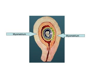
7-Amniyon ve amniyotikkese Önceleri embriyo ile trofoblastlar arasında yer alan amniyon kesesi daha sonra embriyo kaynaklı sıvılar ile büyüyerek embriyoyu tamamen çevreler,Yolk kesesini koryona doğru iter ve gebeliğin 2. trimesterinde koryonik tabaka ile birleşir İlk görülme zamanı 7. haftadır (49.gün).Etraftaki koryonik sıvı villuslardan kaynaklanmaktadır ve amniyondan daha ekojeniktir [13].Kollaps ve düzensiz sınır kötü prognoz belirtisidir
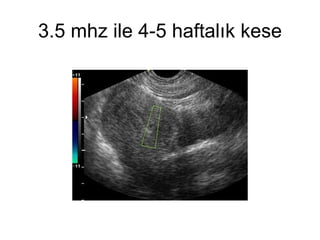
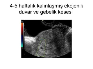
4. Hafta: Gebeliğin4 hafta 2.gününden itibaren transvaginal ultrasonografi ile,kalınlaşmış desidua içinde 2-3 mm'lik hipoekojen gestasyonel kese,çevresindeki ekojen halkası ile birlikte görülür [12].
5. Hafta: İlkgörünen yapı Yolk kesesidir.Beşinci haftanın 4.gününde mutlaka görülmesi gereklidir. Embriyoner kutup (2-5 mm) Yolk kesesinin hemen yakınındadır.Bu kompleks yapı sölomik boşluk içinde yer alır.Birkaç gün içinde,koryonik kese 10 mm olduktan sonra kalp aktivitesi görülür.Kalp atım hızı dakikada 100 civarındadır [23].
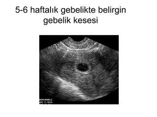
6.Hafta: Embriyoner kutup,Yolkkesesi ve kalp hareketi görülür.Kese 15 mm, embriyo 4-8 mm olmuştur.Embriyonun çevresinde amniyon da varsa sölomdan ayrı,gestasyonel keseye daha yakın duran bu oluşuma çift kabarcık belirtisi de denmektedir. Kalp atım hızı dakikada 135 civarındadır.Bu haftanın sonuna doğru embriyonun kranyal bölümünde, ön beyin:Telensefalon ve diensefalon olarak bölünür.Rombensefalon hipoekojen olarak belirir.Bu günlerde amniyon zarı embriyo etrafında görünür hale gelmiştir.Kalp hızı dakikada 130 civarındadır.
49.
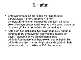
7. Hafta: Koryoniktabaka desidua ile yapışır ve plasentayı oluşturmaya başlar.Embriyo uzunluğu (CRL) 9-14 mm arasındadır Embriyo bu dönemde sagittal planda bir üçgene benzer.Koronal planda ise silindir şeklinde görülür. Ekstremiteler çok kısa olarak görülebilir. Kranyumda önden arkaya doğru hemisferler,diensefalon (ilerideki 3. ventrikül),mezensefalon (ilerideki aquaductus Sylvii) ve rombensefalon (ilerideki 4.ventrikül) oluşmuştur Bu haftada başın hemen altında büyük,parlak ve hareket halindeki kalp görülür. Dakikadaki atım sayısı 130'dan 160'a yükselmiştir,ancak kalbin anatomik ayrıntıları çok zor ayırt edilir.İntestinal traktusta, umbilikal kordonun abdominal insersiyonunda ekojenite artışı ve kalınlaşma ile birlikte fizyolojik barsak herniasyonunun ilk belirtileri oluşur.
50.
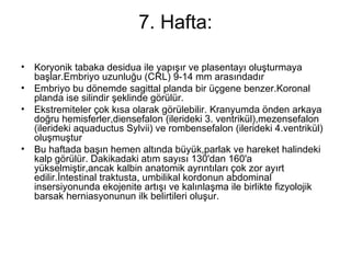
7. Hafta: sonogram-gkesesi ovoid şekilde belirgin dopler ile kardiak aktivitesi belli olan fetus
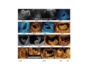
8. Hafta: Embriyo15-22 mm'ye ulaşır.Gövde küboid bir görünüm kazanır. Ekstremiteler belirgindir, parmaklar ayırt edilebilir.Kranyumda geniş kaviteler görülür. Hemisferler önden arkaya doğru genişler, lateral ventrikül içinde koroid pleksusler görülür.Üçüncü ventrikül genişliği en üstteki mezensefalik boşluk kadardır İleride 4. ventriküle dönüşecek olan rombensefalik boşluk,tepesi ponsta bulunan bir piramit şeklinde kaudal bölümde yerini alır.Kalp atım hızı dakikada 160'a ulaşır.Bu haftanın sonlarında atrium ve ventrikül duvarları ayırt edilebilir. Atrium bölümü ventriküllerin kapladığı alandan daha genişçedir ve kalbin 4 odacık görüntüsü bu haftada alınabilir.Kalp,toraksa boşluğunun %50'sini kaplar.Embriyoların %31'inde abdomen içinde sol üstte mide hipoekojen bir alan olarak görülür.
58.
9. Hafta: Embriyo23-31 mm'ye ulaşır ve embriyo hareketleri izlenir.Gövde büyük bir kafaya sahip olan eliptik bir yapıya dönüşmüştür. Abdominal duvar belirgindir,ağız görülebilir,profilden görüntü alınabilir.Ekstremiteler uzamıştır.Eller ve ayaklar birbirlerine çok yakındır ve temas halindedirler.Ayak tabanları sagital planda birbirine bakarlar.Lateral ventriküller belirgindir,içlerini dolduran koroid pleksuslar ise en iyi olarak 9 hafta 4 günde parasagital planda görüntülenebilirler Hemisferler ile eş zamanlı olarak çok hızlı büyürler ve ventrikülün tamamını doldururlar. Korteks düzgün ve hipoekojendir.Diensefalon boşluğu daralırken, mezensefalon hala geniştir.Rombensefalonun lateral genişliği artar ve burayı iki ayrı parça halindeki serebellum hemisferleri doldururlar.Dokuzuncu haftada kalp hızı artar ve dakikada 175'e ulaşır.Sekizinci hafta ortalarından 10. hafta ortalarına kadar tüm embriyolar fizyolojik barsak herniasyonu gösterirler (Resim-6). Bu da en iyi olarak 9.haftada geniş hiperekojen bir kitle olarak görülür. Herniasyonun çapı genelde 6.3 mm'den daha fazla değildir.Dokuzuncu haftanın sonuna kadar embriyoların %75'inde mide görülebilir.Henüz yutma işlemi başlamadığından,görülen hipoekojen alan midenin fizyolojik sekresyonudur.Aşırı dilatasyon,özofagus ve barsak atrezilerini düşündürür.
![11-14 haftalarında anensefali,ensefalosel,hidrosefali,holoprozensefali,Meckel-Gruber sendromu,iniensefali,spina bifida,major kalp anomalileri,omfalosel, gastroşisiz,diyafragmatik herni,renal agenezi,infantil polikistik böbrek,hidronefroz,megasistis,iskelet sistemi anomalileri,kaudal regresyon sendromu gibi fetal defekt,anomali ve sendromların tanısı konulabilmektedir[11,12].](https://image.slidesharecdn.com/1-trimesterultrasonografisi-110821124759-phpapp01/85/1-trimester-ultrasonografisi-7-320.jpg)
![Normal Erken Gebelik Ultrasonografi Bulguları 1-Desidua Beklenen adet dönemine yakın günlerde (24-28 günler) daha ödemli, kalın desidualı (> 18 mm),ekojen bir endometriyum tanı koydurmamakla birlikte gebeliği düşündürür.Bu durumda hassas gebelik testlerinin değeri daha yüksektir[13].Yeh ve arkadaşlarına göre, ultrasonografide ilk belirti (23.gün) implantasyon tarafında desidual kalınlaşma ile birlikte fokal ekojenik bir zonun görülmesidir](https://image.slidesharecdn.com/1-trimesterultrasonografisi-110821124759-phpapp01/85/1-trimester-ultrasonografisi-10-320.jpg)
.](https://image.slidesharecdn.com/1-trimesterultrasonografisi-110821124759-phpapp01/85/1-trimester-ultrasonografisi-11-320.jpg)
![Transvaginal ultrasonografide kese 8 mm'den büyük ve Yolk kesesi yoksa veya kese 20 mm'den büyük ve embriyo yoksa o gebelik bozulmuştur.Eğer halka yapısı yok,köşeli,irregüler,berrak,hidropik bir kese görünümü mevcutsa buna “Psödogestasyonel kese” denir,ektopik veya oluşmamış gebelikte görülür [13,15].](https://image.slidesharecdn.com/1-trimesterultrasonografisi-110821124759-phpapp01/85/1-trimester-ultrasonografisi-18-320.jpg)
![2-Çift desidual kese belirtisi: Bu bulgu intrauterin gebeliğin bir belirtisidir.Son adetin ilk gününden sonraki 39-42.günler arasında,Yolk kesesinin ilk görüldüğü günlerde,desidua vera içinde desidua kapsularis ve koryon leave ile sınırlanmış iki ayrı kesenin görülmesi ile karakterizedir.Desidua bazalis eksantrik ekojen bir kalınlaşma olarak görülebilir [16].](https://image.slidesharecdn.com/1-trimesterultrasonografisi-110821124759-phpapp01/85/1-trimester-ultrasonografisi-24-320.jpg)
.](https://image.slidesharecdn.com/1-trimesterultrasonografisi-110821124759-phpapp01/85/1-trimester-ultrasonografisi-27-320.jpg)
.](https://image.slidesharecdn.com/1-trimesterultrasonografisi-110821124759-phpapp01/85/1-trimester-ultrasonografisi-28-320.jpg)
![4- Çift kabarcık belirtisi: Postmenstruel 39. gün (5.5 hafta) civarında geçici bir bulgudur. Üç ile yedi gün sonra kalp atımları görülür.Embriyonun görülmesi ile bu belirti kaybolur.İkiz ile karıştırılmamalıdır[18].](https://image.slidesharecdn.com/1-trimesterultrasonografisi-110821124759-phpapp01/85/1-trimester-ultrasonografisi-31-320.jpg)
![5-Embriyo ve kalp atımı: Postmenstruel 39-42.günlerde (5.5-6 hafta),Yolk kesesinden 3-7 gün sonra,dikkatli bakıldığnda görülür.Embriyo 2-3 mm olarak belirir. Gebeliğin 6.5 haftasından önce 80-100 vuru/dak, 9.haftasında 160-190 vuru/dak iken,daha sonra normal ritmi olan 120-160 vuru/dak'ya düşer. Kalp aktivitesinin ilk görünümünden sonra kayıp riski %10'dur. Gebeliğin 8.haftasından sonra bu risk %2-4'e düşer[13].Embriyo 4-5 mm'ye ulaşana kadar kalp aktivitesi görülmeyebilir](https://image.slidesharecdn.com/1-trimesterultrasonografisi-110821124759-phpapp01/85/1-trimester-ultrasonografisi-32-320.jpg)
![5-Embriyo ve kalp atımı: hatta transabdominal incelemelerde 9 mm'de bile negatif olabilir[19].Kalp atım hızı 85'in altında ise gebelik prognozunun kötü olduğu bildirilmiştir [20].](https://image.slidesharecdn.com/1-trimesterultrasonografisi-110821124759-phpapp01/85/1-trimester-ultrasonografisi-33-320.jpg)
![6-CRL: Embriyoner kutup olarak da adlandırılan CRL,ilk önce desidua bazalis ile Yolk kesesi arasında küçük ekojen bir yapı olarak (2-3 mm) görülür. Her gün 1 mm büyür[13]. CRL ölçümüne göre gebelik gününün hesaplanması: "CRL (mm)+ 42=Gebelik günü±3gün " formülü ile yapılır[21]. Gebelik kesesi çapı ile CRL arasındaki fark 5 mm'den fazla ise gebelik akıbeti kötüdür. Goldstein ve ark'na göre abortus riski CRL 5 mm'den küçükse:%7.1,5-10 mm arasında ise:%3 ve 10 mm'den büyükse %0.5'tir.Ancak tekrarlayan abortuslarda bu oranlar kullanılmaz [22].](https://image.slidesharecdn.com/1-trimesterultrasonografisi-110821124759-phpapp01/85/1-trimester-ultrasonografisi-37-320.jpg)

![7-Amniyon ve amniyotik kese Önceleri embriyo ile trofoblastlar arasında yer alan amniyon kesesi daha sonra embriyo kaynaklı sıvılar ile büyüyerek embriyoyu tamamen çevreler,Yolk kesesini koryona doğru iter ve gebeliğin 2. trimesterinde koryonik tabaka ile birleşir İlk görülme zamanı 7. haftadır (49.gün).Etraftaki koryonik sıvı villuslardan kaynaklanmaktadır ve amniyondan daha ekojeniktir [13].Kollaps ve düzensiz sınır kötü prognoz belirtisidir](https://image.slidesharecdn.com/1-trimesterultrasonografisi-110821124759-phpapp01/85/1-trimester-ultrasonografisi-41-320.jpg)
![4. Hafta: Gebeliğin 4 hafta 2.gününden itibaren transvaginal ultrasonografi ile,kalınlaşmış desidua içinde 2-3 mm'lik hipoekojen gestasyonel kese,çevresindeki ekojen halkası ile birlikte görülür [12].](https://image.slidesharecdn.com/1-trimesterultrasonografisi-110821124759-phpapp01/85/1-trimester-ultrasonografisi-43-320.jpg)
![5. Hafta: İlk görünen yapı Yolk kesesidir.Beşinci haftanın 4.gününde mutlaka görülmesi gereklidir. Embriyoner kutup (2-5 mm) Yolk kesesinin hemen yakınındadır.Bu kompleks yapı sölomik boşluk içinde yer alır.Birkaç gün içinde,koryonik kese 10 mm olduktan sonra kalp aktivitesi görülür.Kalp atım hızı dakikada 100 civarındadır [23].](https://image.slidesharecdn.com/1-trimesterultrasonografisi-110821124759-phpapp01/85/1-trimester-ultrasonografisi-46-320.jpg)