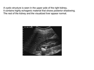
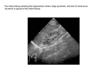
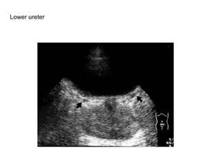
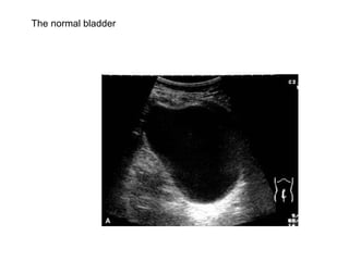
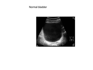
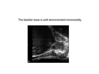
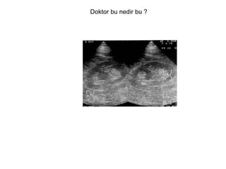
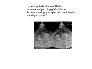
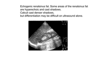
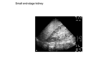
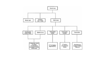
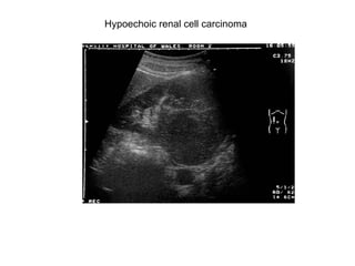
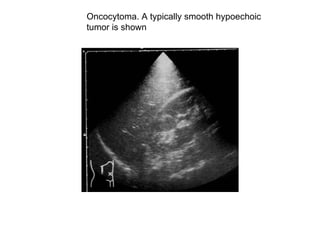
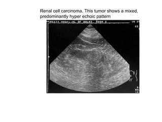
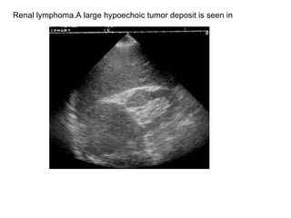
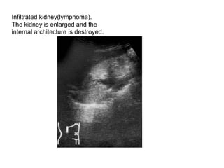
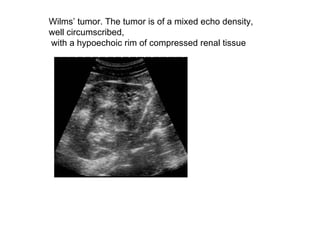
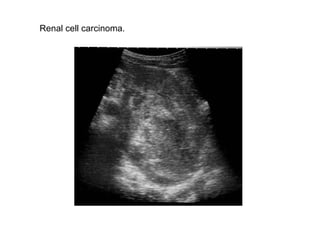
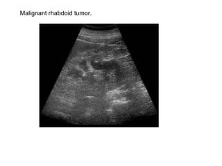
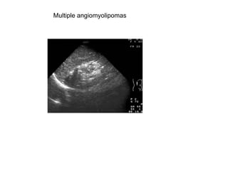
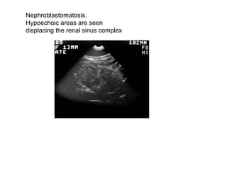
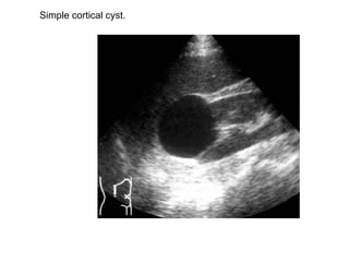
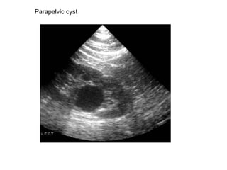
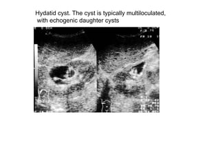
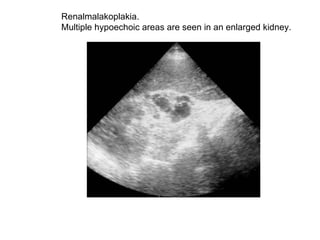
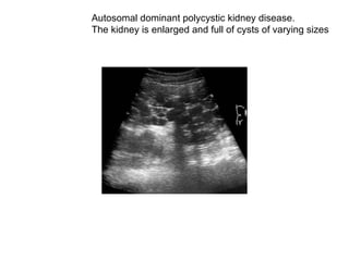
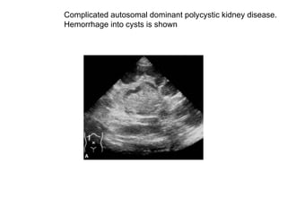
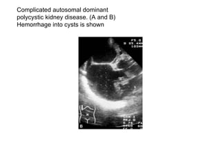
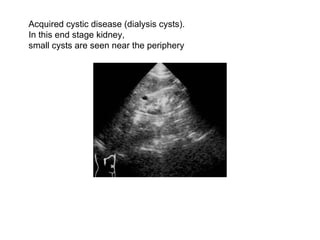
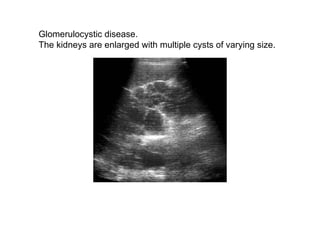
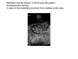
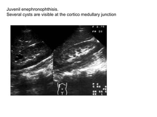
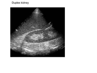
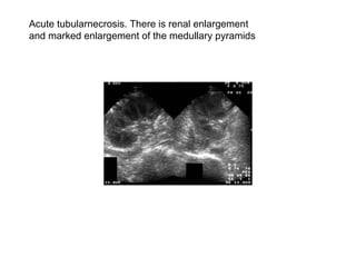
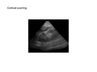
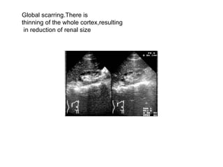
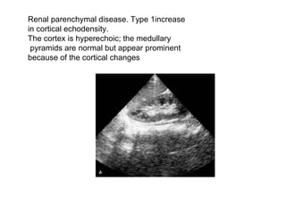
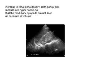
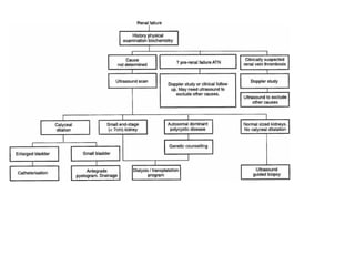
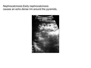
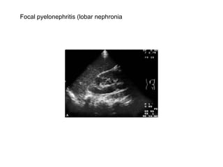
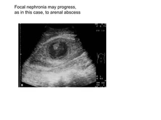
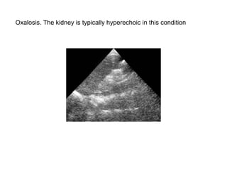
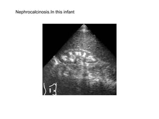
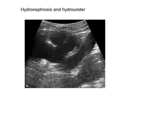
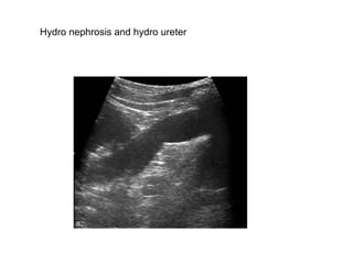
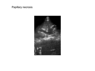
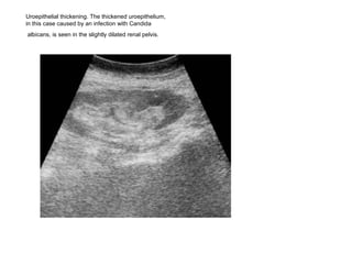
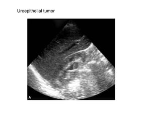
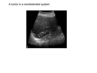
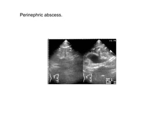
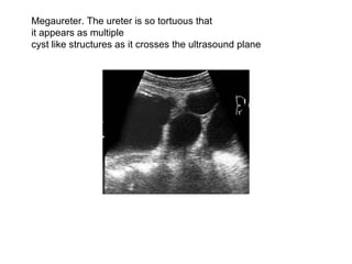
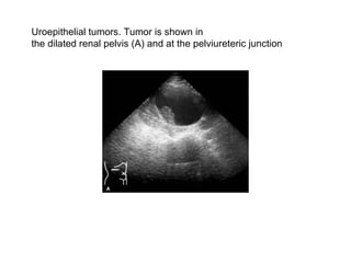
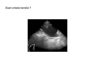
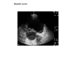
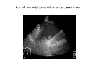
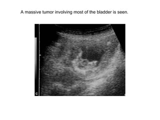
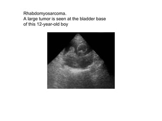
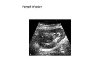
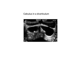
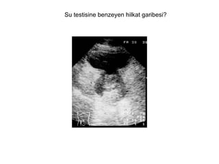
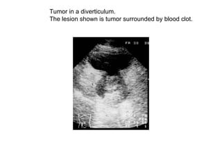
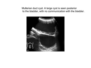
Lectures on Diagnostic Ürosonography Uzm.Dr. BAHRİ YILDIZ Uzm.Dr.M.EROL YAYLA
 
The left kidney is also normal in size and demonstrates extensive,  curvilinear parenchymal calcification with posterior shadowing  in the region of the medulla.
 
The right kidney is normal in size, but exhibits increased echogenicity of the medullary pyramids. No obvious acoustic shadowing is noted from the echogenic foci
A cystic structure is seen in the upper pole of the right kidney.  It contains highly echogenic material that shows posterior shadowing.  The rest of the kidney and the visualized liver appear normal.
Normal Kidneys.The right kidney has been scanned from an antero lateral  approach through the liver. There is good differentiation of the renal capsule, cortex, medulla, and renal sinus complex. The longitudinal plane.
The transverse plane.
 
The infant kidney,showing the hyperechoic cortex, large pyramids, and lack of renal sinus fat,which is typical of the infant kidney
Ureteric orifices.
An atomic diagram of the renal arteries and veins.
Upper ureter.
Lower ureter
The normal bladder
Normal bladder
The bladder base is well demonstrated transrectally.
Doktor bu nedir bu ?
Hypertrophied column of Bertin.  (adamlar radyoizotop görünteleme  ile bu tanıyı doğrulamışlar peki nedir bertin  Hatırlayan varmı ?
 
Echogenic renalsinus fat. Some areas of the renalsinus fat  are hyperechoic and cast shadows.  Calculi cast denser shadows,  but differentiation may be difficult on ultrasound alone.
Small end-stage kidney
 
Hypoechoic renal cell carcinoma
Oncocytoma. A typically smooth hypoechoic  tumor is shown
Renal cell carcinoma. This tumor shows a mixed,  predominantly hyper echoic pattern
Angiomyolipoma
Angiomyolipoma confirmed by ct
Small angiomyolipoma. Small angiomyolipmas are often of an even,high echo density
Ve şimdi buna taş diyeceksiniz öyle mi?
Renal cell carcinoma with calcification. Dense hyperechoic  areas of calcification are seen with in the tumor.
Renal lymphoma.A large hypoechoic tumor deposit is seen in
Infiltrated kidney(lymphoma).  The kidney is enlarged and the  internal architecture is destroyed.
Wilms’ tumor. The tumor is of a mixed echo density,  well circumscribed, with a hypoechoic rim of compressed renal tissue
Renal cell carcinoma.
Malignant rhabdoid tumor.
Multiple angiomyolipomas
Nephroblastomatosis.  Hypoechoic areas are seen  displacing the renal sinus complex
Simple cortical cyst.
Parapelvic cyst
Hydatid cyst. The cyst is typically multiloculated, with echogenic daughter cysts
Renalmalakoplakia.  Multiple hypoechoic areas are seen in an enlarged kidney.
Autosomal dominant polycystic kidney disease. The kidney is enlarged and full of cysts of varying sizes
Complicated autosomal dominant polycystic kidney disease.  Hemorrhage into cysts is shown
Complicated autosomal dominant  polycystic kidney disease. (A and B)  Hemorrhage into cysts is shown
Acquired cystic disease (dialysis cysts).  In this end stage kidney,  small cysts are seen near the periphery
Glomerulocystic disease.  The kidneys are enlarged with multiple cysts of varying size.
Medullary sponge kidney. In this 9-year-old patient, increased echo density is seen in the medullary pyramids from multiple small cysts
Juvenil enephronophthisis.  Several cysts are visible at the cortico medullary junction
Duplex kidney
Acute tubularnecrosis. There is renal enlargement  and marked enlargement of the medullary pyramids
Cortical scarring
Global scarring.There is  thinning of the whole cortex,resulting in reduction of renal size
Renal parenchymal disease. Type 1increase  in cortical echodensity.  The cortex is hyperechoic; the medullary pyramids are normal but appear prominent  because of the cortical changes
increase in renal echo density. Both cortex and  medulla are hyper echoic so  that the medullary pyramids are not seen  as separate structures.
 
Nephrocalcinosis.Early nephrocalcinosis  causes an echo dense rim around the pyramids.
Focal pyelonephritis (lobar nephronia
Focal nephronia may progress,  as in this case, to arenal abscess
Oxalosis. The kidney is typically hyperechoic in this condition
Nephrocalcinosis.In this infant
Hydronephrosis and hydroureter
Hydro nephrosis and hydro ureter
Papillary necrosis
Uroepithelial thickening. The thickened uroepithelium,  in this case caused by an infection with Candida albicans, is seen in the slightly dilated renal pelvis.
Uroepithelial tumor
A tumor in a nondistended system
Perinephric abscess.
Megaureter. The ureter is so tortuous that it appears as multiple  cyst like structures as it crosses the ultrasound plane
Uroepithelial tumors. Tumor is shown in  the dilated renal pelvis (A) and at the pelviureteric junction
Eseri ortada kendisi ?
Ureterocele.Typical balloon-like  dilatations are seen projecting into the bladder
Schizosomiasis.The typical bladder wall thickening  is shown in this patient who presented with renal failure  and bilateral hydronephros is caused by ureteric strictures
A thick-walled bladder
Tuberculosis. The bladder is thick walled and of  small volume.  The patient felt that the bladder was  full at the time of this scan
Bladder tumor
A small polypoidal tumor with a narrow base is shown.
A massive tumor involving most of the bladder is seen.
Rhabdomyosarcoma.  A large tumor is seen at the bladder base  of this 12-year-old boy
Fungal infection
Typical fungal balls are seen in the bladder
Another case with fungal balls in the renal collecting system.
Bladder diverticulum
Calculus in a diverticulum
Su testisine benzeyen hilkat garibesi?
Tumor in a diverticulum.  The lesion shown is tumor surrounded by blood clot.
Mullerian duct cyst. A large cyst is seen posterior to the bladder, with no communication with the bladder.
?
Pyuria. A fluid filled level is seen caused by layered pus
Residual bladder volume measurement
 

Lectures on diagnostic ürosonography