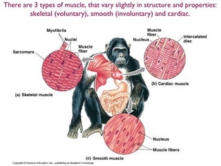
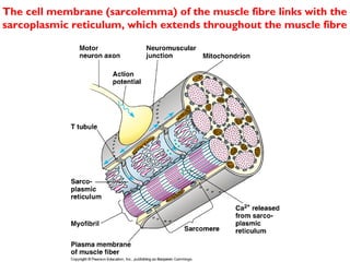
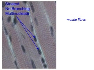
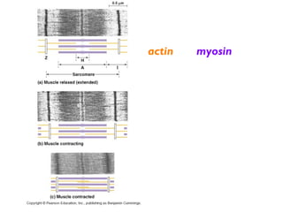
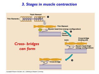
cle
There are 3 types of muscle, that vary slightly in structure and properties:
skeletal (voluntary), smooth (involuntary) and cardiac.
End view of a muscle fibre
muscle fibres are ‘full’ of
long parallel protein
structures
- the myofibrils
The cell membrane (sarcolemma) of the muscle fibre links with the
sarcoplasmic reticulum, which extends throughout the muscle fibre
Muscle tissue is not
made up of
individual ‘cells’, but
giant muscle fibres
As embryonic muscle
tissue differentiates,
individual cells fuse
together, creating
multinucleate
structures, the muscle
fibres
Within a muscle fibre
are many long,
banded structures, the
myofibrils.
These myofibrils
extend the whole
length of a muscle
fibre.
Myofibrils have a
regular repeating
pattern. Each repeating
‘unit’ is called a
sarcomere
A sarcomere is composed of 2 overlapping types of fibrous proteins,
actin and myosin
Muscle fibre
contractions are
controlled from
the CNS by
neurons that
synapse at
neuromuscular
junctions
A neuromuscular junction is a synapse
Acetylcholine (Ach) is the neurotransmitter
The arrival of an impulse releases Calcium ions
allowing myosin/ actin cross links to form
actin
myosin
An action
potential is
transmitted to
the muscle
fibre’s
sarcolemma and
spreads
throughout the
muscle fibre
along its
sarcoplasmic
reticulum
A muscle contraction is
caused by the interlocking
actin and myosin fibres
sliding over one another,
shortening the muscle.
The arrival of a nerve
impulse, and its spread
throughout the muscle
fibre causes this ‘sliding’
contraction
Myosin molecules have a head
and ‘tail’, and occur in
‘bundles’ or filaments
Actin molecules are globular
and occur in chains
In a resting muscle, any reaction between actin & myosin is prevented by
tropomyosin, which blocks actin’s binding site
when a nerve impulse stimulates a muscle
to contract....
The action potential spreads throughout the muscle
fibre, along its sarcoplasmic reticulum
Releasing Calcium ions into the cytoplasm
Calcium ions allow myosin ‘heads’ to form cross
links with actin
The myosin molecule pulls the actin molecule ‘back’,
shortening the overall length of the fibre
ATP provides the energy to release the myosin head
and change its angle, ready to bind again
So long as the actin binding sites are ‘open’, myosin will
continue to bind, contract and move the actin fibres along.
This process requires energy as ATP
1. Stages in muscle contraction
The muscle fibre is at rest;
Myosin is prevented from forming cross links with
actin
2. Stages in muscle contraction
When Calcium ions
are present, actin sites
are ‘unblocked’
3. Stages in muscle contraction
Cross- bridges
can form
4. Stages in muscle contraction
The myosin head pulls the actin ‘back’
Summary
• Energy provided by ATP is needed for any contraction to occur
• a muscle is always “ready” to contract, but this is prevented (or ’inhibited’) by
a lack of Ca2+
ions
• Ca2+
must be present to unblock actin’s binding sites.
• AFTER a contraction, Ca2+
is pumped back into the sarcoplasmic reticulum
• So, in the absence of Ca2+
, the muscle relaxes
Animations
•Sliding filaments
•Actin and myosin binding model

11.2 muscle contraction

  • 1.
  • 2.
    There are 3types of muscle, that vary slightly in structure and properties: skeletal (voluntary), smooth (involuntary) and cardiac.
  • 3.
    End view ofa muscle fibre muscle fibres are ‘full’ of long parallel protein structures - the myofibrils
  • 4.
    The cell membrane(sarcolemma) of the muscle fibre links with the sarcoplasmic reticulum, which extends throughout the muscle fibre
  • 5.
    Muscle tissue isnot made up of individual ‘cells’, but giant muscle fibres As embryonic muscle tissue differentiates, individual cells fuse together, creating multinucleate structures, the muscle fibres
  • 6.
    Within a musclefibre are many long, banded structures, the myofibrils. These myofibrils extend the whole length of a muscle fibre. Myofibrils have a regular repeating pattern. Each repeating ‘unit’ is called a sarcomere
  • 7.
    A sarcomere iscomposed of 2 overlapping types of fibrous proteins, actin and myosin
  • 8.
    Muscle fibre contractions are controlledfrom the CNS by neurons that synapse at neuromuscular junctions
  • 9.
    A neuromuscular junctionis a synapse Acetylcholine (Ach) is the neurotransmitter
  • 11.
    The arrival ofan impulse releases Calcium ions allowing myosin/ actin cross links to form actin myosin
  • 12.
    An action potential is transmittedto the muscle fibre’s sarcolemma and spreads throughout the muscle fibre along its sarcoplasmic reticulum
  • 13.
    A muscle contractionis caused by the interlocking actin and myosin fibres sliding over one another, shortening the muscle. The arrival of a nerve impulse, and its spread throughout the muscle fibre causes this ‘sliding’ contraction
  • 14.
    Myosin molecules havea head and ‘tail’, and occur in ‘bundles’ or filaments Actin molecules are globular and occur in chains In a resting muscle, any reaction between actin & myosin is prevented by tropomyosin, which blocks actin’s binding site
  • 15.
    when a nerveimpulse stimulates a muscle to contract.... The action potential spreads throughout the muscle fibre, along its sarcoplasmic reticulum Releasing Calcium ions into the cytoplasm Calcium ions allow myosin ‘heads’ to form cross links with actin The myosin molecule pulls the actin molecule ‘back’, shortening the overall length of the fibre ATP provides the energy to release the myosin head and change its angle, ready to bind again
  • 16.
    So long asthe actin binding sites are ‘open’, myosin will continue to bind, contract and move the actin fibres along. This process requires energy as ATP
  • 17.
    1. Stages inmuscle contraction The muscle fibre is at rest; Myosin is prevented from forming cross links with actin
  • 18.
    2. Stages inmuscle contraction When Calcium ions are present, actin sites are ‘unblocked’
  • 19.
    3. Stages inmuscle contraction Cross- bridges can form
  • 20.
    4. Stages inmuscle contraction The myosin head pulls the actin ‘back’
  • 21.
    Summary • Energy providedby ATP is needed for any contraction to occur • a muscle is always “ready” to contract, but this is prevented (or ’inhibited’) by a lack of Ca2+ ions • Ca2+ must be present to unblock actin’s binding sites. • AFTER a contraction, Ca2+ is pumped back into the sarcoplasmic reticulum • So, in the absence of Ca2+ , the muscle relaxes
  • 22.