The document discusses excitation-contraction coupling in skeletal muscle. It describes:
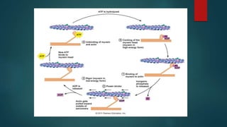
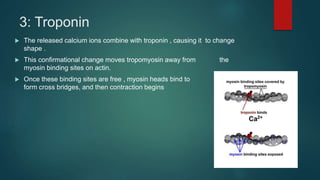
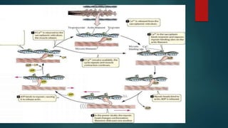
1) The contraction cycle which involves calcium ions binding to troponin and exposing myosin binding sites on actin, allowing cross bridges to form between actin and myosin.
2) Excitation-contraction coupling where an action potential in the transverse tubule causes calcium release from the sarcoplasmic reticulum, binding to troponin and initiating contraction.
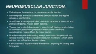
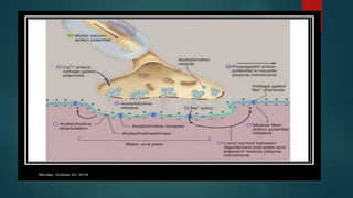
3) The neuromuscular junction where a nerve impulse triggers acetylcholine release, starting a muscle action potential and calcium release from the sarcoplasmic reticulum.