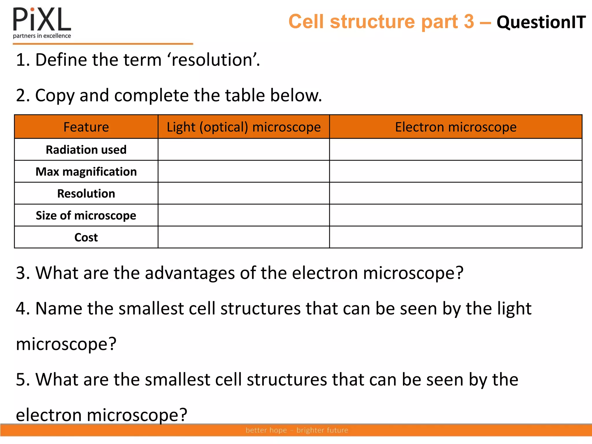
Here are the answers to the questions:
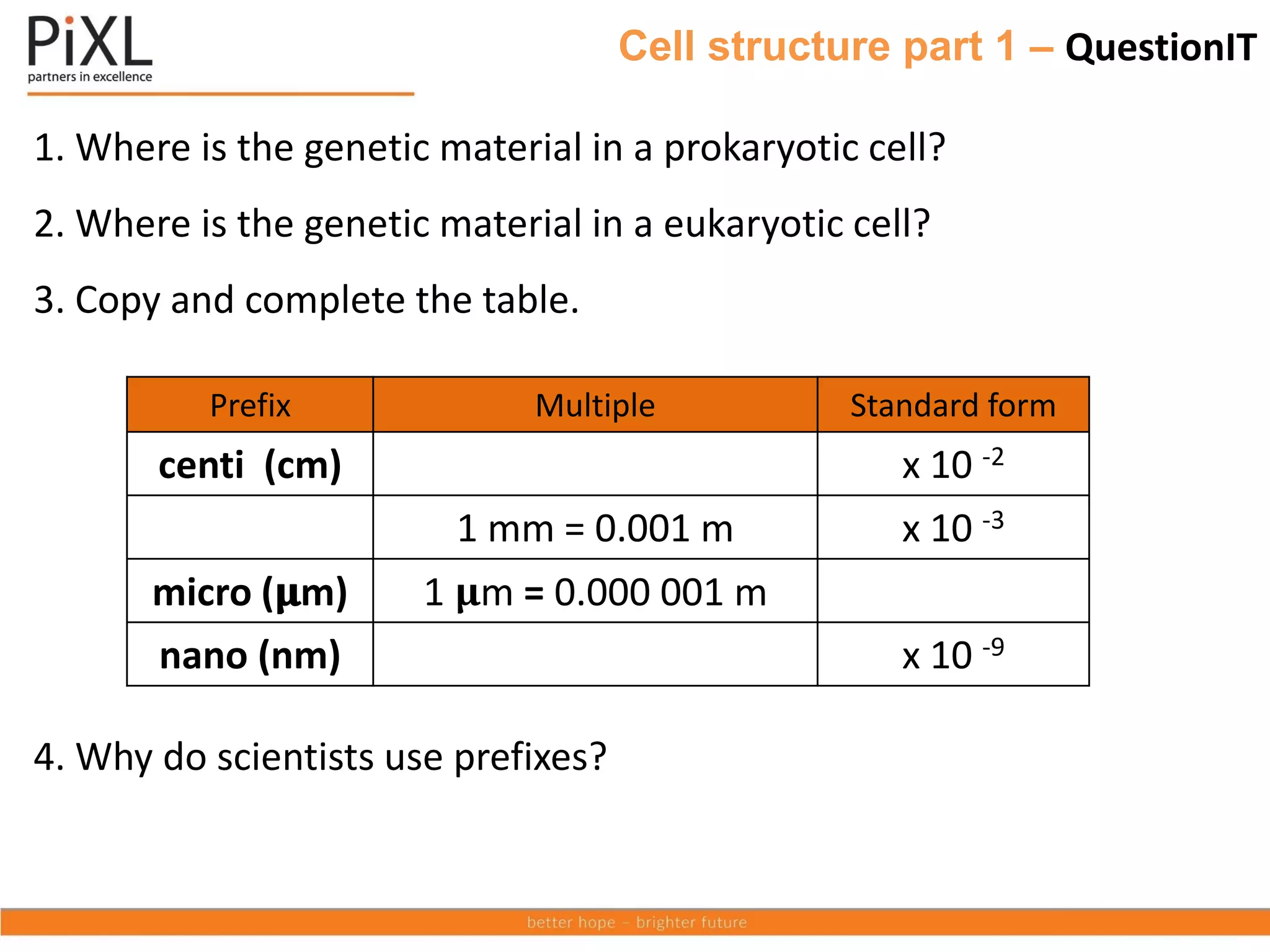
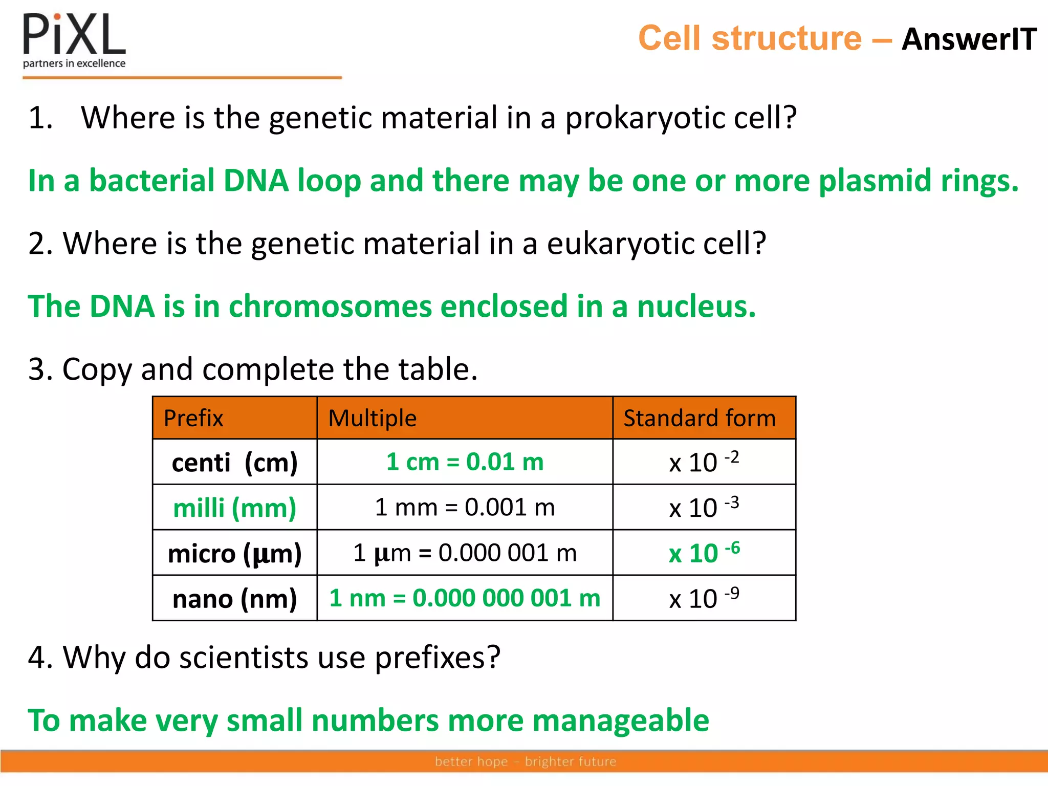
1. In a prokaryotic cell, the genetic material is a single loop of DNA not enclosed in a nucleus.
2. In a eukaryotic cell, the genetic material (DNA) is enclosed within a nucleus.
3. See table below
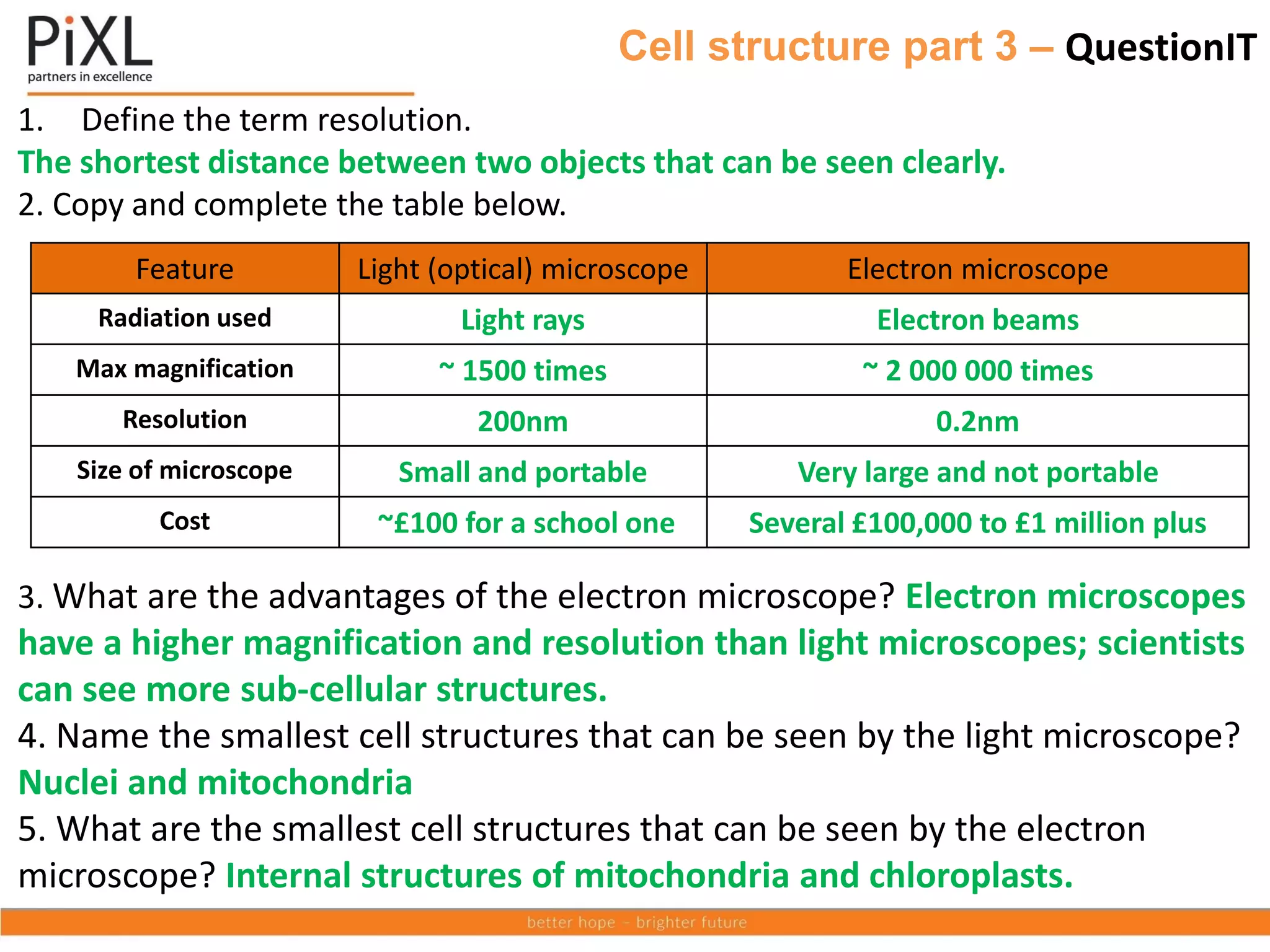
Prefix Multiple Standard form
centi (cm) 1 cm = 0.01 m x 10-2
milli (mm) 1 mm = 0.001 m x 10-3
micro (μm) 1 μm = 0.000 001 m x 10-6
nano (nm) 1 nm = 0.000 000 001 m x 10-9
4