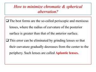
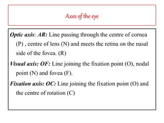
If we think eye as an optical device, mathematically,
it is not a perfect optical device, but functionally it is
an unique optical device.
Obviously there are some physiological optical
defects:
What are the physiological defects of eye?
1) Diffraction of light
2) Chromatic aberration
3) Spherical aberration
4) Decentring
5) Peripheral aberration
 Ref: Duke-Elders p: 35
6/5/2020 3anjumk38dmc@gmail.com
1) Diffraction of light
• The diffraction of light was first explained by
the English scientist Thomas Young in the
early 1800s.
• He made a pin-hole in his window shutter
and observed diffraction of the sunlight on
the board.
Thomas Young Experiment: Diffraction
1) Diffraction of light
Diffraction is the bending of waves around obstacles,
or the spreading of waves by passing them through an
aperture, or opening.
Smaller the pupil more diffraction will be occur
https://www.vocabulary.com/dictionary/diffraction
1) Diffraction of light: The Airy disc
When light passes through a circular aperture, a
circular diffraction pattern is produced.
• This consists of a bright central disc surrounded
by alternate dark and light rings. The central
bright zone is known as the Airy disc.
• (Elkington: 10)
1) Diffraction of light: Applied aspect
a) Diffraction effects are most marked with small
apertures, and occur in all optical systems
including lenses, optical instruments and the
eye.
In the case of lenses and instruments, the
diffraction effect at the apertures used is negligible
compared with the other errors or aberrations of
the system (Elkington).
Diffraction of light: Applied aspect
b) In the case of the eye, diffraction is the main
source of image imperfection when the pupil is
small. (Elkington). The image formed on the retina
from a distant small source has the form a blur
circle the Airy disc (Khurana)
c) The principle of diffraction is used in the design
of some multifocal intraocular lenses (Elkington)
Diffraction of light: Further reading
a) Diffractive multifocal intraocular lenses
b) Second-generation diffractive multifocal
intraocular lenses
(AAO: Vol: 3. P 303-304 2016-17)
Types of diffraction: 2 Types (Khurana P: 6)
1) Fresnel diffraction
2) Fraunhofer diffraction
2. Chromatic aberration
When white light is refracted at an optical
interface, it is dispersed into its component
wavelengths or colours.
The shorter the wavelength of the light, the more it
is deviated on refraction.
Thus a series of coloured images are formed when
white light is incident upon a spherical lens
Chromatic aberration:
RED> GREEN>BLUE
When lenses are used in instruments, it is desirable
to eliminate chromatic aberration.
BUT HOW?
Do you know about Achromatic lenses?
By combination of crown & flint glass
Before that we have to know the Dispersion of Light & dispersive power
White light is composed of various wavelengths.
Light of shorter wavelength is deviated more than
light of longer wavelength, e.g. blue light is
deviated more than red.
• The refractive index of a material is normally
taken to mean that for the yellow sodium flame.
• The angle formed between the red and blue light
around the yellow (Fig. 3.12) indicates the
dispersive power of the medium (cf. chromatic
aberration).
Diagram to show dispersion of light. (The angles
involved are exaggerated.) (Fig: 3.12 Elkington)
Dispersive power
The ratio of angular dispersion to the angle of
deviation for the mean wavelength (yellow colour)
is called dispersive power of the material.
The difference in the angle of deviation of extreme
colours i.e. red and violet is known as
angular dispersion.
How to overcome chromatic aberration in glass?
Flint glass gives a dispersion nearly double that of the
crown glass, and its R,I is 1.7, while the RI of crown glass
is 1.5; hence if we combine a convex lens of crown glass
with a concave lens of half the strength composed of flint
glass, the dispersion will be neutralized, while a
considerable part of the refractivity of the crown glass will
still remain. Such a combination forms an achromatic lens.
Achromatic lens
• Flint glass:
• Refractive Index = 1.7
• Dispersive power: 2
• Crown glass:
• Refractive Index = 1.5
• Dispersive power: 1
• Suppose we take
• +10 D (Crown)
• - 5 (Flint)
• Ref Duke- Elder 33
Duochrome Test
In clinical practice the chromatic aberration of the
eye is made use of in the duochrome test.
The patient views the letters by means of red and
green light respectively, and can easily tell which
appear clearer. The test is sensitive to an alteration
in refraction of 0.25 D or less.
A myopic eye
sees the red
letters more
clearly than
the green (Fig.
8.3) while a
hypermetropic
eye sees the
green letters
more
distinctly
3) Spherical aberration
 What is spherical aberration in lenses?
• The shape of a spherical lens causes a problem
called spherical aberration. In spherical aberration,
parallel light rays that pass through the central
region of the lens focus farther away than light rays
that pass through the edges of the lens. The result is
many focal points, which produce a blurry image
• https://www.google.com/search?q=what+is+spherical+aberration%3F&rlz=1C1CH
BF_enBD875BD875&oq=what+is+spherical+aberration%3F&aqs=chrome..69i57j0l
7.18166j0j8&sourceid=chrome&ie=UTF-8
Aberration of Spherical lens
How to minimize chromatic & spherical
aberration?
The error is diminished by making the curvature of the
anterior surface greater than that of the posterior, it is for
this reason that the objectives of the opera glasses bulges
in front.
Definition of opera glass. : a small low-power binocular
without prisms for use at the opera or theater.
Image of opera glass
How to minimize chromatic & spherical
aberration?
The best forms are the so-called periscopic and meniscus
lenses, where the radius of curvature of the posterior
surface is greater than that of the anterior surface.
This error can be eliminated by grinding lenses so that
their curvature gradually decreases from the center to the
periphery. Such lenses are called Aplantic lenses.
What is “Best formed lens?”
To eliminate the spherical aberration, curvature of the lens
is reduced periphery by grinding. In other way in
periphery of the lens is attached by a plane lens which
eliminate the peripheral ray.
By combining a flint and crown glass in appropriate
power an achromatic and Aplantic combination can
reduced both chromatic and spherical aberration which is
also called the “ Best formed lens”.
6/5/2020 33anjumk38dmc@gmail.com
To form an ideal image by eye, the optical system
have to be accurately centered (cornea & two surfaces
of lens)
But virtually the center of the cornea is 0.25 mm
below the axis of the lens.
On the other hand the fovea is not situated on the
optical axis. 1.25 mm below & downward (temporal)
6/5/2020 35anjumk38dmc@gmail.com
 When we look at an object, we do not look directly
along optic axis (line passing through corneal center,
lens & meets retina on nasal side of fovea. ANR)
 But along a line joining the object with the fovea and
passing through the nodal point. This line is called the
visual axis. (ONF)
Decentering
• FIXATION AXIS: line joining fixation point and
centre of rotation
•  VISUALAXIS: line joining fixation point,
nodal point and fovea
•  OPTICAL AXIS: line passing through centre of
cornea, lens and meets retina on nasal side of
fovea
Axes of the eye
Optic axis: AR: Line passing through the centre of cornea
(P) , centre of lens (N) and meets the retina on the nasal
side of the fovea. (R)
Visual axis: OF: Line joining the fixation point (O), nodal
point (N) and fovea (F).
Fixation axis: OC: Line joining the fixation point (O) and
the centre of rotation (C)
Visual angle
• Angle alpha ONA: Angle form between optic axis
and visual axis at the nodal point.
• Angle gamma OCA: Angle formed optic axis and
fixation axis at the centre of rotation of the eyeball.
• Angle kappa OPA: Line joining the fixation point (O)
and the centre of rotation (C)
Angle alpha: The angle formed at the nodal point
between these two axes, that is the angle ONA is called
the angle alpha, it’s average size is 5 degree.
When the visual axis cuts the cornea, as it usually does,
on the nasal side of the optic axis, the angle is designated
positive, when the two axes coincide it is nil, sometimes
the visual axis cuts the cornea on the temporal side of the
optic axis, in which case the angle alpha is negative.
Peripheral aberration
Peripheral aberration causes:
 Coma
 Oblique astigmatism
 Distortion of images
It is neutralize by peculiar shape of the eye.
6/5/2020 42anjumk38dmc@gmail.com

03 physiological optical defects

  • 2.
    If we thinkeye as an optical device, mathematically, it is not a perfect optical device, but functionally it is an unique optical device. Obviously there are some physiological optical defects:
  • 3.
    What are thephysiological defects of eye? 1) Diffraction of light 2) Chromatic aberration 3) Spherical aberration 4) Decentring 5) Peripheral aberration  Ref: Duke-Elders p: 35 6/5/2020 3anjumk38dmc@gmail.com
  • 4.
    1) Diffraction oflight • The diffraction of light was first explained by the English scientist Thomas Young in the early 1800s. • He made a pin-hole in his window shutter and observed diffraction of the sunlight on the board.
  • 5.
  • 6.
    1) Diffraction oflight Diffraction is the bending of waves around obstacles, or the spreading of waves by passing them through an aperture, or opening. Smaller the pupil more diffraction will be occur https://www.vocabulary.com/dictionary/diffraction
  • 7.
    1) Diffraction oflight: The Airy disc When light passes through a circular aperture, a circular diffraction pattern is produced.
  • 8.
    • This consistsof a bright central disc surrounded by alternate dark and light rings. The central bright zone is known as the Airy disc. • (Elkington: 10)
  • 9.
    1) Diffraction oflight: Applied aspect a) Diffraction effects are most marked with small apertures, and occur in all optical systems including lenses, optical instruments and the eye. In the case of lenses and instruments, the diffraction effect at the apertures used is negligible compared with the other errors or aberrations of the system (Elkington).
  • 10.
    Diffraction of light:Applied aspect b) In the case of the eye, diffraction is the main source of image imperfection when the pupil is small. (Elkington). The image formed on the retina from a distant small source has the form a blur circle the Airy disc (Khurana) c) The principle of diffraction is used in the design of some multifocal intraocular lenses (Elkington)
  • 11.
    Diffraction of light:Further reading a) Diffractive multifocal intraocular lenses b) Second-generation diffractive multifocal intraocular lenses (AAO: Vol: 3. P 303-304 2016-17) Types of diffraction: 2 Types (Khurana P: 6) 1) Fresnel diffraction 2) Fraunhofer diffraction
  • 13.
    2. Chromatic aberration Whenwhite light is refracted at an optical interface, it is dispersed into its component wavelengths or colours. The shorter the wavelength of the light, the more it is deviated on refraction. Thus a series of coloured images are formed when white light is incident upon a spherical lens
  • 14.
  • 15.
    When lenses areused in instruments, it is desirable to eliminate chromatic aberration. BUT HOW? Do you know about Achromatic lenses?
  • 16.
    By combination ofcrown & flint glass
  • 17.
    Before that wehave to know the Dispersion of Light & dispersive power White light is composed of various wavelengths. Light of shorter wavelength is deviated more than light of longer wavelength, e.g. blue light is deviated more than red.
  • 18.
    • The refractiveindex of a material is normally taken to mean that for the yellow sodium flame. • The angle formed between the red and blue light around the yellow (Fig. 3.12) indicates the dispersive power of the medium (cf. chromatic aberration).
  • 20.
    Diagram to showdispersion of light. (The angles involved are exaggerated.) (Fig: 3.12 Elkington)
  • 21.
    Dispersive power The ratioof angular dispersion to the angle of deviation for the mean wavelength (yellow colour) is called dispersive power of the material. The difference in the angle of deviation of extreme colours i.e. red and violet is known as angular dispersion.
  • 22.
    How to overcomechromatic aberration in glass? Flint glass gives a dispersion nearly double that of the crown glass, and its R,I is 1.7, while the RI of crown glass is 1.5; hence if we combine a convex lens of crown glass with a concave lens of half the strength composed of flint glass, the dispersion will be neutralized, while a considerable part of the refractivity of the crown glass will still remain. Such a combination forms an achromatic lens.
  • 23.
    Achromatic lens • Flintglass: • Refractive Index = 1.7 • Dispersive power: 2 • Crown glass: • Refractive Index = 1.5 • Dispersive power: 1 • Suppose we take • +10 D (Crown) • - 5 (Flint) • Ref Duke- Elder 33
  • 25.
    Duochrome Test In clinicalpractice the chromatic aberration of the eye is made use of in the duochrome test. The patient views the letters by means of red and green light respectively, and can easily tell which appear clearer. The test is sensitive to an alteration in refraction of 0.25 D or less.
  • 26.
    A myopic eye seesthe red letters more clearly than the green (Fig. 8.3) while a hypermetropic eye sees the green letters more distinctly
  • 27.
    3) Spherical aberration What is spherical aberration in lenses? • The shape of a spherical lens causes a problem called spherical aberration. In spherical aberration, parallel light rays that pass through the central region of the lens focus farther away than light rays that pass through the edges of the lens. The result is many focal points, which produce a blurry image • https://www.google.com/search?q=what+is+spherical+aberration%3F&rlz=1C1CH BF_enBD875BD875&oq=what+is+spherical+aberration%3F&aqs=chrome..69i57j0l 7.18166j0j8&sourceid=chrome&ie=UTF-8
  • 28.
  • 30.
    How to minimizechromatic & spherical aberration? The error is diminished by making the curvature of the anterior surface greater than that of the posterior, it is for this reason that the objectives of the opera glasses bulges in front. Definition of opera glass. : a small low-power binocular without prisms for use at the opera or theater.
  • 31.
  • 32.
    How to minimizechromatic & spherical aberration? The best forms are the so-called periscopic and meniscus lenses, where the radius of curvature of the posterior surface is greater than that of the anterior surface. This error can be eliminated by grinding lenses so that their curvature gradually decreases from the center to the periphery. Such lenses are called Aplantic lenses.
  • 33.
    What is “Bestformed lens?” To eliminate the spherical aberration, curvature of the lens is reduced periphery by grinding. In other way in periphery of the lens is attached by a plane lens which eliminate the peripheral ray. By combining a flint and crown glass in appropriate power an achromatic and Aplantic combination can reduced both chromatic and spherical aberration which is also called the “ Best formed lens”. 6/5/2020 33anjumk38dmc@gmail.com
  • 35.
    To form anideal image by eye, the optical system have to be accurately centered (cornea & two surfaces of lens) But virtually the center of the cornea is 0.25 mm below the axis of the lens. On the other hand the fovea is not situated on the optical axis. 1.25 mm below & downward (temporal) 6/5/2020 35anjumk38dmc@gmail.com
  • 37.
     When welook at an object, we do not look directly along optic axis (line passing through corneal center, lens & meets retina on nasal side of fovea. ANR)  But along a line joining the object with the fovea and passing through the nodal point. This line is called the visual axis. (ONF)
  • 38.
    Decentering • FIXATION AXIS:line joining fixation point and centre of rotation •  VISUALAXIS: line joining fixation point, nodal point and fovea •  OPTICAL AXIS: line passing through centre of cornea, lens and meets retina on nasal side of fovea
  • 39.
    Axes of theeye Optic axis: AR: Line passing through the centre of cornea (P) , centre of lens (N) and meets the retina on the nasal side of the fovea. (R) Visual axis: OF: Line joining the fixation point (O), nodal point (N) and fovea (F). Fixation axis: OC: Line joining the fixation point (O) and the centre of rotation (C)
  • 40.
    Visual angle • Anglealpha ONA: Angle form between optic axis and visual axis at the nodal point. • Angle gamma OCA: Angle formed optic axis and fixation axis at the centre of rotation of the eyeball. • Angle kappa OPA: Line joining the fixation point (O) and the centre of rotation (C)
  • 41.
    Angle alpha: Theangle formed at the nodal point between these two axes, that is the angle ONA is called the angle alpha, it’s average size is 5 degree. When the visual axis cuts the cornea, as it usually does, on the nasal side of the optic axis, the angle is designated positive, when the two axes coincide it is nil, sometimes the visual axis cuts the cornea on the temporal side of the optic axis, in which case the angle alpha is negative.
  • 42.
    Peripheral aberration Peripheral aberrationcauses:  Coma  Oblique astigmatism  Distortion of images It is neutralize by peculiar shape of the eye. 6/5/2020 42anjumk38dmc@gmail.com