This document provides guidelines for reporting the results of mammography examinations using the BI-RADS assessment and reporting system. It describes the standardized structure for mammography reports, including sections for the indication, breast composition, findings, comparison to prior exams, assessment, and management. The breast composition section provides definitions and illustrations for the four breast composition categories based on fibroglandular density. The assessment category section matches each BI-RADS assessment category with its corresponding management recommendation and likelihood of cancer.

![ACR BI-RADS® ATLAS — MAMMOGRAPHY
American College of Radiology 123
MAMMOGRAPHY
A. REPORT ORGANIZATION (Guidance chapter, see page 147)
The reporting system should be concise and organized using the following structure. A statement
indicating that the current examination has been compared to previous examination(s) should be
included (specify date[s]). If this is not included, it should be assumed that no comparison has been
made, although it is preferable to indicate that no comparison was made.
Table 4. Report Organization
Report Structure
1. Indication for examination
2. Succinct description of the overall breast composition
3. Clear description of any important findings
4. Comparison to previous examination(s), if deemed appropriate by the interpreting physician
5. Assessment
6. Management
1. INDICATION FOR EXAMINATION
Provide a brief description of the indication for examination. This may be screening for an
asymptomatic woman, recall of a screening-detected finding, evaluation of a clinical finding
(specify the finding and its location), or follow-up of either a probably benign lesion or cancer
treatedwithbreastconservation.Ifanimplantispresent,bothstandardandimplant-displaced
views should be performed, and this should be stated in the mammography report.
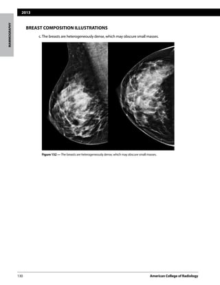
2. SUCCINCT DESCRIPTION OF THE OVERALL BREAST COMPOSITION
This is an overall assessment of the volume of attenuating tissues in the breast, to help in-
dicate the relative possibility that a lesion could be obscured by normal tissue and that the
sensitivity of examination thereby may be compromised by dense breast tissue. A few co-
alescent areas of dense tissue may be present in breasts with as little as 10% dense tissue,
whereas primarily fatty areas may be present in breasts with as much as 90% dense tissue.
Since mammography does not depict all breast cancers, clinical breast examination is a
complementary element of screening. Findings at clinical breast examination should not be
ignored and may have increased importance in the dense breast.
The available data do not support the use of mammographic breast density for determining
screening frequency.
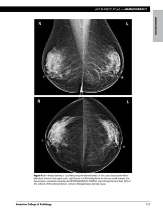
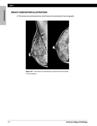
The following four categories of breast composition are defined by the visually estimated con-
tentoffibroglandular-densitytissuewithinthebreasts.Pleasenotethatthecategoriesarelisted
as a,b,c, and d so as not to be confused with the numbered BI-RADS® assessment categories.
If the breasts are not of apparently equal density, the denser breast should be used to catego-
rize breast density. The sensitivity of mammography for noncalcified lesions decreases as the
BI-RADS® breast density category increases. The denser the breast, the larger the lesion(s) that
may be obscured. There is considerable intra- and inter-observer variation in visually estimat-
ing breast density between any two adjacent density categories. Furthermore, there is only](https://image.slidesharecdn.com/02biradsmammographyreporting-150530060816-lva1-app6891/85/02-birads-mammography-reporting-3-320.jpg)