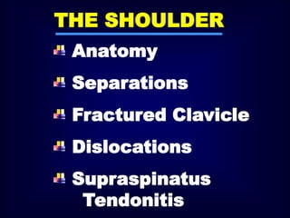
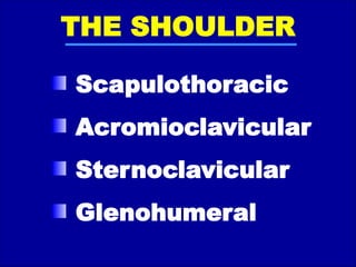
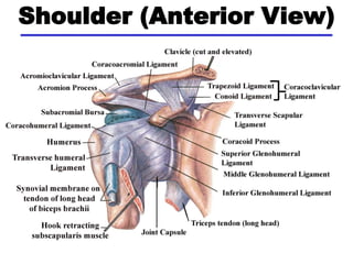
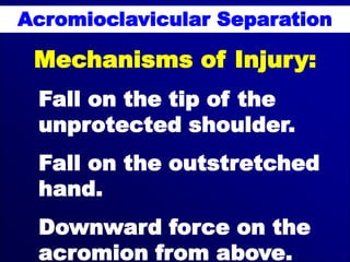
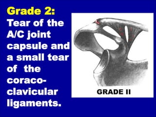
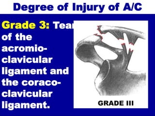
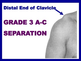
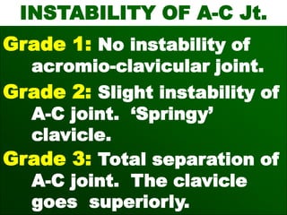
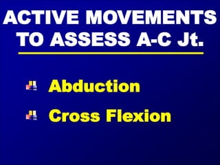
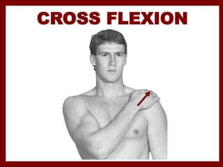
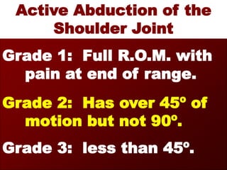
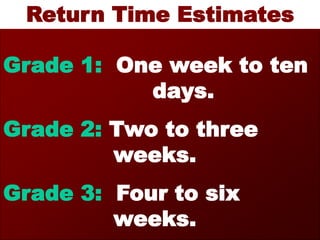
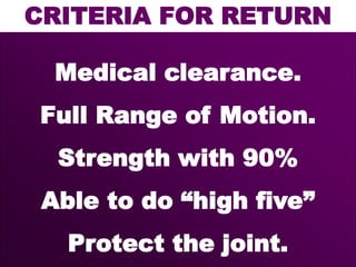
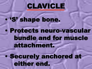
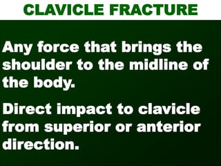
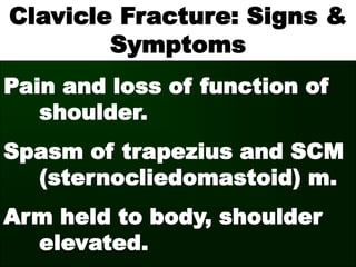
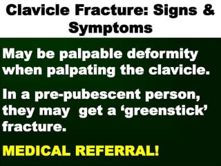
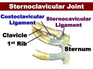
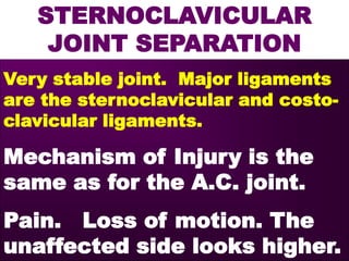
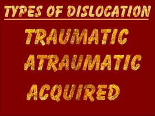
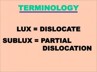
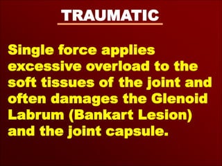
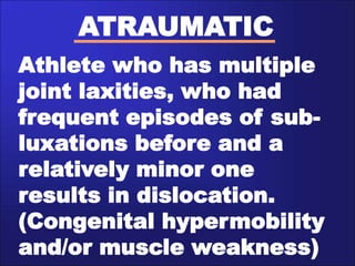
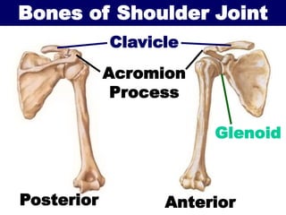
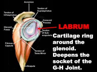
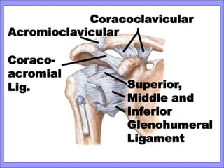
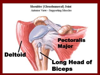
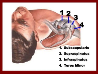
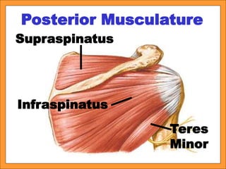
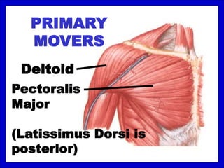
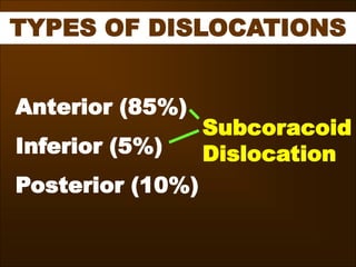
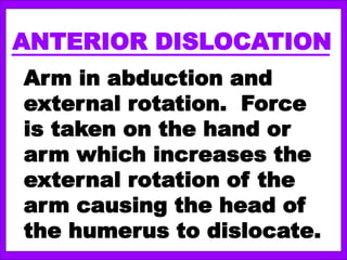
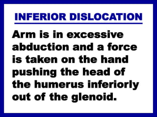
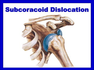
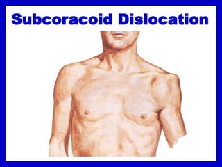
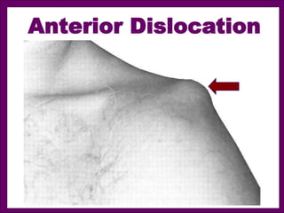
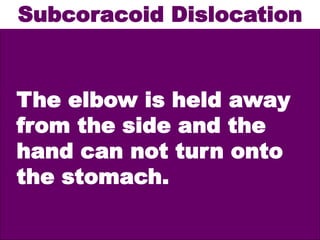
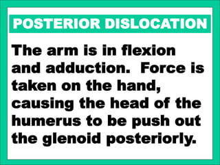
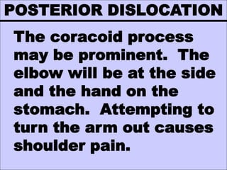
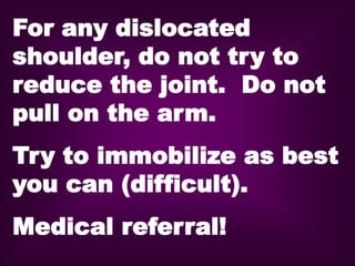
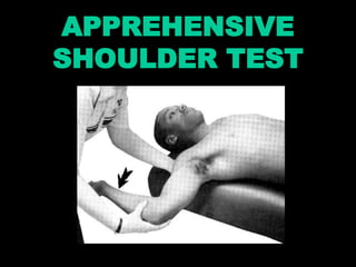
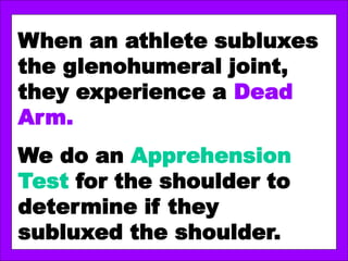
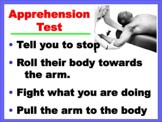
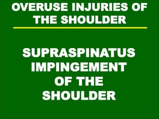
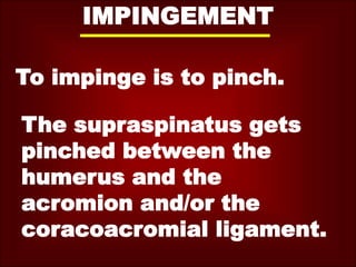
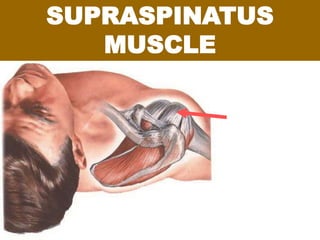
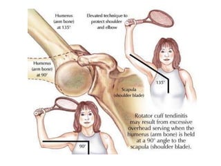
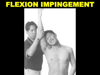
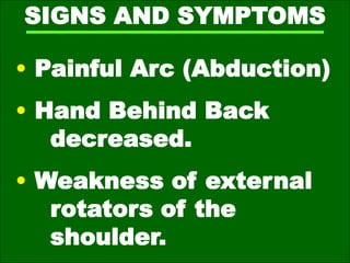
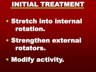
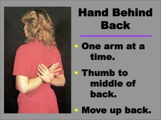
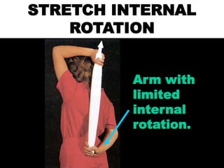
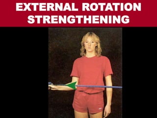
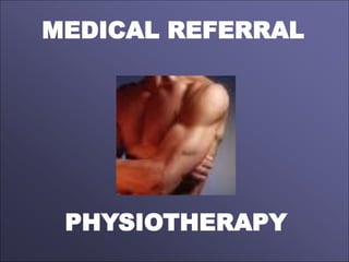
This document discusses various injuries and conditions related to the shoulder. It covers topics like shoulder anatomy, types of separations and fractures, mechanisms of different injuries, grades of injuries, signs and symptoms, treatment approaches, and more. Specific conditions covered include acromioclavicular separations, clavicle fractures, sternoclavicular joint separations, shoulder dislocations, supraspinatus tendonitis, and impingement syndrome. Diagrams are included to illustrate shoulder anatomy and various injuries.
Acromioclavicular (AC) joint injury is a term used to describe an injury to the top of the shoulder, where the front of the shoulder blade (acromion) attaches to the collarbone (clavicle).
The upper limb consists of various joints that enable movement and provide flexibility. These joints can be classified into different types based on their structure and function.
Understanding the anatomy and function of these joints is crucial for assessing and managing conditions related to the upper limb, as well as for rehabilitation and therapeutic interventions. Joint injuries, arthritis, and other disorders may affect the functionality of these joints, and appropriate medical care may be necessary for optimal outcomes.
Seminar clinical anatomy of upper limb joints and musclesQuan Fu Gan
This is not all, there are many more clinical anatomy in terms of condition such as Popeye Deformity with are not included here and Special Test such as Neer's Impingement and Hawkins Kennedy etc... with touches on the upper limb muscles and joints. Also not forgotten Long tendon test and so forth. In general, this is just a simplified slides. Tq
Can read freely here
https://sethiortho.blogspot.com/
Examination of Knee Joint Ligaments
SethiNet Presentations
Introduction
Proper use of the examination techniques requires
An understanding of the anatomy
Pathophysiology of knee ligament injuries
Advanced imaging - Augment a history and examination when necessary
Imaging should not replace a thorough history and physical examination
History taking
A description of the mechanism of injury
The patient should be queried about previous injuries
The current injury may be the sequela of a previous injury
Common ligament Injuries
Anterior Cruciate Ligament
Anatomy
The ACL originates at posteromedial aspect of the lateral femoral condyle
Wide tibial insertion at the lateral aspect of the anterior tibial spine
The ACL has two fiber bundles
The anteromedial
Posterolateral bundles
Which provide varying tension from flexion through extension
Functions
Primary restraint against anterior tibial translation
Provides rotational stability, especially in extension
ACL - Mechanism of Injury
Injury to the knee ligaments is typically the result of
A non contact change in direction
Twisting injury
Landing from a jump.
The patient often describes a “pop” that is felt or heard at injury
The appearance of swelling (hemarthrosis) within a few hours
ACL -Examinations
Examinations
The Anterior drawer test
The Lachman Test
The Pivot Shift Test
Novel Tests
ACL - Anterior drawer test
Patient with patient supine position
The hip flexed at 45° / knee flexed at 90°
The foot is fixed to the table - often by sitting on it
The clinician applies an anterior force to the proximal tibia, palpating the joint line for anterior translation.
Increased anterior translation indicates ACL insufficiency.
Sensitivity – only 50% with the patient under anesthesia
because the posterior horn of the medial meniscus may act as a so-called doorstop that prevents anterior translation, even in the presence of a torn ACL.
ACL - Lachman Test
It was designed to overcome three identified limitations of the anterior drawer test
Acute effusion that often precludes flexion to 90°
Protective spasm of the hamstring muscles that can prevent anterior translation of the tibia
The articulation of the relatively acute convexity of the posterior medial femoral condyle and the posterior horn of the medial meniscus that buttresses and prevents anterior translation of the tibia.
These limitations can lead to false-negative findings
The Lachman test is typically done with the knee flexed 20° to 30°.
The examiner places one hand laterally on the patient’s thigh to stabilize the femur
while the other hand grasps the proximal and more subcutaneous medial tibia and applies anterior stres
The test is positive
In the presence of anterior translation
A soft or mushy end point.
When the ACL is intact, the end point is hard
ACL - Grading - Lachman test
Elbow is the most common joint to dislocate in children. Posterior dislocation is most common.
Simple dislocations are those without fracture.
Complex dislocations are those that occur with an associated fracture
How to Make a Field invisible in Odoo 17Celine George
It is possible to hide or invisible some fields in odoo. Commonly using “invisible” attribute in the field definition to invisible the fields. This slide will show how to make a field invisible in odoo 17.
How to Create Map Views in the Odoo 17 ERPCeline George
The map views are useful for providing a geographical representation of data. They allow users to visualize and analyze the data in a more intuitive manner.
Synthetic Fiber Construction in lab .pptxPavel ( NSTU)
Synthetic fiber production is a fascinating and complex field that blends chemistry, engineering, and environmental science. By understanding these aspects, students can gain a comprehensive view of synthetic fiber production, its impact on society and the environment, and the potential for future innovations. Synthetic fibers play a crucial role in modern society, impacting various aspects of daily life, industry, and the environment. ynthetic fibers are integral to modern life, offering a range of benefits from cost-effectiveness and versatility to innovative applications and performance characteristics. While they pose environmental challenges, ongoing research and development aim to create more sustainable and eco-friendly alternatives. Understanding the importance of synthetic fibers helps in appreciating their role in the economy, industry, and daily life, while also emphasizing the need for sustainable practices and innovation.
Welcome to TechSoup New Member Orientation and Q&A (May 2024).pdfTechSoup
In this webinar you will learn how your organization can access TechSoup's wide variety of product discount and donation programs. From hardware to software, we'll give you a tour of the tools available to help your nonprofit with productivity, collaboration, financial management, donor tracking, security, and more.
Students, digital devices and success - Andreas Schleicher - 27 May 2024..pptxEduSkills OECD
Andreas Schleicher presents at the OECD webinar ‘Digital devices in schools: detrimental distraction or secret to success?’ on 27 May 2024. The presentation was based on findings from PISA 2022 results and the webinar helped launch the PISA in Focus ‘Managing screen time: How to protect and equip students against distraction’ https://www.oecd-ilibrary.org/education/managing-screen-time_7c225af4-en and the OECD Education Policy Perspective ‘Students, digital devices and success’ can be found here - https://oe.cd/il/5yV
Unit 8 - Information and Communication Technology (Paper I).pdfThiyagu K
This slides describes the basic concepts of ICT, basics of Email, Emerging Technology and Digital Initiatives in Education. This presentations aligns with the UGC Paper I syllabus.
The Indian economy is classified into different sectors to simplify the analysis and understanding of economic activities. For Class 10, it's essential to grasp the sectors of the Indian economy, understand their characteristics, and recognize their importance. This guide will provide detailed notes on the Sectors of the Indian Economy Class 10, using specific long-tail keywords to enhance comprehension.
For more information, visit-www.vavaclasses.com
5. Acromioclavicular Separation
Mechanisms of Injury:
Fall on the tip of the
unprotected shoulder.
Fall on the outstretched
hand.
Downward force on the
acromion from above.
6. Grade of Injury of A/C
Grade 1:
Small tear of
the capsule of
the AC joint.
No instability
of joint.
P.O.P.
7. Grade 2:
Tear of the
A/C joint
capsule and
a small tear
of the
coraco-
clavicular
ligaments.
8. Degree of Injury of A/C
Grade 3: Tear
of the
acromio-
clavicular
ligament and
the coraco-
clavicular
ligament.
10. INSTABILITY OF A-C Jt.
Grade 1: No instability of
acromio-clavicular joint.
Grade 2: Slight instability of
A-C joint. ‘Springy’
clavicle.
Grade 3: Total separation of
A-C joint. The clavicle
goes superiorly.
13. Active Abduction of the
Shoulder Joint
Grade 1: Full R.O.M. with
pain at end of range.
Grade 2: Has over 45º of
motion but not 90º.
Grade 3: less than 45º.
14. Return Time Estimates
Grade 1: One week to ten
days.
Grade 2: Two to three
weeks.
Grade 3: Four to six
weeks.
15. CRITERIA FOR RETURN
Medical clearance.
Full Range of Motion.
Strength with 90%
Able to do “high five”
Protect the joint.
16. CLAVICLE
• ‘S’ shape bone.
• Protects neuro-vascular
bundle and for muscle
attachment.
• Securely anchored at
either end.
17. CLAVICLE FRACTURE
Any force that brings the
shoulder to the midline of
the body.
Direct impact to clavicle
from superior or anterior
direction.
18. Clavicle Fracture: Signs &
Symptoms
Pain and loss of function of
shoulder.
Spasm of trapezius and SCM
(sternocliedomastoid) m.
Arm held to body, shoulder
elevated.
19. Clavicle Fracture: Signs &
Symptoms
May be palpable deformity
when palpating the clavicle.
In a pre-pubescent person,
they may get a ‘greenstick’
fracture.
MEDICAL REFERRAL!
21. STERNOCLAVICULAR
JOINT SEPARATION
Very stable joint. Major ligaments
are the sternoclavicular and costo-
clavicular ligaments.
Mechanism of Injury is the
same as for the A.C. joint.
Pain. Loss of motion. The
unaffected side looks higher.
30. ATRAUMATIC
Athlete who has multiple
joint laxities, who had
frequent episodes of sub-
luxations before and a
relatively minor one
results in dislocation.
(Congenital hypermobility
and/or muscle weakness)
31. ACQUIRED
Sports such as swimming,
gymnastics and baseball
where repetitive micro-
trauma, poor stretching and
motion lead to capsular
stretching. Eventual
feeling of instability.
32. Bones of Shoulder Joint
Acromion
Process
Clavicle
Posterior Anterior
Glenoid
40. ANTERIOR DISLOCATION
Arm in abduction and
external rotation. Force
is taken on the hand or
arm which increases the
external rotation of the
arm causing the head of
the humerus to dislocate.
41. INFERIOR DISLOCATION
Arm is in excessive
abduction and a force
is taken on the hand
pushing the head of
the humerus inferiorly
out of the glenoid.
46. POSTERIOR DISLOCATION
The arm is in flexion
and adduction. Force is
taken on the hand,
causing the head of the
humerus to be push out
the glenoid posteriorly.
47. POSTERIOR DISLOCATION
The coracoid process
may be prominent. The
elbow will be at the side
and the hand on the
stomach. Attempting to
turn the arm out causes
shoulder pain.
48. For any dislocated
shoulder, do not try to
reduce the joint. Do not
pull on the arm.
Try to immobilize as best
you can (difficult).
Medical referral!
49. Recurrent dislocations
have nothing to do with
the treatment after the
first dislocation.
Recurrent dislocations are
dependent upon the
damage that happens
during the first dislocation.
51. When an athlete subluxes
the glenohumeral joint,
they experience a Dead
Arm.
We do an Apprehension
Test for the shoulder to
determine if they
subluxed the shoulder.
52. Apprehension
Test
• Tell you to stop
• Roll their body towards
the arm.
• Fight what you are doing
• Pull the arm to the body