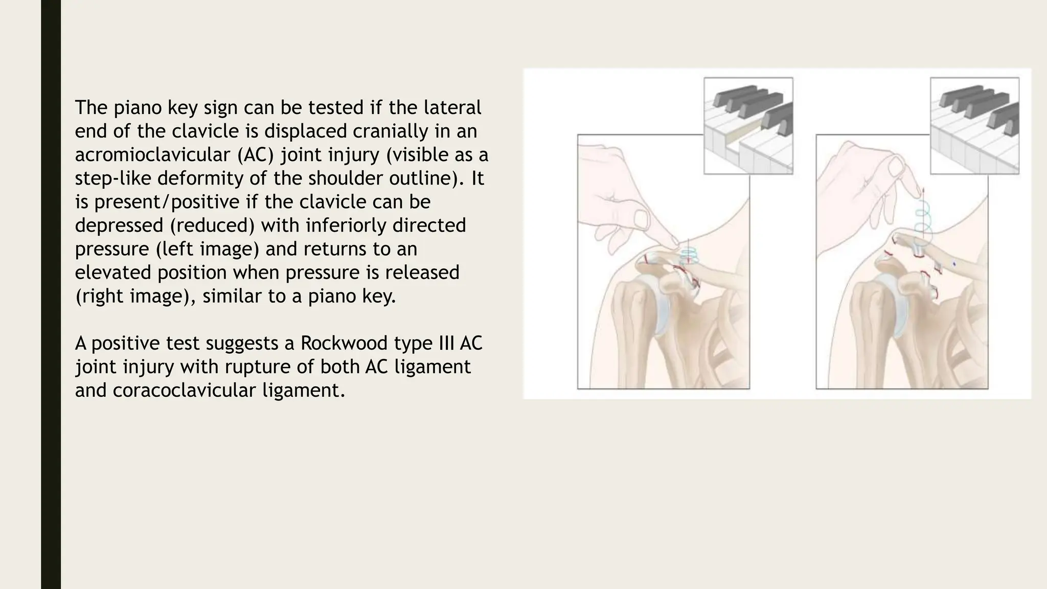
2. Acromioclavicular joint injury
■ ETIOLOGY
■ Most common: direct force injury to the superior aspect of the acromion while
the arm is adducted (e.g., a fall while cycling or riding a horse)
■ Less common: indirect injury via falling on an outstretched hand, which
transmits force up the arm through the humerus to the acromion, causing
displacement that distresses the AC ligaments
3. ■ Photograph of a patient who
sustained a fall on her right
shoulder (ventral view)
■ The lateral end of the right
clavicle is displaced cranially
and is visible as a step-like
deformity of the shoulder
outline.
■ Examination revealed a positive
piano key sign (spring test), in
which the displaced clavicle was
reducible with the application
of pressure and returned to this
elevated position when the
pressure was released.
■ These are the typical clinical
features of type III
acromioclavicular injury.
4. ■ Photograph of a patient who
sustained a fall on her right
shoulder (dorsal view)
■ The lateral end of the right
clavicle is displaced cranially
and is visible as a step-like
deformity of the shoulder
outline.
■ Examination revealed a
positive piano key sign
(spring test), in which the
displaced clavicle was
reducible with the
application of pressure and
returned to this elevated
position when the pressure
was released.
■ These are the typical clinical
features of type III
acromioclavicular injury.
5. ■ Clinical features
■ Local tenderness, swelling, and/or bruising
■ Pain is elicited by the cross-body adduction test, in which the patient elevates
their arm to 90° and actively adducts it across their body.
■ Limited range of motion of the shoulder joint
■ Visible deformity of the lateral aspect of the clavicle may be seen in types III
and above
6. ■ Diagnostics
■ Approach: AC joint injury is a clinical diagnosis that can be classified according
to the Rockwood classification via x-ray. If x-ray findings are questionable, an
MRI, CT, or possibly ultrasound may be considered.
■ X-ray (anterior-posterior view, oblique view, axillary view) of the shoulder
joint, acromion, and scapula:
■ Subluxation of the AC joint space
■ Widening of the CC space
■ Displacement of the clavicle
■ Accompanying injuries (e.g., clavicle fracture)
■ Chronic cases: features of AC arthritis, distal clavicle osteolysis
7.
8.
9. ■ • Strained ligaments = red area
■ • Ruptured ligaments = serrated red lines
■ • Detached deltoid and trapezius muscle = serrated blue lines
■ • Normal position of the clavicle = red line
■ • Dislocation of the clavicle = red arrow (Type II → not above the superior border of the acromion, type III →
coracoclavicular distance < 25 mm, type IV → posteriorly displaced clavicle, type V → coracoclavicular
distance > 25 mm, type VI → clavicle is inferiorly displaced below the coracoid)
■ Findings for each type
■ • Type I: Mild sprain of the acromioclavicular and coracoclavicular ligaments (red shaded area).
■ • Type II: Ruptured acromioclavicular ligament and sprain of the coracoclavicular ligament (serrated lines);
clavicle elevated, but does not extend above the superior border of the acromion (red arrow).
■ • Type III: The acromioclavicular ligament, coracoclavicular ligament, and joint capsule are ruptured
(serrated lines); clavicle elevated above the superior border of the acromion (red arrow) and can be
repositioned with minimal pressure, which is known as the piano key sign.
■ • Type IV: The acromioclavicular ligament, coracoclavicular ligament, and joint capsule are ruptured (red
serrated lines). The trapezius and deltoid are detached (serrated blue lines). Posteriorly displaced clavicle
(red arrow).
■ • Type V: Type IV plus marked elevation of the clavicle; coracoclavicular distance is > 25 mm. The piano
key sign is present.
■ • Type VI: Clavicle is inferiorly displaced behind the coracobrachialis and biceps tendons; all ligaments are
ruptured; the original position of the clavicle is indicated by the red lines.
10. The piano key sign can be tested if the lateral
end of the clavicle is displaced cranially in an
acromioclavicular (AC) joint injury (visible as a
step-like deformity of the shoulder outline). It
is present/positive if the clavicle can be
depressed (reduced) with inferiorly directed
pressure (left image) and returns to an
elevated position when pressure is released
(right image), similar to a piano key.
A positive test suggests a Rockwood type III AC
joint injury with rupture of both AC ligament
and coracoclavicular ligament.
11.
12. ■ Treatment
■ Acute management
■ Conservative treatment
■ Indications: types I and II
■ Methods
■ Sling for comfort: 1–3 weeks (e.g., Desault or Gilchrist bandage)
■ Avoid heavy lifting
■ Analgesia (e.g., NSAIDs)
■ Surgical treatment
■ Indications
■ Types III and above
■ Management of type III is controversial and determined on an individual
basis
■ All patients with type III and above should be referred to an orthopedist
■ Open fractures
■ Neurovascular injury
■ Failed conservative treatment
■ Objective: ligament repair and reconstruction
■ Methods
■ Arthroscopic (all or assisted): preferred as less invasive
■ Open surgery
• Long-term management
• Indications
• Persistent pain after healing of
initial ligamentous injury
• Repeated minor injury without
instability but persistent AC joint
arthralgia
• Methods
• Avoiding painful movement and
analgesia
• Intraarticular glucocorticoid
injections
13. Elbow dislocation
■ Etiology
■ Trauma
■ Fall on an outstretched hand (most common) → posterior elbow dislocation
■ A posterior, direct trauma to a flexed elbow → anterior elbow dislocation
■ Medial/lateral trauma to the elbow → medial/lateral elbow dislocation
■ High impact trauma to the elbow → divergent elbow dislocation
15. ■ Clinical features
■ Pain, swelling of the elbow
■ Limited range of motion: inability to flex or extend the elbow
■ Elbow deformity
■ Limb length discrepancy
■ Nerve injury (up to 10% of cases)
■ Ulnar nerve palsy
■ Median nerve palsy
■ Radial nerve palsy or posterior interosseous neuropathy (depending on site of
injury)
■ Brachial artery injury (very rare)
16. ■ Diagnostics
■ Physical examination
■ Signs of fracture
■ Neurovascular deficits
■ X-ray of the elbow joint: anteroposterior and lateral views to confirm dislocation
and exclude fracture
■ Posterior fat pad sign: seen in patients with concomitant fractures (usually of the
humerus/radial head) [4]
■ Radiocapitellar line
■ On a lateral x-ray of the elbow joint, an imaginary line drawn through the center
of the neck of the radius should pass through the center of the capitellum of the
humerus.
■ If an elbow dislocation is present, the line does not intersect the capitellum.
■ CT scan of the elbow joint: indicated only if a complex elbow dislocation is
suspected to evaluate the extent of associated fractures
17. ■ X-ray elbow (left: AP view; right:
lateral view)
■ A posterolateral dislocation is seen
(white arrows), with elevation of
the posterior fat pad (red overlay)
caused by a joint effusion. The
dislocation is considered simple
since there is no associated
fracture.
■ Elbow dislocations may be
posterolateral, posteromedial,
posterior, anterior, medial, or
lateral. The majority of dislocations
are posterolateral or posterior. In
anatomical position, the radius is
located lateral to the ulna.
■ Green overlay: trochlear notch
18. ■ X-ray of the elbow joint, lateral
view
■ 13-year-old girl who fell on an
outstretched hand. The bones
show no fracture, but a fat pad
sign can be seen ventral and
dorsal to the humeral shaft. The
fat pad sign indicates elbow joint
effusion. It arises when the fatty
tissue usually attached to the
distal humerus is lifted off the
bone because of the joint
effusion; while the fatty tissue is
usually visible as a lucency
parallel to the humerus, it
becomes crescent-shaped if there
is an effusion.
19.
20. ■ There are various techniques that can be used in a
number of combinations. Administration of analgesics
prior to the procedure is advisable.
■ This illustration shows a common approach in which
the patient lies down on a stretcher in a supine
position with one physician on each side (A).
■ One of the physicians raises the affect arm, clasping
the elbow from both sides with both thumbs placed
on the olecranon. The elbow is flexed to 90 degrees.
The second physician then gently applies axial
traction in the distal direction. At the same time, the
first physician carefully applies pressure on the
olecranon (to guide it back into position) and
simultaneously applies gentle counter-traction to
stabilize the position of the humerus (B).
■ Depending on the injury, the patient’s condition, and
possible concomitant injuries, this procedure can also
be conducted with the forearm supinated rather than
a pronated.
■ If neurovascular damage and/or fractures are present
or suspected, additional diagnostic steps and
different therapeutic measures are necessary.
21.
22. Forearm Fractures
■ Initial management
■ The following are indicated irrespective of the fracture type and bones involved:
[3][2]
■
■ Perform neurovascular exam.
■ Assess radial and ulnar artery pulses and capillary refill time.
■ Evaluate for median nerve injury , radial nerve injury , and ulnar nerve injury.
■ Consider indications for orthopedic consultation for fractures.
■ Obtain imaging of the forearm and consider adding imaging of the elbow and wrist to
check for associated injuries.
■ Evaluate for signs of compartment syndrome in any patient with high-energy impact
trauma.
■ Provide analgesia for acute fractures.
■ Continue with management specific to the injury identified on imaging, e.g., complete
forearm fracture, Monteggia fracture, Galeazzi fracture
23.
24.
25. Complete forearm fractures
■ Definition: fracture of both the radial shaft and ulnar shaft
■ Epidemiology: more common in children [2]
■ Etiology: FOOSH injury (common in children), high-energy trauma (e.g., MVC)
[2]
■ Clinical features [2]
■ Pain and swelling of the mid-forearm
■ Gross deformity
■ Nerve injury is uncommon with closed fractures.
■ Diagnostics: x-ray [2][5]
■ May show nondisplaced, displaced, or greenstick fractures of both shafts of
the radius and ulna
■ Injury from high-energy trauma: may show angulation > 10° and/or
26. ■ Management: Begin general management of forearm fractures.
■ Fracture-dislocations with signs of skin tenting or neurovascular compromise
■ Consult orthopedics for emergency reduction.
■ If timely access to orthopedics is unavailable, consider closed reduction by an
experienced emergency physician.
■ Open fractures
■ Begin irrigation and IV antibiotics for open fractures.
■ Consult orthopedics for emergency operative management.
■ Closed fractures without reasons for urgent orthopedic consultation for fractures
■ All patients
■ Immobilize with a long-arm AP splint or sugar tong splint.
■ Refer for prompt follow-up with orthopedics, e.g., within 3–5 days.
■ Children: closed reduction under sedation and immobilization
■ Adults
■ Nondisplaced (uncommon): long-term immobilization
■ Displaced : typically requires ORIF
27. ■ X-ray distal forearm (left: AP view;
right: lateral view) of a child
■ Angulated mid-diaphyseal
fractures of the radius (green
arrowheads) and ulna (red
arrowheads) are seen on the
lateral view. The cortex of the
radius is disrupted on the convex
side, but intact on the concave
side (greenstick fracture). The
fracture on the convex side of the
ulna is conspicuous, but the
concave side of the ulna is not
well-evaluated due to overlap with
the radius. The fractures are well-
aligned on the AP view.
28. ■ X-ray right wrist (A:
lateral view; B: PA view)
■ Comminuted fractures of
the distal radius and ulna
are accompanied by
dorsal angulation and
displacement.
Intraarticular extension
of the radial fracture is
seen on the PA view.
29. ■ Monteggia fracture
■ Definition: proximal (or middle) ulnar fracture with concomitant dislocation of
the radial head
■ Etiology
■ Fall on outstretched and pronated forearm (low-energy trauma)
■ Direct blow to the forearm, high-energy trauma (e.g., MVC)
■ Clinical features [2][5]
■ Pain, crepitus, and limited range of motion at the elbow
■ Radial head palpable in antecubital fossa Shortened forearm
■ Posterior interosseous nerve injury can occur. Paresthesias to the dorsal
aspects of the thumb, second, and third fingers
■ Loss of thumb extension
30.
31. ■ Diagnostics:
■ x-ray [2]
■ Shows a fracture of the proximal (or middle) ulna with dislocation of the radial
head (dislocation can be anterior, posterior, or lateral)
■ Lateral view: The radiocapitellar line does not intersect the middle of the
capitellum, suggesting elbow dislocation.
■ Treatment Begin general management of forearm fractures. [2]
■ Children with uncomplicated fractures: Closed reduction by an orthopedic
surgeon is often successful.
■ Adults and patients with complicated fractures
■ Initial: Immobilize in a posterior long arm splint.
■ Definitive: ORIF (e.g., plating, K-wire fixation) required for most injuries
■ Disposition: Consult orthopedics urgently.
32. ■ Radial head fracture
■ Definition: fracture of the radial head
■ Epidemiology: more common in adults than radial head subluxation or dislocation [8]
■ Etiology
■ FOOSH with the elbow partially flexed and pronated [9]
■ Stress fracture (e.g., in throwing sports)
■ Clinical evaluation [9]
■ Perform a neurovascular exam. [10]
■ Radial head region is tender to touch.
■ Pronation and supination of the forearm are painful.
■ Effusion or hemarthrosis of the elbow joint may be present.
33. ■ Diagnostics: x-ray elbow (AP, lateral and oblique) [3][9][10]
■ Fracture through the radial head is not always visible.Evidence of effusion (sail
sign and/or posterior fat pad sign) may be the only finding.
■ Comminuted fractures: Consider imaging the wrist, as these fractures may be
associated with additional injuries. [10]
■ Treatment: Begin general management of forearm fractures.Fractures with >
60 degrees of angulation often require open reduction and orthopedic consult. [3]
■ Nondisplaced fractures: conservative treatment
■ Immobilize in a sling or posterior long arm splint for 24–72 hours. [3][11]
■ Start early ROM exercises.
■ Complex fractures: typically surgical treatment
■ Pain management
■ Consider hemarthrosis aspiration only as an adjunct to splinting in select patients.
■ Avoid intraarticular local anesthetic infiltration.
■ Disposition: typically outpatient management with short-term orthopedic follow-
up
■ Complication: cubitus valgus
34. ■ X-ray left elbow (left: AP
view; right: lateral view)
■ The radial head fracture
shows intraarticular
extension and slight
distraction (widening) at
the fracture line (red
overlay).