thoracic cage, rib cage, thoracic cavity by dr shahid alam
•
3 likes•815 views

thoracic cage, rib cage, thoracic cavity by dr shahid alam

Recommended
More Related Content
What's hot
What's hot (20)
Similar to thoracic cage, rib cage, thoracic cavity by dr shahid alam
Similar to thoracic cage, rib cage, thoracic cavity by dr shahid alam (20)
More from Dr Shahid Alam
More from Dr Shahid Alam (7)
Recently uploaded
Recently uploaded (20)
thoracic cage, rib cage, thoracic cavity by dr shahid alam
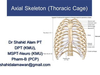
- 1. Axial Skeleton (Thoracic Cage) Dr Shahid Alam PT DPT (KMU), MSPT-Neuro (KMU) Pharm-B (PCP) shahidalamawan@gmail.com
- 2. The Thoracic Cage The skeleton of the chest Supports the thoracic cavity Consists of: – thoracic vertebrae – ribs – sternum (breastbone) The Rib Cage Formed of ribs and sternum
- 3. The Thoracic Cage Functions of the Thoracic Cage Protects organs of the thoracic cavity Heart, lungs, trachea, and esophagus. Attaches muscles For respiration Of the vertebral column Of the pectoral girdle Of the upper limbs
- 4. The Thoracic Cage Figure 7–23a The Thoracic Cage.
- 5. The Thoracic Cage Figure 7–23b The Thoracic Cage.
- 7. The Thoracic Cage The sternum A flat bone In the midline of the anterior chest wall Three parts of the sternum The manubrium The sternal body The xiphoid process
- 8. The Thoracic Cage Manubrium The manubrium is the superior portion of the sternum. It articulates with the body of the sternum at the manubriosternal joint. Broad, triangular shape Articulates with clavicles (collarbones) Articulates with cartilages of first rib pair
- 9. The Thoracic Cage The sternal angle (angle of Louis), formed by the articulation of the manubrium with the body of the sternum, can be recognized by the presence of a transverse ridge on the anterior aspect of the sternum. The transverse ridge lies at the level of the 2nd costal cartilage, the point from which all costal cartilages and ribs are counted. The sternal angle lies opposite the intervertebral disc between the 4th and 5th thoracic vertebrae.
- 10. The Thoracic Cage The sternal body Is tongue-shaped Attaches to the manubrium Attaches to costal cartilages of ribs 2–7 The xiphoid process Is the smallest part of the sternum Attaches to the sternal body at the xiphisternal joint. Attaches to diaphragm and rectus abdominis muscles
- 12. Sternum.
- 13. The Thoracic Cage Development of the Sternum The developing sternal body Consists of four unfused bones Completes fusion about age 25 Leaving transverse lines The xiphoid process Is the last part of sternum to fuse Can easily be broken away
- 14. The Thoracic Cage Ribs Are mobile Can absorb shock Functions of ribs Rib movements (breathing): – affect width and depth of thoracic cage – changing its volume
- 15. The Thoracic Cage Ribs (costae) Are 12 pairs of long, curved, flat bones Attached posteriorly to the thoracic vertebrae. Extending from the thoracic vertebrae Ribs are divided into two types True ribs False ribs
- 16. The Thoracic Cage Ribs 1–7 (true ribs) Vertebro-sternal ribs Attached anteriorly to the sternum by costal cartilages Ribs 8–12 (false ribs) Do not attach directly to the sternum. Vertebro-chondral ribs (ribs 8–10) The 8th, 9th, and 10th pairs of ribs are attached anteriorly to each other and to the 7th rib by means of their costal cartilages and small synovial joints.
- 17. The Thoracic Cage Floating ribs: The 11th and 12th pairs have no anterior attachment. Floating or vertebral ribs (ribs 11–12) Connect only to the vertebrae and back muscles Have no connection with the sternum
- 18. Rib Anatomy 1. Typical Rib A typical rib is a long, twisted, flat bone having a rounded, smooth superior border and a sharp, thin inferior border. The inferior border overhangs and forms the costal groove, which accommodates the intercostal vessels and nerve. The anterior end of each rib is attached to the corresponding costal cartilage. 18
- 19. Rib Anatomy The head has two facets for anterior and posterior articulation. The neck is a constricted portion situated between the head and the tubercle. The tubercle is a prominence on the outer surface of the rib at the junction of the neck with the shaft. The shaft is thin and flattened and twisted on its long axis. Its inferior border has the costal groove. The angle is where the shaft of the rib bends sharply forward.
- 20. Rib Anatomy 2. Atypical Rib The rib which is short and does not have a proper costal grove, angle and shaft. The 1st rib is important clinically because of its close relationship to the lower nerves of the brachial plexus and the main vessels to the arm, namely, the subclavian artery and vein.
- 21. Rib Anatomy Typical Ribs must have a Head Neck Tubercle Angle Shaft Subcostal Groove •Atypical Ribs •No.1-short, flat, wide, Supports Subclavian vessels. •No.11, 12 don’t articulate with transverse processes, or anteriorly at all. 21
- 22. The Thoracic Cage Figure 7–24b Typical Ribs.
- 24. Atypical ribs.
- 25. Costal Cartilages Costal cartilages are bars of cartilage connecting the upper seven ribs to the lateral edge of the sternum and the 8th, 9th, and 10th ribs to the cartilage immediately above. cartilages of the 11th and 12th ribs end in the abdominal musculature. The costal cartilages contribute significantly to the elasticity and mobility of the thoracic walls. In old age, the costal cartilages tend to lose some of their flexibility as the result of superficial calcification