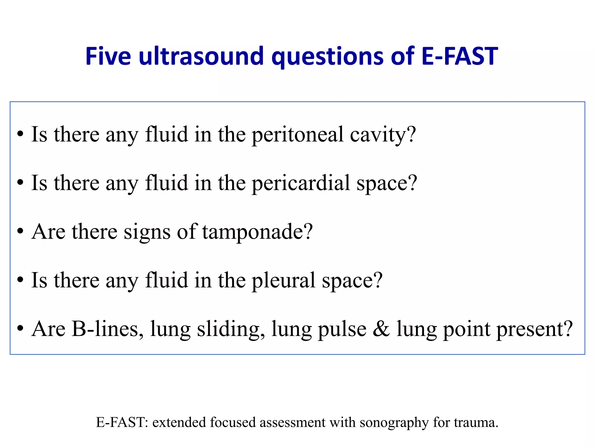
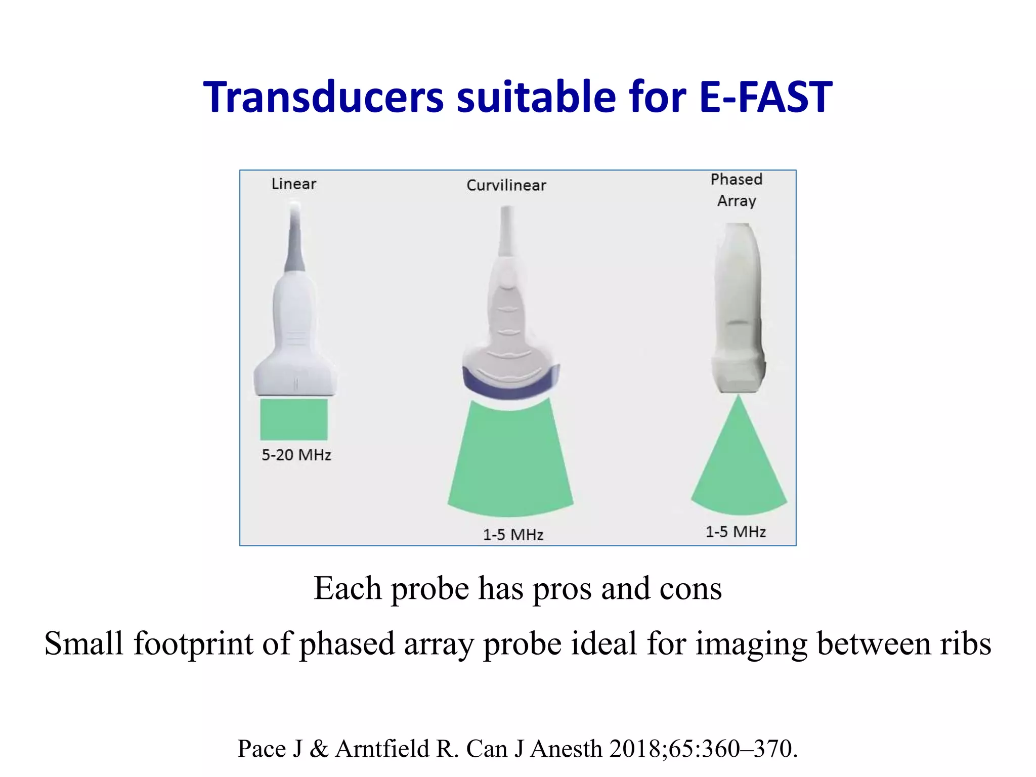
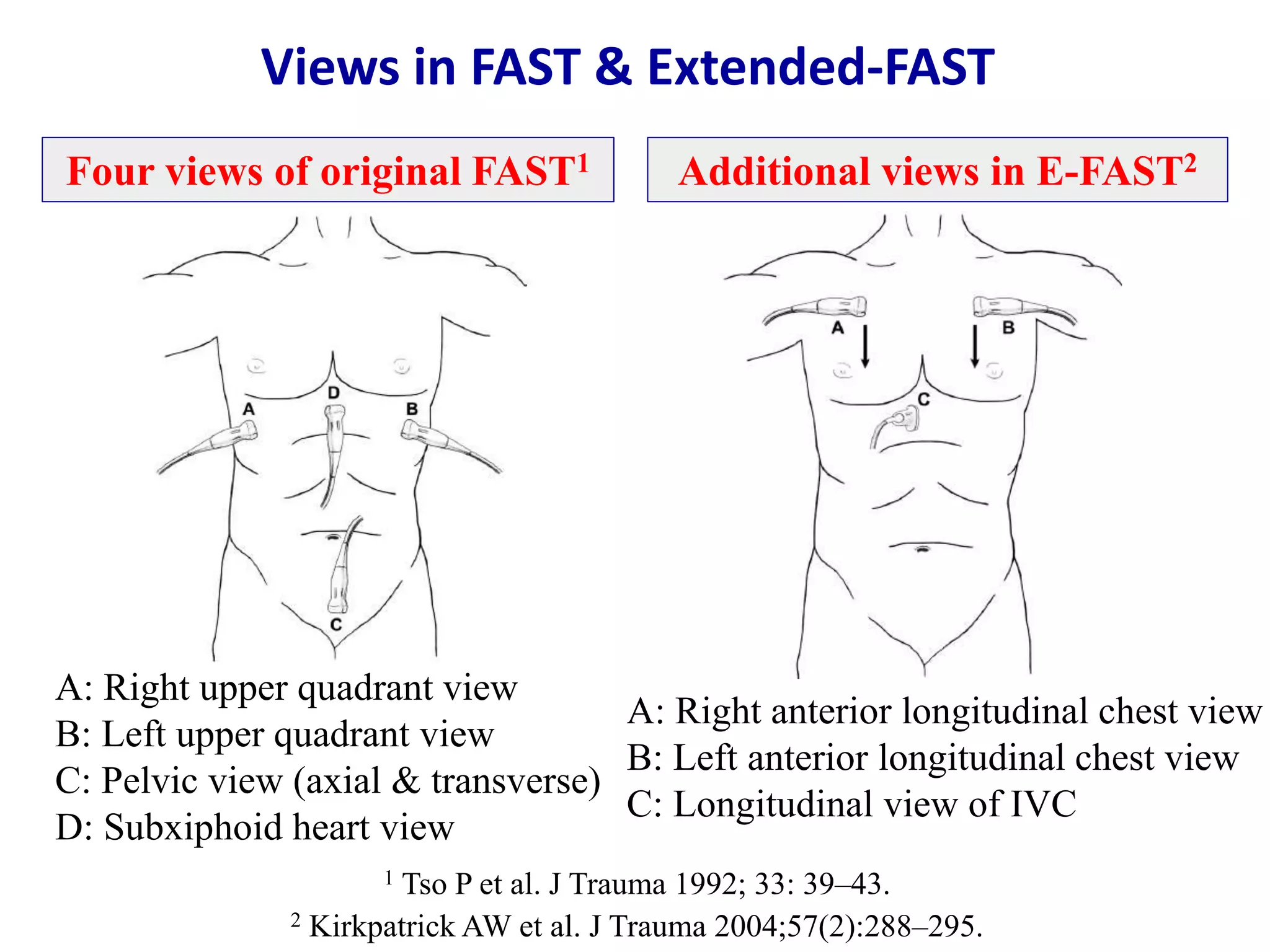
Samir Haffar MD
Gastroenterologist & Sonographer
Damascus – Syrian Arab Republic
Extended focused assessment with
sonography for trauma
E-FAST
Indications and contraindications of E-FAST
• Urgent evaluation of traumatic patients for abdominal,
thoracic or cardiac bleeding or pneumothorax
• May be used to evaluate traumatic patients serially
Indications
Contraindications
• Primary survey should precede E-FAST
• E-FAST exam should not delay definitive diagnostic or
therapeutic interventions
Four clinical questions of E-FAST
• Does the patient have blood in abdomen?
• Does the patient have blood in thorax?
• Does the patient have a pericardial blood/tamponade?
• Does the patient have a pneumothorax?
E-FAST: extended focused assessment with sonography for trauma.
Five ultrasound questions of E-FAST
• Is there any fluid in the peritoneal cavity?
• Is there any fluid in the pericardial space?
• Are there signs of tamponade?
• Is there any fluid in the pleural space?
• Are B-lines, lung sliding, lung pulse & lung point present?
E-FAST: extended focused assessment with sonography for trauma.
Diagnostic modalities in blunt abdominal trauma
• Diagnostic peritoneal lavage (DPL)
• CT scan
• Ultrasound (FAST exam)
Comparison of DPL, E-FAST, and CT
DPL E-FAST CT
Time 10 – 15 min 3 – 5 min variable
Repeatability possible & rarely done easy & frequently done yes
Reliability not organ specific operator dependent obesity
Sensibility high medium high
Specificity low high high
Advantages inexpensive,
detects bowel injury
noninvasive, rapid,
portable, no radiation
noninvasive,
highly accurate
Disadvantages invasive limited by gas & obesity,
operator dependent
radiation, expensive,
may miss small bowel
& pancreatic injuries
CT: computed tomography – DPL: diagnostic peritoneal lavage
E-FAST: extended focused assessment with sonography for trauma
Advantages of ultrasound over other techniques
• Simple
• Non-invasive without radiation
• Cost-effective
• Performed by attending physician at bedside
• Can be repeated as often as necessary
• Needs minimal amount of training
Pace J & Arntfield R. Can J Anesth 2018;65:360–370.
Transducers suitable for E-FAST
Each probe has pros and cons
Small footprint of phased array probe ideal for imaging between ribs
Transducers suitable for E-FAST
Pace J & Arntfield R. Can J Anesth 2018;65:360–370.
Phased array probeCurvilinear probe
Good penetration into abdomen & thorax (low frequency 2 – 5 MHz)
Small footprint of phased array probe (ideal for imaging between ribs)
Training in E-FAST
• Number of required exams to achieve competency in E-FAST varies
• Physicians practicing in Canada must perform 50 directly
supervised scans before they are deemed to have competency
• Authors discourage this kind of numeric, boilerplate approaches to
credentialing and certification and favor incorporating point-of-care
ultrasonography skills into core competencies of residency training
Pace J et al. Can J Anesth 2018: 65:360–370.
Views in FAST & Extended-FAST
1 Tso P et al. J Trauma 1992; 33: 39–43.
2 Kirkpatrick AW et al. J Trauma 2004;57(2):288–295.
A: Right upper quadrant view
B: Left upper quadrant view
C: Pelvic view (axial & transverse)
D: Subxiphoid heart view
Four views of original FAST1
A: Right anterior longitudinal chest view
B: Left anterior longitudinal chest view
C: Longitudinal view of IVC
Additional views in E-FAST2
Extended-FAST
• Right & left anterior longitudinal chest:
Assess for pneumothorax
• Right & left pericolic gutter views
Free fluid adjacent to bowel along flanks
• Inferior vena cava view:
Intravascular volume status
1. Right upper quadrant view (RUQ)
2. Left upper quadrant view (LUQ)
3. Pelvic view: sagittal & transverse axis
4. Subxyphoid cardiac view: long & short axis
5. Right & left anterior longitudinal chest view
6. Inferior vena cava view (intravascular volume)
Six standard views
Extended focused assessment with sonography for trauma
E-FAST
How fast is E-FAST
• Each view 30 – 60 seconds
• Number of views dependent on clinical questions
and findings on initial views
• Total exam time usually 3 – 5 minutes
Not all fluid are blood
• Ascites
• Ruptured ovarian cyst
• Lavage fluid
• Urine from ruptured bladder
Ultrasound appearance of blood
• Fresh blood
Anechoic (black)
• Coagulating blood
First hypoechoic
Later hyperechoic
Fresh & coagulated blood
Anechogenic fresh blood between liver & right kidney
Hypoechogenic clot anterior to liver
McGahan JP et al. Radiol Clin N Am 2004;42:417–425.
Right upper quadrant view
• Longitudinal scan of liver, hepatorenal space & right kidney
• Cephalad probe movement to visualize right pleural space
Area between dome of liver & diaphragm
• Caudal probe movement to visualize inferior pole of right
kidney & right paracolic gutter
American Institute of Ultrasound in Medicine. J Ultrasound Med 2014;33:2047–2056.
Probe positioning in RUQ view
Focused assessment with sonography for trauma.
In: Manual of emergency and critical care ultrasound. Noble VE, Nelson BP & Sutingco N Eds,
Cambridge University Press, 2007.
Right upper quadrant view
Longitudinal liver kidney scan
Normal Morison pouch Fluid in Morison pouch
If mean thickness of fluid in Morison’s pouch > 1 cm,
it can be assumed that up to 1 liter of intraperitoneal fluid is present
Right upper quadrant view
Craniodorsal scan at posterior axillary line
Right hemothorax & atelectasis
Volume in ml in supine position:
width of fluid in cross-section from interior thoracic wall
to dorsal margin of lung in ml x 20
Richards JR et al. Radiology 2017;283: 30–48.
Right paracolic gutter
Echogenic region inferior to kidney in right paracolic gutter
that corresponds to hematoma (arrow)
McGahan JP et al. Radiol Clin N Am 2004;42:417–425.
• Scan of spleen, perisplenic space below diaphragm,
splenorenal recess, and left kidney
• Cephalad probe movement to visualize left pleural space
• Caudal probe movement to visualize inferior pole of left
kidney and left paracolic gutter
American Institute of Ultrasound in Medicine. J Ultrasound Med 2014;33:2047–2056.
Left upper quadrant view
Perisplenic view
Probe positioning in LUQ view
Focused assessment with sonography for trauma.
In: Manual of emergency and critical care ultrasound. Noble VE, Nelson BP & Sutingco N Eds,
Cambridge University Press, 2007.
Left upper quadrant view
Spleen kidney scan
Normal Koller pouch Free fluid in Koller pouch
Osterwalder J et al. Extended focused assessment with sonography for trauma.
In: EFSUMB course book, CF Dietrich editor, 2014.
spleen
left kidney
Koller pouch
spleen
left kidney
free fluid
Left upper quadrant view
Peri-splenic free fluid
Peri-splenic fluid not always present in Koller pouch
Sometimes occurs in subcapsular, subphrenic regions or at caudal pole
Osterwalder J et al. Extended focused assessment with sonography for trauma.
In: EFSUMB course book, CF Dietrich editor, 2014.
left kidneyspleen
free fluid
D
Left upper quadrant view
Craniodorsal scan at posterior axillary line
Left pleural effusion
Focused assessment with sonography for trauma (FAST).
In: Manual of emergency and critical care ultrasound. Noble VE, Nelson BP & Sutingco N Eds,
Cambridge University Press, 2007.
left pleural
effusion spleen
diaphragm
Probe positioning for pelvic view
Transverse and longitudinal scan
Focused assessment with sonography for trauma (FAST).
In: Manual of emergency and critical care ultrasound. Noble VE, Nelson BP & Sutingco N Eds,
Cambridge University Press, 2007.
Pelvic view in woman
Normal pelvic sagittal scan
Focused assessment with sonography for trauma (FAST).
In: Manual of emergency and critical care ultrasound. Noble VE, Nelson BP & Sutingco N Eds,
Cambridge University Press, 2007.
Pelvic view in woman
Pelvic sagittal scan
Free fluid in Douglas pouch & around the uterus
Focused assessment with sonography for trauma (FAST).
In: Manual of emergency and critical care ultrasound. Noble VE, Nelson BP & Sutingco N Eds,
Cambridge University Press, 2007.
Pelvic view in man
Normal pelvic sagittal scan Normal pelvic transverse scan
bladder
prostate
rectum
bladder
prostate
seminal vesicule
Osterwalder J et al. Extended focused assessment with sonography for trauma.
In: EFSUMB course book, CF Dietrich editor, 2014.
Free fluid & clot in rectovesical excavation
Pelvic view in man
Pelvic transverse scan Pelvic sagittal scan
free fluid
clot
Osterwalder J et al. Extended focused assessment with sonography for trauma.
In: EFSUMB course book, CF Dietrich editor, 2014.
free fluid
clot
bladder
Folley
intestine
• Transducer on upper abdomen & pointing superiorly
toward left shoulder using left liver as acoustic window
• If adequate subxiphoid view cannot be obtained:
Parasternal long-axis view of heart
Apical four chamber view
Subxyphoid cardiac view
American Institute of Ultrasound in Medicine. J Ultrasound Med 2014;33:2047–2056.
Probe positioning for subxyphoid cardiac view
Focused assessment with sonography for trauma (FAST).
In: Manual of emergency and critical care ultrasound. Noble VE, Nelson BP & Sutingco N Eds,
Cambridge University Press, 2007.
Subxyphoid cardiac view
Four chamber view of the heart
Normal longitudinal view
LV
RV
liver
LA
RA
Subxyphoid cardiac view
Pericardial effusion
Right ventricular is seen: no cardiac temponade
Until proven otherwise, any amount of pericardial fluid must
be considered to represent evolving hemorrhage due to cardiac injury
Richards JR et al. Radiology 2017;283: 30–48.
Pace J & Arntfield R. Can J Anesth 2018;65:360–370.
LV
RV
PE
Cardiac temponade
Right ventricule is collapsed
Cardiac temponade: diastolic collapse of right atrium or right ventricule
Richards JR et al. Radiology 2017;283: 30–48.
Subxyphoid cardiac view
no
Subxiphoid cardiac view
Pericardiac clot
Focused assessment with sonography for trauma (FAST).
In: Manual of emergency and critical care ultrasound. Noble VE, Nelson BP & Sutingco N Eds,
Cambridge University Press, 2007.
It is important to recognize pericardiac clot
This can be lifesaving (not all fluid are anechoic)
Anterior longitudinal lung view
Right & left
• Longitudinal plane of anterior chest wall
2nd or 3rd intercostal space
Mid-clavicle line (MCL)
• Other intercostal spaces may be used for lung analysis
• M-mode can aid in evaluation for pneumothorax
American Institute of Ultrasound in Medicine. J Ultrasound Med 2014;33:2047–2056.
Probe positioning for anterior longitudinal lung view
Focused assessment with sonography for trauma (FAST).
In: Manual of emergency and critical care ultrasound. Noble VE, Nelson BP & Sutingco N Eds,
Cambridge University Press, 2007.
Pleural line
Anterior longitudinal lung view
Shrestha GS et al. Rev Recent Clin Trials 2018;13:15–26.
R1: upper rib – R2: lower rib – Vertical arrow: pleural line
All signs in lung ultrasound arise from pleural line
Bat sign
ribs are the wings
Normal A-lines
Anterior longitudinal lung view
Bat sign Schematic view
Present in normal subjects
Reverberation hyperchoic artifacts from visceral & parietal pleura
Always equally distributed (same distance between skin & pleura)
Present in normal subjects
Reverberation hyperchoic artifacts from visceral & parietal pleura
Always equally distributed (same distance between skin & pleura)
Normal A-lines
Anterior longitudinal lung view
Touw HRW et al. Netherlands J Med 2015;7 3:100–107.
Bat sign
ribs are the wings
Normal A-lines in B and M mode
Anterior longitudinal lung view
B mode M mode
Wongwaisayawan et al. RadioGraphics 2016; 36:640–659
Normal B-lines
Anterior longitudinal lung view
Present in normal subjects – Extend from pleural line – Erase A-lines
Focused assessment with sonography for trauma (FAST).
In: Manual of emergency and critical care ultrasound. Noble VE, Nelson BP & Sutingco N Eds,
Cambridge University Press, 2007.
Laser-like
Normal B lines (comet tail artifact)
anterior longitudinal lung view
Presence of B Lines exclude the diagnosis of pneumothorax
Characteristics of B-lines
• Arise from pleural line
• Long vertical hyperechoic lines (laser-like)
• Continue to lower edge of screen without fading
• Look like comet tails (old name for them)
• Erase A lines
• Move with lung sliding
Miller A. BJA Education 2016;16(2):39–45.
Normal Z-lines
Anterior longitudinal lung view
Wongwaisayawan et al. RadioGraphics 2016; 36:640–659
White arrow: pleural line
Arrowheads: B line
Black arrows: Z-lines
• Arise from pleural line
• Short ill-defined vertical hyperechoic lines
• Do not reach the edge of the screen
• Do not erase A-lines
• Do not move with lung sliding
Wongwaisayawan et al. RadioGraphics 2016; 36:640–659
Characteristics of B-lines
Abnormal numerous B-Lines (B pattern)
Lung rockets
More than 2 B-lines in a lung field indicates interstitial syndrome
Pulmonary edema is the commonest cause
Number of B-lines or distance between B-lines assess severity
More than 10 per screen or only 3 mm apart favors diagnosis of ARDS
Richards JR et al. Radiology 2017;283:30–48.
B- and M-mode image
Normal lung sliding (seashore sign)
Anterior longitudinal lung view
Miller A. BJA Education 2016;16(2):39–45.
Subcutaneous tissue above pleural line generates horizontal straight lines
Sandy appearance below pleural line created by lung sliding movement
Normal lung sliding in M mode
Seashore sign or wavy/sandy beach sign
Normal lung sliding in power Doppler
Anterior longitudinal lung view
Normal to-and-fro sliding movement of pleura can be seen in real time
Normal lung sliding visualized as lighting up in power Doppler
Probe must be steady to avoid unwanted color artifacts
Wongwaisayawan et al. RadioGraphics 2016; 36:640–659
B mode image Power doppler image
rib
pleural line
subcutaneous fat
muscle
Lung sliding
Miller A. BJA Education 2016;16(2):39–45.
Reduced lung sliding Low tidal volumes
Hyper-inflated lung
Absence of lung sliding Pleura not directly opposed:
Pleura stuck together:
Absent respiration:
Pneumothorax
Effusion
Pneumonia
ARDS
Pleurodesis
Peumonectomy
Lung intubation
Normal lung pulse
Rhythmic movement of pleura in synchrony with cardiac rhythm
M mode
Osterwalder J et al. Extended focused assessment with sonography for trauma.
In: EFSUMB course book, CF Dietrich editor, 2014.
Pneumothorax
Pneumothorax is caused by one of the followings
1. Air leaking from airways or alveolar space
2. Gas-producing organisms within pleura
3. Air communication between pleural space & atmosphere
Oveland NP. Pneumothorax.
In: Chest ultrasound, Laursen CB, Rahman NM & Volpicelli G Editors.
European Respiratory Society, 2018.
Normal lung & pneumothorax
Normal longitudinal lung view
Normal lung Pneumothorax
Sonographic evaluation of pneumothorax
Volpicelli G. Intensive Care Med 2011;37:224–232.
Probe begins from this area Probe moved toward lateral chest
Supine position
3rd intercostal space
Mid-clavicle line
Check for lung sliding & lung point
at different locations (white crosses)
Lung point confirms pneumothorax
& evaluates its extension
Progression of sonographic probe
towards lateral chest (white crosses)
to check for lung point (black cross)
Volpicelli G. Intensive Care Med 2011;37:224–232.
Points checked on chest wall by US
correspond to CT scan image (black
& white triangles) & predict extension
of pneumothorax on chest wall
Sonographic evaluation of pneumothorax
Ultrasound in pneumothorax
• Absence of B-lines
• Absence of lung sliding
• Absence of lung pulse
• Presence of lung point sign (B and M mode)
Confirm pneumothorax & evaluate its extension
Four ultrasonographic signs
B- and M-mode image
Absence of lung sliding
Anterior longitudinal lung view
Miller A. BJA Education 2016;16(2):39–45.
Stratosphere or barcode sign
Absence of lung sliding in M mode
Anterior longitudinal lung view
Stratosphere or barcode sign
McGahan JP et al. Radiol Clin N Am 2004;42:417–425.
Right side of image:
Echogenic line (parietal & visceral pleura)
Distal comet tail artifacts (B Lines)
Left side of image:
Loss of this pattern (pneumothorax)
Lung point sign in B-mode
Interface between normal lung & pneumothorax
Richards JR et al. Radiology 2017;283: 30–48.
Lung point sign in M-mode
Interface between normal lung & pneumothorax
Right side of image:
Echogenic line (parietal & visceral pleural)
Seashore sign: granular normal lung
Left side of image:
Barcode sign: representing pneumothorax
Lung point sign in pneumothrorax
• Sensitivity:
Depends on setting
Hemodynamic instability: low sensibility – collapsed lung
Stable patient: high sensibility
• Specificity:
Presence of lung point 100% specific for ruling in pneumothorax
Volpicelli G. Intensive Care Med 2011;37:224–232.
Extension of pneumothorax
• Chest radiography: used in clinical practice – low accuracy
• CT scan: highly accurate if available
• Lung ultrasound: if CT not available
The more lateral the lung point is on the chest wall in supine
position, the greater is the extension of the air layer
Volpicelli G. Intensive Care Med 2011;37:224–232.
Clavicle fracture
Longitudinal US images of clavicle
Discontinuity of left clavicle (arrow)
Focal reverberation artifact deep
to site of fracture (arrowhead)
Normal right clavicle
Wongwaisayawan et al. RadioGraphics 2016; 36:640–659
Rib fracture
Displaced rib fracture (arrows)
Adjacent hematoma (*)
Minimally displaced rib fracture
(arrow)
Wongwaisayawan et al. RadioGraphics 2016; 36:640–659
Longitudinal scan over rib
Rib fracture
Anterior & posterior echogenic lines correspond with
two anterior rib margins & gap (displaced rib fracture)
Richards JR et al. Radiology 2017;283: 30–48.
Longitudinal scan over rib
Longitudinal view of IVC
Richards JR et al. Radiology 2017;283: 30–48.
Measurement of IVC diameter 2 cm below
cavo-atrial junction (arrows)
IVC
liver
Variation of IVC diameter with spontaneous
breathing in a healthy patient
Richards JR et al. Radiology 2017;283: 30–48.
Normal IVC diameter: 1,5 – 2.5 cm
IVC collapses during inspiration to < 50% of its expiratory diameter
Volume depletion: IVC <1.5 cm & >50% inspiratory collapse
Volume overload: IVC >2.5 cm & <50% inspiratory collapse
Caval index
Caval index close to 100% Almost complete collapse
Likely volume depletion
Caval index close to 0% Minimal collapse
Suggesting volume overload
IVC expiratory diameter – IVC inspiratory diameter
IVC expiratory diameter
x 100
Goldflam K et al. ACEP News 2011. http://www.acep.org/Content. Published June 2011.
Pitfalls in E-FAST
Double line sign in Morison’s pouch
Hyperechoic line along the liver (2)
Hyperechoic line along along the kidney (3)
Hypoechoic fat in the middle
Hypo-echoic fat in Morison pouch can simulate fluid
Double-line sign
Osterwalder J et al. Extended focused assessment with sonography for trauma.
In: EFSUMB course book, CF Dietrich editor, 2014.
right kidney
liver
Patient positioning
Trendelenburg position and right lateral decubitus position
significantly increase detection rate of fluid in Morison pouch
Renal cortical cysts
Cysts should be completely visualized and in two planes
Osterwalder J et al. Extended focused assessment with sonography for trauma.
In: EFSUMB course book, CF Dietrich editor, 2014.
liver
renal cyst
right kidney
Gastric fluid sign in LUQ
Fluid in stomach or free intra-peritoneal fluid
Gastric fluid
Free fluid Subphrenically or between spleen & kidney
Oval shape
Surrounded by echo-rich edge (stomach wall)
Contains food residues (floating internal echoes)
Osterwalder J et al. Extended focused assessment with sonography for trauma.
In: EFSUMB course book, CF Dietrich editor, 2014.
spleen
gastric content
gastric wall
Kissing liver in LUQ
If left hepatic lobe touches the spleen, it can resemble free fluid
Increase gain so liver tissue is easier to identify
Color Doppler to visualize liver vessels
Position the probe anteriorly
Differentiation
spleen
diaphragm
liver
Osterwalder J et al. Extended focused assessment with sonography for trauma.
In: EFSUMB course book, CF Dietrich editor, 2014.
Seminal vesicle in pelvic view
Large seminal vesicle can simulate fluid
Free peritoneal fluid never found beside or directly above prostate
but more cranially and beside or above the bladder
Transverse scan Longitudinal scan
bladder
prostate
seminal vesicule
bladder
prostate
seminal vesicule
Osterwalder J et al. Extended focused assessment with sonography for trauma.
In: EFSUMB course book, CF Dietrich editor, 2014.
Physiological fluid in childbearing age women
Normal Douglas free fluid in 30–40% of childbearing age women
Anterior-posterior diameter > 3 cm
Presence of internal echoes
Fluid spreads above or beside bladder
Pathological findings
bladder
uterusintestine
ovary
Osterwalder J et al. Extended focused assessment with sonography for trauma.
In: EFSUMB course book, CF Dietrich editor, 2014.
Epicardial fat
Typically anterior to right ventricle & rarely tracks around left ventricle
Fat is isodense & has granular echotexture
Fat in M-mode: wave-like movement in whole width of band
(1) Parietal pericardium, (2) visceral pericardium
Epicardial fat can be mistaken for pericardial effusion
Osterwalder J et al. Extended focused assessment with sonography for trauma.
In: EFSUMB course book, CF Dietrich editor, 2014.
Pericardial effusion
(1) parietal pericardium, (2) visceral pericardium
Systolic interval (yellow arrow), diastolic interval (red arrow)
Fluid in M mode: increase in systole & decrease in diastole
Osterwalder J et al. Extended focused assessment with sonography for trauma.
In: EFSUMB course book, CF Dietrich editor, 2014.
B mode M mode
Pleural or pericardial effusion
Parasternal long-axis view
Fluid between atrium & transversally descending aorta is
pathognomic for pericardial effusion
If it spreads dorsally from descending aorta, pleural effusion is present
Osterwalder J et al. Extended focused assessment with sonography for trauma.
In: EFSUMB course book, CF Dietrich editor, 2014.
Pleural effusion Pericardial effusion
RV
LV
ascending aorta
pleural effusion
descending aorta
RV
anterior PE
ascending aorta
descending aorta
posterior PE
LV
Pitfalls in pneumothorax
No pneumothorax Pneumothorax
B-lines
Lung sliding
Lung pulse
No lung point
No B-lines
No lung sliding
No lung pulse
Lung point
Lung point is the only firm evidence of pneumothorax
Large bulla
Thick adherent pleura
No B-lines
No lung sliding
No lung pulse
No lung point
Osterwalder J et al. Extended focused assessment with sonography for trauma.
In: EFSUMB course book, CF Dietrich editor, 2014.
Subcutaneous emphysema
E-lines
43-year-old man with blunt thoracic trauma
Muliple hyperechoic artifacts (arrows) in subcutaneous tissue
Casting dirty shadows across depth of the image
Pleural line & ribs not depicted
Wongwaisayawan et al. RadioGraphics 2016; 36:640–659
Serial E-FAST
• Give examiner more time for a comprehensive exam
• Amount of free fluid in active bleeding may increase with time
• Serial FAST exam increases sensitivity for free fluid detection
• Additional view of interloop space (triangular hypoechoic area
between bowel) improved sensitivity in primary & secondary exams
• Logical alternative to CT for stable trauma patients, patients with
sudden change in hemodynamic status or physical examination,
and pregnant patients to mitigate radiation exposure
Richards JR et al. Radiology 2017;283: 30–48.
Free fluid in interloop space
Triangular hypoechoic area between bowel loops
John Ma, James Mateer, Robert Reardon, Scott Joing.
Ma and Mateer's Emergency Ultrasound, 3rd edition.
• Visualize small amounts of free fluid: peritoneal, pleural & pericardial
• Not intended to detect type of fluid or source of bleeding:
Puncture under ultrasound control in case of doubt
• Aim is to answer 5 definite questions:
If the fluid is blood, is there indirect evidence of organ injury?
Is immediate surgical necessary, can patient wait, or is CT scan needed?
If surgery is necessary, what should be opened first, abdomen or chest?
In hemopericardium: temporary drainage or pericardiotomy
Is chest X-ray/CT needed for surgery in hemothorax/pneumothorax?
Osterwalder J et al. Extended focused assessment with sonography for trauma.
In: EFSUMB course book, CF Dietrich editor, 2014.
Positive E-FAST
Negative E-FAST
• Negative E-FAST does not exclude intraabdominal
or intrathoracic injuries
• Negative E-FAST does not exclude retroperitoneal or
mediastinal lesions
Osterwalder J et al. Extended focused assessment with sonography for trauma.
In: EFSUMB course book, CF Dietrich editor, 2014.
FAST protocol
International consensus conference
CT: computed tomography – DPL: diagnostic peritoneal lavage
FAST: focused assessment with sonography for trauma – OR: operating room
Scalea TM et al. J Trauma 1999;46: 466–72.
Diagnostic algorithm of FAST in abdominal trauma
Richards JR et al. Radiology 2017;283: 30–48.
E-FAST protocol in pneumothorax
Osterwalder J et al. Extended focused assessment with sonography for trauma.
In: EFSUMB course book, CF Dietrich editor, 2014.
Lung point is the only firm evidence of pneumothorax
Diagnostic performance of E-FAST
Sensibility Specificity
Hemoperitonium 63 – 99% 88 – 93%
Hemothorax 96 – 98%* 99 – 100%
Hemopericardium 56 – 100%** 87 – 100%
Pneumothorax 91%*
* Far superior to chest X-ray
** Low sensitivity of 56% because blood from myocardial injuries leaks into pleural space
Osterwalder J et al. Extended focused assessment with sonography for trauma.
In: EFSUMB course book, CF Dietrich editor, 2014.
Use of E-FAST in prehospital setting
Richards JR et al. Radiology 2017;283: 30–48.
Natural disorders: Earthquake in Armenia 1988
Earthquake in Turkey 1999
Floods in Guatemala 2005
Cyclone in Australia 2007
Earthquake in China 2008
Earthquake in Haiti 2010
Terrorist attacks Madrid 2004
London 2005
Additional traumatic injuries
depicted by ultrasound
Solid organ evaluation
FAST originally intended to detect intraperitoneal free fluid
• Limited sensitivity for solid organ injury by US (41 – 44%)
• Splenic laceration: diffuse heterogenous pattern in splenic laceration
hypoechoic over few days
• Subcapsular splenic hematoma: hyper- or hypoechoic rim
• Hepatic laceration: discrete hyperechoic pattern
• Renal trauma: mixed echogenicity with disorganized pattern
• Bladder trauma: hematoma frequently echogenic
Richards JR et al. Radiology 2017;283: 30–48.
Right upper quadrant view
Longitudinal liver kidney scan
Well marginated echogenic region in liver (arrows)
corresponds to liver laceration
McGahan JP et al. Radiol Clin N Am 2004;42:417–425.
Longitudinal right upper quadrant view
McGahan JP et al. Radiol Clin N Am 2004;42:417–425.
Ill-defined region without reniform shape
corresponds to severe renal laceration (shattered kidney) (arrows)
Left upper quadrant view
Echogenic subcapsular hematoma of the spleen (arrow)
Richards JR et al. Radiology 2017;283: 30–48.
Bowel and mesenteric injury
Difficult detection by ultrasound
• Small volume of bleeding/extravasated bowel contents after injury
• Fluid-filled bowel not to be confused with free intraperitoneal fluid
• Bowel distinguished by round shape & peristalsis
• Pneumoperitoneum mimics air within small & large bowel loops
• Free air shifts to least dependent areas of peritoneal cavity with
change in patient position “shifting phenomenon”
• If both free fluid and air present in the peritoneal cavity,
“enhancement of peritoneal stripe sign” may be visualized
Richards JR et al. Radiology 2017;283: 30–48.
Chang-Chien CS et al. J Clin Ultrasound 1989;17:95-100.
Braccini G et al. Abdom Imaging 1996;21(5):404–412.
Lower portion of liver obscured by
interference echo pattern (arrowheads)
Upper portion of liver & ascites identified
Supine position Left lateral decubitus
Interference echo pattern moved to
upper portion of liver (arrowheads)
Lower portion of liver & GB appear
Pneumoperitoneum
Shifting phenomenon
Pneumoperitoneum
Enhancement of peritoneal stripe sign (EPSS)
Arsani A. Emerg Radiol 2007;14:29–39.
Transverse scan of RLQ
Double-layered peritoneal stripe (arrows)
Echogenic bowel echoes (double arrows)
Normal peritoneal stripe Enhancement of peritoneal stripe
Longitudinal scan of RLQ
EPSS (arrow)
Perforation of ileum
Diagnosis of pneumoperitoneum by ultrasound
• First described in 19841
• US compared to plain radiography for pneumoperitoneum2
More sensitive: 93% vs 79%
Comparable specificity: 64% vs 64%
Comparable positive predictive value 97% vs 96%
US: ultrasound
1 Nirapathpongporn S et al. Radiology 1984; 150: 831–2.
2 Jones R. Am J Emerg Med 2007; 25: 838–41.
Conclusion
• E-FAST is a focused, rapid and easy to learn ultrasound examination
• Performed at bedside to answer 4 clinical questions
• Transformed into 5 sonographic questions
• Using information from 6 standard ultrasound views
• Main purpose: know if patient has haemodynamically bleeding in
peritoneal cavity, pleural or pericardial space, or pneumothorax
• Together with the overall clinical assessment, this information helps
to assess whether there is ongoing bleeding demanding operation
Thank You

Extended focus assessment with sonography for trauma

  • 1.
    Samir Haffar MD Gastroenterologist& Sonographer Damascus – Syrian Arab Republic Extended focused assessment with sonography for trauma E-FAST
  • 2.
    Indications and contraindicationsof E-FAST • Urgent evaluation of traumatic patients for abdominal, thoracic or cardiac bleeding or pneumothorax • May be used to evaluate traumatic patients serially Indications Contraindications • Primary survey should precede E-FAST • E-FAST exam should not delay definitive diagnostic or therapeutic interventions
  • 3.
    Four clinical questionsof E-FAST • Does the patient have blood in abdomen? • Does the patient have blood in thorax? • Does the patient have a pericardial blood/tamponade? • Does the patient have a pneumothorax? E-FAST: extended focused assessment with sonography for trauma.
  • 4.
    Five ultrasound questionsof E-FAST • Is there any fluid in the peritoneal cavity? • Is there any fluid in the pericardial space? • Are there signs of tamponade? • Is there any fluid in the pleural space? • Are B-lines, lung sliding, lung pulse & lung point present? E-FAST: extended focused assessment with sonography for trauma.
  • 5.
    Diagnostic modalities inblunt abdominal trauma • Diagnostic peritoneal lavage (DPL) • CT scan • Ultrasound (FAST exam)
  • 6.
    Comparison of DPL,E-FAST, and CT DPL E-FAST CT Time 10 – 15 min 3 – 5 min variable Repeatability possible & rarely done easy & frequently done yes Reliability not organ specific operator dependent obesity Sensibility high medium high Specificity low high high Advantages inexpensive, detects bowel injury noninvasive, rapid, portable, no radiation noninvasive, highly accurate Disadvantages invasive limited by gas & obesity, operator dependent radiation, expensive, may miss small bowel & pancreatic injuries CT: computed tomography – DPL: diagnostic peritoneal lavage E-FAST: extended focused assessment with sonography for trauma
  • 7.
    Advantages of ultrasoundover other techniques • Simple • Non-invasive without radiation • Cost-effective • Performed by attending physician at bedside • Can be repeated as often as necessary • Needs minimal amount of training
  • 8.
    Pace J &Arntfield R. Can J Anesth 2018;65:360–370. Transducers suitable for E-FAST Each probe has pros and cons Small footprint of phased array probe ideal for imaging between ribs
  • 9.
    Transducers suitable forE-FAST Pace J & Arntfield R. Can J Anesth 2018;65:360–370. Phased array probeCurvilinear probe Good penetration into abdomen & thorax (low frequency 2 – 5 MHz) Small footprint of phased array probe (ideal for imaging between ribs)
  • 10.
    Training in E-FAST •Number of required exams to achieve competency in E-FAST varies • Physicians practicing in Canada must perform 50 directly supervised scans before they are deemed to have competency • Authors discourage this kind of numeric, boilerplate approaches to credentialing and certification and favor incorporating point-of-care ultrasonography skills into core competencies of residency training Pace J et al. Can J Anesth 2018: 65:360–370.
  • 11.
    Views in FAST& Extended-FAST 1 Tso P et al. J Trauma 1992; 33: 39–43. 2 Kirkpatrick AW et al. J Trauma 2004;57(2):288–295. A: Right upper quadrant view B: Left upper quadrant view C: Pelvic view (axial & transverse) D: Subxiphoid heart view Four views of original FAST1 A: Right anterior longitudinal chest view B: Left anterior longitudinal chest view C: Longitudinal view of IVC Additional views in E-FAST2
  • 12.
    Extended-FAST • Right &left anterior longitudinal chest: Assess for pneumothorax • Right & left pericolic gutter views Free fluid adjacent to bowel along flanks • Inferior vena cava view: Intravascular volume status
  • 13.
    1. Right upperquadrant view (RUQ) 2. Left upper quadrant view (LUQ) 3. Pelvic view: sagittal & transverse axis 4. Subxyphoid cardiac view: long & short axis 5. Right & left anterior longitudinal chest view 6. Inferior vena cava view (intravascular volume) Six standard views Extended focused assessment with sonography for trauma E-FAST
  • 14.
    How fast isE-FAST • Each view 30 – 60 seconds • Number of views dependent on clinical questions and findings on initial views • Total exam time usually 3 – 5 minutes
  • 15.
    Not all fluidare blood • Ascites • Ruptured ovarian cyst • Lavage fluid • Urine from ruptured bladder
  • 16.
    Ultrasound appearance ofblood • Fresh blood Anechoic (black) • Coagulating blood First hypoechoic Later hyperechoic
  • 17.
    Fresh & coagulatedblood Anechogenic fresh blood between liver & right kidney Hypoechogenic clot anterior to liver McGahan JP et al. Radiol Clin N Am 2004;42:417–425.
  • 18.
    Right upper quadrantview • Longitudinal scan of liver, hepatorenal space & right kidney • Cephalad probe movement to visualize right pleural space Area between dome of liver & diaphragm • Caudal probe movement to visualize inferior pole of right kidney & right paracolic gutter American Institute of Ultrasound in Medicine. J Ultrasound Med 2014;33:2047–2056.
  • 19.
    Probe positioning inRUQ view Focused assessment with sonography for trauma. In: Manual of emergency and critical care ultrasound. Noble VE, Nelson BP & Sutingco N Eds, Cambridge University Press, 2007.
  • 20.
    Right upper quadrantview Longitudinal liver kidney scan Normal Morison pouch Fluid in Morison pouch If mean thickness of fluid in Morison’s pouch > 1 cm, it can be assumed that up to 1 liter of intraperitoneal fluid is present
  • 21.
    Right upper quadrantview Craniodorsal scan at posterior axillary line Right hemothorax & atelectasis Volume in ml in supine position: width of fluid in cross-section from interior thoracic wall to dorsal margin of lung in ml x 20 Richards JR et al. Radiology 2017;283: 30–48.
  • 22.
    Right paracolic gutter Echogenicregion inferior to kidney in right paracolic gutter that corresponds to hematoma (arrow) McGahan JP et al. Radiol Clin N Am 2004;42:417–425.
  • 23.
    • Scan ofspleen, perisplenic space below diaphragm, splenorenal recess, and left kidney • Cephalad probe movement to visualize left pleural space • Caudal probe movement to visualize inferior pole of left kidney and left paracolic gutter American Institute of Ultrasound in Medicine. J Ultrasound Med 2014;33:2047–2056. Left upper quadrant view Perisplenic view
  • 24.
    Probe positioning inLUQ view Focused assessment with sonography for trauma. In: Manual of emergency and critical care ultrasound. Noble VE, Nelson BP & Sutingco N Eds, Cambridge University Press, 2007.
  • 25.
    Left upper quadrantview Spleen kidney scan Normal Koller pouch Free fluid in Koller pouch Osterwalder J et al. Extended focused assessment with sonography for trauma. In: EFSUMB course book, CF Dietrich editor, 2014. spleen left kidney Koller pouch spleen left kidney free fluid
  • 26.
    Left upper quadrantview Peri-splenic free fluid Peri-splenic fluid not always present in Koller pouch Sometimes occurs in subcapsular, subphrenic regions or at caudal pole Osterwalder J et al. Extended focused assessment with sonography for trauma. In: EFSUMB course book, CF Dietrich editor, 2014. left kidneyspleen free fluid D
  • 27.
    Left upper quadrantview Craniodorsal scan at posterior axillary line Left pleural effusion Focused assessment with sonography for trauma (FAST). In: Manual of emergency and critical care ultrasound. Noble VE, Nelson BP & Sutingco N Eds, Cambridge University Press, 2007. left pleural effusion spleen diaphragm
  • 28.
    Probe positioning forpelvic view Transverse and longitudinal scan Focused assessment with sonography for trauma (FAST). In: Manual of emergency and critical care ultrasound. Noble VE, Nelson BP & Sutingco N Eds, Cambridge University Press, 2007.
  • 29.
    Pelvic view inwoman Normal pelvic sagittal scan Focused assessment with sonography for trauma (FAST). In: Manual of emergency and critical care ultrasound. Noble VE, Nelson BP & Sutingco N Eds, Cambridge University Press, 2007.
  • 30.
    Pelvic view inwoman Pelvic sagittal scan Free fluid in Douglas pouch & around the uterus Focused assessment with sonography for trauma (FAST). In: Manual of emergency and critical care ultrasound. Noble VE, Nelson BP & Sutingco N Eds, Cambridge University Press, 2007.
  • 31.
    Pelvic view inman Normal pelvic sagittal scan Normal pelvic transverse scan bladder prostate rectum bladder prostate seminal vesicule Osterwalder J et al. Extended focused assessment with sonography for trauma. In: EFSUMB course book, CF Dietrich editor, 2014.
  • 32.
    Free fluid &clot in rectovesical excavation Pelvic view in man Pelvic transverse scan Pelvic sagittal scan free fluid clot Osterwalder J et al. Extended focused assessment with sonography for trauma. In: EFSUMB course book, CF Dietrich editor, 2014. free fluid clot bladder Folley intestine
  • 33.
    • Transducer onupper abdomen & pointing superiorly toward left shoulder using left liver as acoustic window • If adequate subxiphoid view cannot be obtained: Parasternal long-axis view of heart Apical four chamber view Subxyphoid cardiac view American Institute of Ultrasound in Medicine. J Ultrasound Med 2014;33:2047–2056.
  • 34.
    Probe positioning forsubxyphoid cardiac view Focused assessment with sonography for trauma (FAST). In: Manual of emergency and critical care ultrasound. Noble VE, Nelson BP & Sutingco N Eds, Cambridge University Press, 2007.
  • 35.
    Subxyphoid cardiac view Fourchamber view of the heart Normal longitudinal view LV RV liver LA RA
  • 36.
    Subxyphoid cardiac view Pericardialeffusion Right ventricular is seen: no cardiac temponade Until proven otherwise, any amount of pericardial fluid must be considered to represent evolving hemorrhage due to cardiac injury Richards JR et al. Radiology 2017;283: 30–48. Pace J & Arntfield R. Can J Anesth 2018;65:360–370. LV RV PE
  • 37.
    Cardiac temponade Right ventriculeis collapsed Cardiac temponade: diastolic collapse of right atrium or right ventricule Richards JR et al. Radiology 2017;283: 30–48. Subxyphoid cardiac view no
  • 38.
    Subxiphoid cardiac view Pericardiacclot Focused assessment with sonography for trauma (FAST). In: Manual of emergency and critical care ultrasound. Noble VE, Nelson BP & Sutingco N Eds, Cambridge University Press, 2007. It is important to recognize pericardiac clot This can be lifesaving (not all fluid are anechoic)
  • 39.
    Anterior longitudinal lungview Right & left • Longitudinal plane of anterior chest wall 2nd or 3rd intercostal space Mid-clavicle line (MCL) • Other intercostal spaces may be used for lung analysis • M-mode can aid in evaluation for pneumothorax American Institute of Ultrasound in Medicine. J Ultrasound Med 2014;33:2047–2056.
  • 40.
    Probe positioning foranterior longitudinal lung view Focused assessment with sonography for trauma (FAST). In: Manual of emergency and critical care ultrasound. Noble VE, Nelson BP & Sutingco N Eds, Cambridge University Press, 2007.
  • 41.
    Pleural line Anterior longitudinallung view Shrestha GS et al. Rev Recent Clin Trials 2018;13:15–26. R1: upper rib – R2: lower rib – Vertical arrow: pleural line All signs in lung ultrasound arise from pleural line Bat sign ribs are the wings
  • 42.
    Normal A-lines Anterior longitudinallung view Bat sign Schematic view Present in normal subjects Reverberation hyperchoic artifacts from visceral & parietal pleura Always equally distributed (same distance between skin & pleura)
  • 43.
    Present in normalsubjects Reverberation hyperchoic artifacts from visceral & parietal pleura Always equally distributed (same distance between skin & pleura) Normal A-lines Anterior longitudinal lung view Touw HRW et al. Netherlands J Med 2015;7 3:100–107. Bat sign ribs are the wings
  • 44.
    Normal A-lines inB and M mode Anterior longitudinal lung view B mode M mode Wongwaisayawan et al. RadioGraphics 2016; 36:640–659
  • 45.
    Normal B-lines Anterior longitudinallung view Present in normal subjects – Extend from pleural line – Erase A-lines Focused assessment with sonography for trauma (FAST). In: Manual of emergency and critical care ultrasound. Noble VE, Nelson BP & Sutingco N Eds, Cambridge University Press, 2007. Laser-like
  • 46.
    Normal B lines(comet tail artifact) anterior longitudinal lung view Presence of B Lines exclude the diagnosis of pneumothorax
  • 47.
    Characteristics of B-lines •Arise from pleural line • Long vertical hyperechoic lines (laser-like) • Continue to lower edge of screen without fading • Look like comet tails (old name for them) • Erase A lines • Move with lung sliding Miller A. BJA Education 2016;16(2):39–45.
  • 48.
    Normal Z-lines Anterior longitudinallung view Wongwaisayawan et al. RadioGraphics 2016; 36:640–659 White arrow: pleural line Arrowheads: B line Black arrows: Z-lines
  • 49.
    • Arise frompleural line • Short ill-defined vertical hyperechoic lines • Do not reach the edge of the screen • Do not erase A-lines • Do not move with lung sliding Wongwaisayawan et al. RadioGraphics 2016; 36:640–659 Characteristics of B-lines
  • 50.
    Abnormal numerous B-Lines(B pattern) Lung rockets More than 2 B-lines in a lung field indicates interstitial syndrome Pulmonary edema is the commonest cause Number of B-lines or distance between B-lines assess severity More than 10 per screen or only 3 mm apart favors diagnosis of ARDS Richards JR et al. Radiology 2017;283:30–48.
  • 51.
    B- and M-modeimage Normal lung sliding (seashore sign) Anterior longitudinal lung view Miller A. BJA Education 2016;16(2):39–45. Subcutaneous tissue above pleural line generates horizontal straight lines Sandy appearance below pleural line created by lung sliding movement
  • 52.
    Normal lung slidingin M mode Seashore sign or wavy/sandy beach sign
  • 53.
    Normal lung slidingin power Doppler Anterior longitudinal lung view Normal to-and-fro sliding movement of pleura can be seen in real time Normal lung sliding visualized as lighting up in power Doppler Probe must be steady to avoid unwanted color artifacts Wongwaisayawan et al. RadioGraphics 2016; 36:640–659 B mode image Power doppler image rib pleural line subcutaneous fat muscle
  • 54.
    Lung sliding Miller A.BJA Education 2016;16(2):39–45. Reduced lung sliding Low tidal volumes Hyper-inflated lung Absence of lung sliding Pleura not directly opposed: Pleura stuck together: Absent respiration: Pneumothorax Effusion Pneumonia ARDS Pleurodesis Peumonectomy Lung intubation
  • 55.
    Normal lung pulse Rhythmicmovement of pleura in synchrony with cardiac rhythm M mode Osterwalder J et al. Extended focused assessment with sonography for trauma. In: EFSUMB course book, CF Dietrich editor, 2014.
  • 56.
    Pneumothorax Pneumothorax is causedby one of the followings 1. Air leaking from airways or alveolar space 2. Gas-producing organisms within pleura 3. Air communication between pleural space & atmosphere Oveland NP. Pneumothorax. In: Chest ultrasound, Laursen CB, Rahman NM & Volpicelli G Editors. European Respiratory Society, 2018.
  • 57.
    Normal lung &pneumothorax Normal longitudinal lung view Normal lung Pneumothorax
  • 58.
    Sonographic evaluation ofpneumothorax Volpicelli G. Intensive Care Med 2011;37:224–232. Probe begins from this area Probe moved toward lateral chest Supine position 3rd intercostal space Mid-clavicle line Check for lung sliding & lung point at different locations (white crosses) Lung point confirms pneumothorax & evaluates its extension
  • 59.
    Progression of sonographicprobe towards lateral chest (white crosses) to check for lung point (black cross) Volpicelli G. Intensive Care Med 2011;37:224–232. Points checked on chest wall by US correspond to CT scan image (black & white triangles) & predict extension of pneumothorax on chest wall Sonographic evaluation of pneumothorax
  • 60.
    Ultrasound in pneumothorax •Absence of B-lines • Absence of lung sliding • Absence of lung pulse • Presence of lung point sign (B and M mode) Confirm pneumothorax & evaluate its extension Four ultrasonographic signs
  • 61.
    B- and M-modeimage Absence of lung sliding Anterior longitudinal lung view Miller A. BJA Education 2016;16(2):39–45. Stratosphere or barcode sign
  • 62.
    Absence of lungsliding in M mode Anterior longitudinal lung view Stratosphere or barcode sign
  • 63.
    McGahan JP etal. Radiol Clin N Am 2004;42:417–425. Right side of image: Echogenic line (parietal & visceral pleura) Distal comet tail artifacts (B Lines) Left side of image: Loss of this pattern (pneumothorax) Lung point sign in B-mode Interface between normal lung & pneumothorax
  • 64.
    Richards JR etal. Radiology 2017;283: 30–48. Lung point sign in M-mode Interface between normal lung & pneumothorax Right side of image: Echogenic line (parietal & visceral pleural) Seashore sign: granular normal lung Left side of image: Barcode sign: representing pneumothorax
  • 65.
    Lung point signin pneumothrorax • Sensitivity: Depends on setting Hemodynamic instability: low sensibility – collapsed lung Stable patient: high sensibility • Specificity: Presence of lung point 100% specific for ruling in pneumothorax Volpicelli G. Intensive Care Med 2011;37:224–232.
  • 66.
    Extension of pneumothorax •Chest radiography: used in clinical practice – low accuracy • CT scan: highly accurate if available • Lung ultrasound: if CT not available The more lateral the lung point is on the chest wall in supine position, the greater is the extension of the air layer Volpicelli G. Intensive Care Med 2011;37:224–232.
  • 67.
    Clavicle fracture Longitudinal USimages of clavicle Discontinuity of left clavicle (arrow) Focal reverberation artifact deep to site of fracture (arrowhead) Normal right clavicle Wongwaisayawan et al. RadioGraphics 2016; 36:640–659
  • 68.
    Rib fracture Displaced ribfracture (arrows) Adjacent hematoma (*) Minimally displaced rib fracture (arrow) Wongwaisayawan et al. RadioGraphics 2016; 36:640–659 Longitudinal scan over rib
  • 69.
    Rib fracture Anterior &posterior echogenic lines correspond with two anterior rib margins & gap (displaced rib fracture) Richards JR et al. Radiology 2017;283: 30–48. Longitudinal scan over rib
  • 70.
    Longitudinal view ofIVC Richards JR et al. Radiology 2017;283: 30–48. Measurement of IVC diameter 2 cm below cavo-atrial junction (arrows) IVC liver
  • 71.
    Variation of IVCdiameter with spontaneous breathing in a healthy patient Richards JR et al. Radiology 2017;283: 30–48. Normal IVC diameter: 1,5 – 2.5 cm IVC collapses during inspiration to < 50% of its expiratory diameter Volume depletion: IVC <1.5 cm & >50% inspiratory collapse Volume overload: IVC >2.5 cm & <50% inspiratory collapse
  • 72.
    Caval index Caval indexclose to 100% Almost complete collapse Likely volume depletion Caval index close to 0% Minimal collapse Suggesting volume overload IVC expiratory diameter – IVC inspiratory diameter IVC expiratory diameter x 100 Goldflam K et al. ACEP News 2011. http://www.acep.org/Content. Published June 2011.
  • 73.
  • 74.
    Double line signin Morison’s pouch Hyperechoic line along the liver (2) Hyperechoic line along along the kidney (3) Hypoechoic fat in the middle Hypo-echoic fat in Morison pouch can simulate fluid Double-line sign Osterwalder J et al. Extended focused assessment with sonography for trauma. In: EFSUMB course book, CF Dietrich editor, 2014. right kidney liver
  • 75.
    Patient positioning Trendelenburg positionand right lateral decubitus position significantly increase detection rate of fluid in Morison pouch
  • 76.
    Renal cortical cysts Cystsshould be completely visualized and in two planes Osterwalder J et al. Extended focused assessment with sonography for trauma. In: EFSUMB course book, CF Dietrich editor, 2014. liver renal cyst right kidney
  • 77.
    Gastric fluid signin LUQ Fluid in stomach or free intra-peritoneal fluid Gastric fluid Free fluid Subphrenically or between spleen & kidney Oval shape Surrounded by echo-rich edge (stomach wall) Contains food residues (floating internal echoes) Osterwalder J et al. Extended focused assessment with sonography for trauma. In: EFSUMB course book, CF Dietrich editor, 2014. spleen gastric content gastric wall
  • 78.
    Kissing liver inLUQ If left hepatic lobe touches the spleen, it can resemble free fluid Increase gain so liver tissue is easier to identify Color Doppler to visualize liver vessels Position the probe anteriorly Differentiation spleen diaphragm liver Osterwalder J et al. Extended focused assessment with sonography for trauma. In: EFSUMB course book, CF Dietrich editor, 2014.
  • 79.
    Seminal vesicle inpelvic view Large seminal vesicle can simulate fluid Free peritoneal fluid never found beside or directly above prostate but more cranially and beside or above the bladder Transverse scan Longitudinal scan bladder prostate seminal vesicule bladder prostate seminal vesicule Osterwalder J et al. Extended focused assessment with sonography for trauma. In: EFSUMB course book, CF Dietrich editor, 2014.
  • 80.
    Physiological fluid inchildbearing age women Normal Douglas free fluid in 30–40% of childbearing age women Anterior-posterior diameter > 3 cm Presence of internal echoes Fluid spreads above or beside bladder Pathological findings bladder uterusintestine ovary Osterwalder J et al. Extended focused assessment with sonography for trauma. In: EFSUMB course book, CF Dietrich editor, 2014.
  • 81.
    Epicardial fat Typically anteriorto right ventricle & rarely tracks around left ventricle Fat is isodense & has granular echotexture Fat in M-mode: wave-like movement in whole width of band (1) Parietal pericardium, (2) visceral pericardium Epicardial fat can be mistaken for pericardial effusion Osterwalder J et al. Extended focused assessment with sonography for trauma. In: EFSUMB course book, CF Dietrich editor, 2014.
  • 82.
    Pericardial effusion (1) parietalpericardium, (2) visceral pericardium Systolic interval (yellow arrow), diastolic interval (red arrow) Fluid in M mode: increase in systole & decrease in diastole Osterwalder J et al. Extended focused assessment with sonography for trauma. In: EFSUMB course book, CF Dietrich editor, 2014. B mode M mode
  • 83.
    Pleural or pericardialeffusion Parasternal long-axis view Fluid between atrium & transversally descending aorta is pathognomic for pericardial effusion If it spreads dorsally from descending aorta, pleural effusion is present Osterwalder J et al. Extended focused assessment with sonography for trauma. In: EFSUMB course book, CF Dietrich editor, 2014. Pleural effusion Pericardial effusion RV LV ascending aorta pleural effusion descending aorta RV anterior PE ascending aorta descending aorta posterior PE LV
  • 84.
    Pitfalls in pneumothorax Nopneumothorax Pneumothorax B-lines Lung sliding Lung pulse No lung point No B-lines No lung sliding No lung pulse Lung point Lung point is the only firm evidence of pneumothorax Large bulla Thick adherent pleura No B-lines No lung sliding No lung pulse No lung point Osterwalder J et al. Extended focused assessment with sonography for trauma. In: EFSUMB course book, CF Dietrich editor, 2014.
  • 85.
    Subcutaneous emphysema E-lines 43-year-old manwith blunt thoracic trauma Muliple hyperechoic artifacts (arrows) in subcutaneous tissue Casting dirty shadows across depth of the image Pleural line & ribs not depicted Wongwaisayawan et al. RadioGraphics 2016; 36:640–659
  • 86.
    Serial E-FAST • Giveexaminer more time for a comprehensive exam • Amount of free fluid in active bleeding may increase with time • Serial FAST exam increases sensitivity for free fluid detection • Additional view of interloop space (triangular hypoechoic area between bowel) improved sensitivity in primary & secondary exams • Logical alternative to CT for stable trauma patients, patients with sudden change in hemodynamic status or physical examination, and pregnant patients to mitigate radiation exposure Richards JR et al. Radiology 2017;283: 30–48.
  • 87.
    Free fluid ininterloop space Triangular hypoechoic area between bowel loops John Ma, James Mateer, Robert Reardon, Scott Joing. Ma and Mateer's Emergency Ultrasound, 3rd edition.
  • 88.
    • Visualize smallamounts of free fluid: peritoneal, pleural & pericardial • Not intended to detect type of fluid or source of bleeding: Puncture under ultrasound control in case of doubt • Aim is to answer 5 definite questions: If the fluid is blood, is there indirect evidence of organ injury? Is immediate surgical necessary, can patient wait, or is CT scan needed? If surgery is necessary, what should be opened first, abdomen or chest? In hemopericardium: temporary drainage or pericardiotomy Is chest X-ray/CT needed for surgery in hemothorax/pneumothorax? Osterwalder J et al. Extended focused assessment with sonography for trauma. In: EFSUMB course book, CF Dietrich editor, 2014. Positive E-FAST
  • 89.
    Negative E-FAST • NegativeE-FAST does not exclude intraabdominal or intrathoracic injuries • Negative E-FAST does not exclude retroperitoneal or mediastinal lesions Osterwalder J et al. Extended focused assessment with sonography for trauma. In: EFSUMB course book, CF Dietrich editor, 2014.
  • 90.
    FAST protocol International consensusconference CT: computed tomography – DPL: diagnostic peritoneal lavage FAST: focused assessment with sonography for trauma – OR: operating room Scalea TM et al. J Trauma 1999;46: 466–72.
  • 91.
    Diagnostic algorithm ofFAST in abdominal trauma Richards JR et al. Radiology 2017;283: 30–48.
  • 92.
    E-FAST protocol inpneumothorax Osterwalder J et al. Extended focused assessment with sonography for trauma. In: EFSUMB course book, CF Dietrich editor, 2014. Lung point is the only firm evidence of pneumothorax
  • 93.
    Diagnostic performance ofE-FAST Sensibility Specificity Hemoperitonium 63 – 99% 88 – 93% Hemothorax 96 – 98%* 99 – 100% Hemopericardium 56 – 100%** 87 – 100% Pneumothorax 91%* * Far superior to chest X-ray ** Low sensitivity of 56% because blood from myocardial injuries leaks into pleural space Osterwalder J et al. Extended focused assessment with sonography for trauma. In: EFSUMB course book, CF Dietrich editor, 2014.
  • 94.
    Use of E-FASTin prehospital setting Richards JR et al. Radiology 2017;283: 30–48. Natural disorders: Earthquake in Armenia 1988 Earthquake in Turkey 1999 Floods in Guatemala 2005 Cyclone in Australia 2007 Earthquake in China 2008 Earthquake in Haiti 2010 Terrorist attacks Madrid 2004 London 2005
  • 95.
  • 96.
    Solid organ evaluation FASToriginally intended to detect intraperitoneal free fluid • Limited sensitivity for solid organ injury by US (41 – 44%) • Splenic laceration: diffuse heterogenous pattern in splenic laceration hypoechoic over few days • Subcapsular splenic hematoma: hyper- or hypoechoic rim • Hepatic laceration: discrete hyperechoic pattern • Renal trauma: mixed echogenicity with disorganized pattern • Bladder trauma: hematoma frequently echogenic Richards JR et al. Radiology 2017;283: 30–48.
  • 97.
    Right upper quadrantview Longitudinal liver kidney scan Well marginated echogenic region in liver (arrows) corresponds to liver laceration McGahan JP et al. Radiol Clin N Am 2004;42:417–425.
  • 98.
    Longitudinal right upperquadrant view McGahan JP et al. Radiol Clin N Am 2004;42:417–425. Ill-defined region without reniform shape corresponds to severe renal laceration (shattered kidney) (arrows)
  • 99.
    Left upper quadrantview Echogenic subcapsular hematoma of the spleen (arrow) Richards JR et al. Radiology 2017;283: 30–48.
  • 100.
    Bowel and mesentericinjury Difficult detection by ultrasound • Small volume of bleeding/extravasated bowel contents after injury • Fluid-filled bowel not to be confused with free intraperitoneal fluid • Bowel distinguished by round shape & peristalsis • Pneumoperitoneum mimics air within small & large bowel loops • Free air shifts to least dependent areas of peritoneal cavity with change in patient position “shifting phenomenon” • If both free fluid and air present in the peritoneal cavity, “enhancement of peritoneal stripe sign” may be visualized Richards JR et al. Radiology 2017;283: 30–48.
  • 101.
    Chang-Chien CS etal. J Clin Ultrasound 1989;17:95-100. Braccini G et al. Abdom Imaging 1996;21(5):404–412. Lower portion of liver obscured by interference echo pattern (arrowheads) Upper portion of liver & ascites identified Supine position Left lateral decubitus Interference echo pattern moved to upper portion of liver (arrowheads) Lower portion of liver & GB appear Pneumoperitoneum Shifting phenomenon
  • 102.
    Pneumoperitoneum Enhancement of peritonealstripe sign (EPSS) Arsani A. Emerg Radiol 2007;14:29–39. Transverse scan of RLQ Double-layered peritoneal stripe (arrows) Echogenic bowel echoes (double arrows) Normal peritoneal stripe Enhancement of peritoneal stripe Longitudinal scan of RLQ EPSS (arrow) Perforation of ileum
  • 103.
    Diagnosis of pneumoperitoneumby ultrasound • First described in 19841 • US compared to plain radiography for pneumoperitoneum2 More sensitive: 93% vs 79% Comparable specificity: 64% vs 64% Comparable positive predictive value 97% vs 96% US: ultrasound 1 Nirapathpongporn S et al. Radiology 1984; 150: 831–2. 2 Jones R. Am J Emerg Med 2007; 25: 838–41.
  • 104.
    Conclusion • E-FAST isa focused, rapid and easy to learn ultrasound examination • Performed at bedside to answer 4 clinical questions • Transformed into 5 sonographic questions • Using information from 6 standard ultrasound views • Main purpose: know if patient has haemodynamically bleeding in peritoneal cavity, pleural or pericardial space, or pneumothorax • Together with the overall clinical assessment, this information helps to assess whether there is ongoing bleeding demanding operation
  • 105.

Editor's Notes

  • #94 This wide variability is due to differences in study design, objectives (fluid, injuries to organs), gold standard (surgery, CT, clinical course etc.), training and experience of the operator, quality and whether the E-FAST was repeated. Only a few studies have had as their objective the actual sense and purpose of performing abdominal E-FAST. The main sense and purpose are that the E-FAST should help the physician with two decisions: 1. Immediate surgery to rescue life – yes or no? and 2. Continue investigations with CT or serial ultrasound investigations?