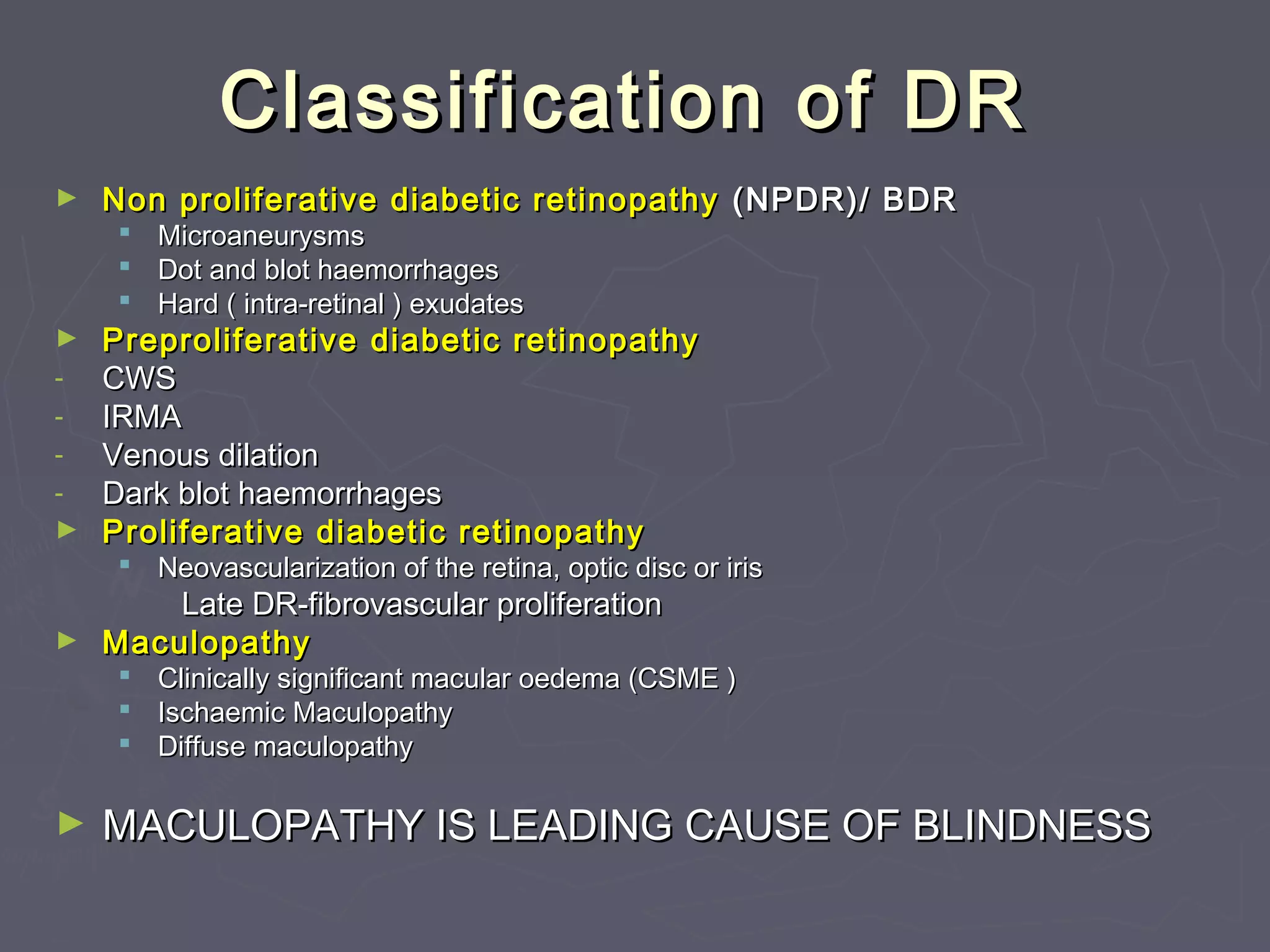
Diabetic RetinopathyDiabetic Retinopathy
St John Eye HospitalSt John Eye Hospital
Diabetic Eye DiseaseDiabetic Eye Disease
ComponentsComponents
►Diabetic Retinopathy
►Cataract
►Refractive changes
►Cranial nerve palsies
►Glaucoma
Diabetic Eye DiseaseDiabetic Eye Disease
significancesignificance
►10% of blind registrations are due to DR
►Commonest cause in 20-65 year old age
group
►Most cases of blindness can be prevented
with implementation of existing technology
Risk factorsRisk factors
►Duration of diabetes is the principle
determinant of the presence of diabetes
related retinopathy
►Poor glycemic control is the most critical risk
factor for the development and progression
of diabetes associated retinopathy
Normal RetinaNormal Retina
Classification of DRClassification of DR
► Non proliferative diabetic retinopathyNon proliferative diabetic retinopathy (NPDR)/ BDR(NPDR)/ BDR
 MicroaneurysmsMicroaneurysms
 Dot and blot haemorrhagesDot and blot haemorrhages
 Hard ( intra-retinal ) exudatesHard ( intra-retinal ) exudates
► PreproliferativePreproliferative diabetic retinopathydiabetic retinopathy
- CWSCWS
- IRMAIRMA
- Venous dilationVenous dilation
- Dark blot haemorrhagesDark blot haemorrhages
► Proliferative diabetic retinopathyProliferative diabetic retinopathy
 Neovascularization of the retina, optic disc or irisNeovascularization of the retina, optic disc or iris
Late DR-fibrovascular proliferationLate DR-fibrovascular proliferation
► MaculopathyMaculopathy
 Clinically significant macular oedema (CSME )Clinically significant macular oedema (CSME )
 Ischaemic MaculopathyIschaemic Maculopathy
 Diffuse maculopathyDiffuse maculopathy
► MACULOPATHY IS LEADING CAUSE OF BLINDNESSMACULOPATHY IS LEADING CAUSE OF BLINDNESS
MicroaneurysmsMicroaneurysms
► Retinal microaneurysms are focal dilatations of retinal capillariesRetinal microaneurysms are focal dilatations of retinal capillaries
► appear as red dots.appear as red dots.
► They are usually seen at the posterior pole, especially temporal to theThey are usually seen at the posterior pole, especially temporal to the
fovea.fovea.
Retinal HaemorrhagesRetinal Haemorrhages
► When the wall of a capillary or microaneurysm is sufficientlyWhen the wall of a capillary or microaneurysm is sufficiently
weakened, it may rupture, giving rise to an intraretinal hemorrhage.weakened, it may rupture, giving rise to an intraretinal hemorrhage.
► If the hemorrhage is deep (i.e., in the inner nuclear layer or outerIf the hemorrhage is deep (i.e., in the inner nuclear layer or outer
plexiform layer), it usually is round or oval ("dot or blot")plexiform layer), it usually is round or oval ("dot or blot")
► Dot haemorrhages appear as bright red dots and are the same size asDot haemorrhages appear as bright red dots and are the same size as
large microaneurysmslarge microaneurysms
► Blot haemorrhages are larger lesions they are located within the midBlot haemorrhages are larger lesions they are located within the mid
retina and often within or surrounding areas of ischaemia.retina and often within or surrounding areas of ischaemia.
Hard exudatesHard exudates
(Intra retinal lipid exudates)(Intra retinal lipid exudates)
 Hard exudates (Intra retinal lipid exudates) are yellowHard exudates (Intra retinal lipid exudates) are yellow
deposits of lipid and protein within the sensory retina.deposits of lipid and protein within the sensory retina.
 Accumulations of lipids leak from surrounding capillariesAccumulations of lipids leak from surrounding capillaries
and microaneurysms, they may form a circinate pattern.and microaneurysms, they may form a circinate pattern.
Cotton Wool SpotsCotton Wool Spots
 Cotton wool spots result from occlusion of retinalCotton wool spots result from occlusion of retinal
pre-capillary arterioles supplying the nerve fiberpre-capillary arterioles supplying the nerve fiber
layer with concomitant swelling of local nerve fiberlayer with concomitant swelling of local nerve fiber
axons.axons.
 Also called "soft exudates" or "nerve fiber layerAlso called "soft exudates" or "nerve fiber layer
infarctions" they are white, fluffy lesions in theinfarctions" they are white, fluffy lesions in the
nerve fiber layer.nerve fiber layer.
Proliferative diabeticProliferative diabetic
retinopathyretinopathy
 Retinal ischaemia due toRetinal ischaemia due to
widespread capillary nonwidespread capillary non
perfusion results in theperfusion results in the
production ofproduction of
vasoproliferative substancesvasoproliferative substances
and to the development ofand to the development of
neovascularizationneovascularization
 Neovascularization canNeovascularization can
involve the retina, optic discinvolve the retina, optic disc
or the iris (or the iris (rubeosis iridis)rubeosis iridis)
Proliferative diabeticProliferative diabetic
retinopathyretinopathy
► Rubeosis iridisRubeosis iridis is a sign of severeis a sign of severe
proliferative disease, it may causeproliferative disease, it may cause
intractable glaucoma.intractable glaucoma.
► Bleeding from fragile new vesselsBleeding from fragile new vessels
involving the retina or optic disc caninvolving the retina or optic disc can
result inresult in vitreous or retinal hemorrhagevitreous or retinal hemorrhage..
Retinal damage can result fromRetinal damage can result from
persistent vitreous hemorrhage.persistent vitreous hemorrhage.
► Pre retinal haemorrhages are oftenPre retinal haemorrhages are often
associated with retinalassociated with retinal
neovascularization, they mayneovascularization, they may
dramatically reduce vision within a fewdramatically reduce vision within a few
minutes.minutes.
Proliferative diabeticProliferative diabetic
retinopathyretinopathy
Late DiseaseLate Disease
 Contraction ofContraction of
associated fibrousassociated fibrous
tissue formed bytissue formed by
proliferative diseaseproliferative disease
tissue can result intissue can result in
deformation of thedeformation of the
retina and tractionalretina and tractional
retinal detachmentretinal detachment
Macular OedemaMacular Oedema
► Macular oedema is an important manifestation ofMacular oedema is an important manifestation of
DR because it is now the leading cause of legalDR because it is now the leading cause of legal
blindness in diabetics.blindness in diabetics.
► The intercellular fluid comes from leakingThe intercellular fluid comes from leaking
microaneurysms or from diffuse capillary leakagemicroaneurysms or from diffuse capillary leakage
Flourescein AngiographyFlourescein Angiography
OCTOCT
►Mainly for macular edemaMainly for macular edema
Good Control of DMGood Control of DM
Laser TherapyLaser Therapy
► LaserLaser
photocoagulationphotocoagulation
causes a retinal burncauses a retinal burn
which is visible onwhich is visible on
fundoscopy.fundoscopy.
► Retinal and optic discRetinal and optic disc
neovascularization canneovascularization can
regress with the use ofregress with the use of
retinal laserretinal laser
photocoagulationphotocoagulation
Panretinal laser photocoagulationPanretinal laser photocoagulation
for proliferative DRfor proliferative DR
► The mainstay of treatment ofThe mainstay of treatment of
diabetic retinopathy is retinaldiabetic retinopathy is retinal
laser photocoagulation.laser photocoagulation.
► Laser therapy is highlyLaser therapy is highly
effective; the rate of severeeffective; the rate of severe
visual loss at 2 years due tovisual loss at 2 years due to
proliferative disease can beproliferative disease can be
reduced by 60%.reduced by 60%.
► Rubeosis iridis requiresRubeosis iridis requires
urgent panretinalurgent panretinal
photocoagulation to preventphotocoagulation to prevent
ocular pain and blindnessocular pain and blindness
from glaucoma.from glaucoma.
Panretinal laser photocoagulationPanretinal laser photocoagulation
Macular laser therapyMacular laser therapy
► The indications for laser therapy now include CSME whichThe indications for laser therapy now include CSME which
is treated with a macular laser grid or treatment of focalis treated with a macular laser grid or treatment of focal
lesions such as microaneurysms.lesions such as microaneurysms.
► Early referral and detection of disease is important asEarly referral and detection of disease is important as
treatment of maculopathy is far more successful iftreatment of maculopathy is far more successful if
undertaken at an early stage of the disease processundertaken at an early stage of the disease process
Anti VEGFAnti VEGF
Vascular endothelial growth factor (VEGF)
► A protein called vascular endothelial growth factor (VEGF)A protein called vascular endothelial growth factor (VEGF)
is one reason why weak abnormal blood vessels may growis one reason why weak abnormal blood vessels may grow
under the retina and lead to vision loss.under the retina and lead to vision loss.
► Anti-VEGF medicines block the effects of VEGF.Anti-VEGF medicines block the effects of VEGF.
► Treatment of macular oedeme, PDRTreatment of macular oedeme, PDR
► Treatment of ARMDTreatment of ARMD
VITRECTOMY IN DIABETICVITRECTOMY IN DIABETIC
PATIENTSPATIENTS
► Vitrectomy, plays a vitalVitrectomy, plays a vital
role in the management ofrole in the management of
severe complications ofsevere complications of
diabetic retinopathy.diabetic retinopathy.
► The major indications areThe major indications are
non-clearing vitreousnon-clearing vitreous
hemorrhage, tractionhemorrhage, traction
retinal detachment, andretinal detachment, and
combined tractioncombined traction
/rhegmatogenous retinal/rhegmatogenous retinal
detachment.detachment.
Thank You

Diabetic retinopathy for medical student

  • 1.
    Diabetic RetinopathyDiabetic Retinopathy StJohn Eye HospitalSt John Eye Hospital
  • 2.
    Diabetic Eye DiseaseDiabeticEye Disease ComponentsComponents ►Diabetic Retinopathy ►Cataract ►Refractive changes ►Cranial nerve palsies ►Glaucoma
  • 3.
    Diabetic Eye DiseaseDiabeticEye Disease significancesignificance ►10% of blind registrations are due to DR ►Commonest cause in 20-65 year old age group ►Most cases of blindness can be prevented with implementation of existing technology
  • 4.
    Risk factorsRisk factors ►Durationof diabetes is the principle determinant of the presence of diabetes related retinopathy ►Poor glycemic control is the most critical risk factor for the development and progression of diabetes associated retinopathy
  • 5.
  • 6.
    Classification of DRClassificationof DR ► Non proliferative diabetic retinopathyNon proliferative diabetic retinopathy (NPDR)/ BDR(NPDR)/ BDR  MicroaneurysmsMicroaneurysms  Dot and blot haemorrhagesDot and blot haemorrhages  Hard ( intra-retinal ) exudatesHard ( intra-retinal ) exudates ► PreproliferativePreproliferative diabetic retinopathydiabetic retinopathy - CWSCWS - IRMAIRMA - Venous dilationVenous dilation - Dark blot haemorrhagesDark blot haemorrhages ► Proliferative diabetic retinopathyProliferative diabetic retinopathy  Neovascularization of the retina, optic disc or irisNeovascularization of the retina, optic disc or iris Late DR-fibrovascular proliferationLate DR-fibrovascular proliferation ► MaculopathyMaculopathy  Clinically significant macular oedema (CSME )Clinically significant macular oedema (CSME )  Ischaemic MaculopathyIschaemic Maculopathy  Diffuse maculopathyDiffuse maculopathy ► MACULOPATHY IS LEADING CAUSE OF BLINDNESSMACULOPATHY IS LEADING CAUSE OF BLINDNESS
  • 7.
    MicroaneurysmsMicroaneurysms ► Retinal microaneurysmsare focal dilatations of retinal capillariesRetinal microaneurysms are focal dilatations of retinal capillaries ► appear as red dots.appear as red dots. ► They are usually seen at the posterior pole, especially temporal to theThey are usually seen at the posterior pole, especially temporal to the fovea.fovea.
  • 8.
    Retinal HaemorrhagesRetinal Haemorrhages ►When the wall of a capillary or microaneurysm is sufficientlyWhen the wall of a capillary or microaneurysm is sufficiently weakened, it may rupture, giving rise to an intraretinal hemorrhage.weakened, it may rupture, giving rise to an intraretinal hemorrhage. ► If the hemorrhage is deep (i.e., in the inner nuclear layer or outerIf the hemorrhage is deep (i.e., in the inner nuclear layer or outer plexiform layer), it usually is round or oval ("dot or blot")plexiform layer), it usually is round or oval ("dot or blot") ► Dot haemorrhages appear as bright red dots and are the same size asDot haemorrhages appear as bright red dots and are the same size as large microaneurysmslarge microaneurysms ► Blot haemorrhages are larger lesions they are located within the midBlot haemorrhages are larger lesions they are located within the mid retina and often within or surrounding areas of ischaemia.retina and often within or surrounding areas of ischaemia.
  • 9.
    Hard exudatesHard exudates (Intraretinal lipid exudates)(Intra retinal lipid exudates)  Hard exudates (Intra retinal lipid exudates) are yellowHard exudates (Intra retinal lipid exudates) are yellow deposits of lipid and protein within the sensory retina.deposits of lipid and protein within the sensory retina.  Accumulations of lipids leak from surrounding capillariesAccumulations of lipids leak from surrounding capillaries and microaneurysms, they may form a circinate pattern.and microaneurysms, they may form a circinate pattern.
  • 10.
    Cotton Wool SpotsCottonWool Spots  Cotton wool spots result from occlusion of retinalCotton wool spots result from occlusion of retinal pre-capillary arterioles supplying the nerve fiberpre-capillary arterioles supplying the nerve fiber layer with concomitant swelling of local nerve fiberlayer with concomitant swelling of local nerve fiber axons.axons.  Also called "soft exudates" or "nerve fiber layerAlso called "soft exudates" or "nerve fiber layer infarctions" they are white, fluffy lesions in theinfarctions" they are white, fluffy lesions in the nerve fiber layer.nerve fiber layer.
  • 11.
    Proliferative diabeticProliferative diabetic retinopathyretinopathy Retinal ischaemia due toRetinal ischaemia due to widespread capillary nonwidespread capillary non perfusion results in theperfusion results in the production ofproduction of vasoproliferative substancesvasoproliferative substances and to the development ofand to the development of neovascularizationneovascularization  Neovascularization canNeovascularization can involve the retina, optic discinvolve the retina, optic disc or the iris (or the iris (rubeosis iridis)rubeosis iridis)
  • 12.
    Proliferative diabeticProliferative diabetic retinopathyretinopathy ►Rubeosis iridisRubeosis iridis is a sign of severeis a sign of severe proliferative disease, it may causeproliferative disease, it may cause intractable glaucoma.intractable glaucoma. ► Bleeding from fragile new vesselsBleeding from fragile new vessels involving the retina or optic disc caninvolving the retina or optic disc can result inresult in vitreous or retinal hemorrhagevitreous or retinal hemorrhage.. Retinal damage can result fromRetinal damage can result from persistent vitreous hemorrhage.persistent vitreous hemorrhage. ► Pre retinal haemorrhages are oftenPre retinal haemorrhages are often associated with retinalassociated with retinal neovascularization, they mayneovascularization, they may dramatically reduce vision within a fewdramatically reduce vision within a few minutes.minutes.
  • 13.
  • 14.
    Late DiseaseLate Disease Contraction ofContraction of associated fibrousassociated fibrous tissue formed bytissue formed by proliferative diseaseproliferative disease tissue can result intissue can result in deformation of thedeformation of the retina and tractionalretina and tractional retinal detachmentretinal detachment
  • 15.
    Macular OedemaMacular Oedema ►Macular oedema is an important manifestation ofMacular oedema is an important manifestation of DR because it is now the leading cause of legalDR because it is now the leading cause of legal blindness in diabetics.blindness in diabetics. ► The intercellular fluid comes from leakingThe intercellular fluid comes from leaking microaneurysms or from diffuse capillary leakagemicroaneurysms or from diffuse capillary leakage
  • 17.
  • 18.
    OCTOCT ►Mainly for macularedemaMainly for macular edema
  • 19.
    Good Control ofDMGood Control of DM
  • 20.
    Laser TherapyLaser Therapy ►LaserLaser photocoagulationphotocoagulation causes a retinal burncauses a retinal burn which is visible onwhich is visible on fundoscopy.fundoscopy. ► Retinal and optic discRetinal and optic disc neovascularization canneovascularization can regress with the use ofregress with the use of retinal laserretinal laser photocoagulationphotocoagulation
  • 21.
    Panretinal laser photocoagulationPanretinallaser photocoagulation for proliferative DRfor proliferative DR ► The mainstay of treatment ofThe mainstay of treatment of diabetic retinopathy is retinaldiabetic retinopathy is retinal laser photocoagulation.laser photocoagulation. ► Laser therapy is highlyLaser therapy is highly effective; the rate of severeeffective; the rate of severe visual loss at 2 years due tovisual loss at 2 years due to proliferative disease can beproliferative disease can be reduced by 60%.reduced by 60%. ► Rubeosis iridis requiresRubeosis iridis requires urgent panretinalurgent panretinal photocoagulation to preventphotocoagulation to prevent ocular pain and blindnessocular pain and blindness from glaucoma.from glaucoma.
  • 22.
  • 23.
    Macular laser therapyMacularlaser therapy ► The indications for laser therapy now include CSME whichThe indications for laser therapy now include CSME which is treated with a macular laser grid or treatment of focalis treated with a macular laser grid or treatment of focal lesions such as microaneurysms.lesions such as microaneurysms. ► Early referral and detection of disease is important asEarly referral and detection of disease is important as treatment of maculopathy is far more successful iftreatment of maculopathy is far more successful if undertaken at an early stage of the disease processundertaken at an early stage of the disease process
  • 24.
    Anti VEGFAnti VEGF Vascularendothelial growth factor (VEGF) ► A protein called vascular endothelial growth factor (VEGF)A protein called vascular endothelial growth factor (VEGF) is one reason why weak abnormal blood vessels may growis one reason why weak abnormal blood vessels may grow under the retina and lead to vision loss.under the retina and lead to vision loss. ► Anti-VEGF medicines block the effects of VEGF.Anti-VEGF medicines block the effects of VEGF. ► Treatment of macular oedeme, PDRTreatment of macular oedeme, PDR ► Treatment of ARMDTreatment of ARMD
  • 25.
    VITRECTOMY IN DIABETICVITRECTOMYIN DIABETIC PATIENTSPATIENTS ► Vitrectomy, plays a vitalVitrectomy, plays a vital role in the management ofrole in the management of severe complications ofsevere complications of diabetic retinopathy.diabetic retinopathy. ► The major indications areThe major indications are non-clearing vitreousnon-clearing vitreous hemorrhage, tractionhemorrhage, traction retinal detachment, andretinal detachment, and combined tractioncombined traction /rhegmatogenous retinal/rhegmatogenous retinal detachment.detachment.
  • 26.