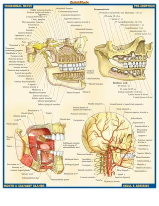
PERMANENT TEETH
MOUTH
PERMANENT TEETH
TOOTH
DECIDUOUS ARCH MARGINAL GINGIVA
** = cut
a. = artery
l.l. = ligaments
m. = muscle
n. = nerve
n.n. = nerves
v. = vein
3rd
molars
3rd
molars
2nd
molars
2nd
molars
1st
molars
1st
molars
2nd
bicuspid
(premolars)
2nd
bicuspid
(premolars)
1st
bicuspid
(premolars)
1st
bicuspid
(premolars)
Cuspids
(canine)
Cuspids
(canine)
Lateral
incisors
Lateral
incisors
Central
incisors
Stria of Retzius
Junctional
epithelium
Neutrophilic
granulocyte
Lymphocyte
Dentinal
tubules
Periodontal
l.l.
Enamel spindle Gingival sulcus
Oral sulcular epithelium
Oral epithelium
Collagen fibers
in cross section
Vessels of the
gingival plexus
Collagen fibers
in long section
Marginal
alveolar
bone
Sharpey’s
fibers
Labial surface Central incisor
Lateral incisor
Cuspid
1st molar
2nd molar
2nd molar
Cingulum
Dental tubercle
Gum
Interproximal
surface
Distal surface
Buccal
surface
Gingival
margin
Mesial surface
Lingual surface
Incisal edge
Cusp
Tomes
granular layer
Acellular
cementum
Central incisor
Facial View
Lateral incisor
Cuspid
1st molar
Central incisor
Lateral incisor
Cuspid
3rd molar
2nd molar
1st molar
2nd bicuspid
1st bicuspid
3rd molar
2nd molar
1st molar
1st bicuspid
(premolar)
Cuspid (canine)
Lateral incisor
Central incisor
Incisal edge
Lingual surface
Gingival papilla
Mesial surface
Gingival margin
Buccal surface
Contingent surface
Cusp
Triangular ridge
Interproximal surface
Distal surface
Gum
Cingulum
Labial surface
2nd bicuspid
(premolar)
Enamel
Crown
Contact area
Dentin
Cusp
Fissure
Tuft
Stria of Retzius Enamel spindle
Enamel prism
Pulp
Pulp chamber
Neck
Gingiva
Dentinal tubules
Root
Interradicular septum
Mandible**
Compact bone
Cancellous bone
Pulp horn
Contour line of Owen
Interdental papilla
Pulpal vessels & n.n.
Dentogingival fibers
Circular fibers
Trans-septal fibers
Cementum
Oblique fibers
of periodontium
Root canal**
Pulpal sensory n.
Apical foramen
Apical fibers
Alveolar n., a. & v.Superior labial frenulum
Gingiva
Palatopharyngeal arch
Posterior
wall of
oropharynx
Gingiva
Inferior labial frenulum Inferior lip
Palatine
tonsil
Uvula
Palatoglossal arch
Soft palate
Palatine raphe
Hard palate
Superior lip
Tongue
Vestibule
Lingual View
Cusp of Carabelli
TRIGEMINAL NERVE PRE-ERUPTION
Permanent teeth:
3rd molar (wisdom tooth) (not illustrated) 17-25 yr.
2nd molar 12-13 yr.
2nd molar 2 yr.
1st molar 6-7 yr.
2nd bicuspid (premolar) 12-13 yr.
1st bicuspid (premolar) 11 yr.
Cuspid (canine) 11 yr.
Pons
Maxillary n. (V2)
Ophthalmic n. (V1)
Tentorial branch
Frontal n.
Lacrimal n.
Nasociliary n.
Pterygopalatine branches
Zygomatic n.
Infraorbital foramen
Communicating branch
Zygomaticotemporal n.
Zygomaticofacial n.
Infraorbital n.
Infraorbital foramen
Anterior superior alveolar n.
Middle superior alveolar n.
Posterior superior alveolar n.
Dental branches
Gingival
branches
Labial branches
Ciliary ganglion
Long & short ciliary n.n.
Trigeminal n. (V)
Mandibular n. (V3)
Trigeminal
(semilunar) ganglion
Medulla oblongata
Auriculotemporal n.
Anterior division
Posterior deep temporal n.
Lateral pterygoid n.
Chorda tympani n.
Buccal n.
Anterior deep temporal n.
Lingual n.
Inferior alveolar n.
Mylohyoid n.
Mandibular foramen
Inferior alveolar n.
Inferior dental branches
Inferior dental plexus
Inferior gingival branches
Mental foramen
Mental branches
Mental n.
Posterior division
Foramen ovale
Canine (eyetooth) 16-20 mo.
1st molar 10-15 mo.
Lateral incisor 8 yr.
Central incisor 7 yr.
Deciduous teeth:
Central incisor 6-8 mo.
Lateral incisor 8-10 mo.
Submandibular glands
Submandibular duct
Salivary pool
Masseter m.**
Buccinator m.**
Parotid
duct
Saliva
Parotid gland
Parotid gland
Uvula**
Palatine glands
Anterior lingual glands
Mylohyoid m.**
Sublingual gland
Tongue
Parotid duct
Buccinator m.**
Sublingual caruncle
with opening of
submandibular duct
Sublingual ducts
Supratrochlear a.
Supraorbital a.
Frontal branch of superficial temporal a.
Deep temporal a.
Angular a.
Anterior superior
alveolar a.
Infraorbital a.
Descending
palatine a.
Maxillary a.
Superior labial a.
Dental branches
Transverse facial a.
Inferior labial a.
Inferior alveolar a.
Mentalis a.
Submentalis a.Deep lingual a.
Lingual a.
Superior thyroid a.
Facial a.
Vertebral a.
Mylohyoid branch
Internal carotid a.
External carotid a.
Ascending
pharyngeal a.
Posterior auricular a.
Occipitalis a.
Posterior superior alveolar a.
Middle temporal a.
Parietal branch of
superficial temporal a.
Infraorbital a.
MOUTH & SALIVARY GLANDS SKULL & ARTERIES

Dental anatomy reference_guide

  • 1.
    PERMANENT TEETH MOUTH PERMANENT TEETH TOOTH DECIDUOUSARCH MARGINAL GINGIVA ** = cut a. = artery l.l. = ligaments m. = muscle n. = nerve n.n. = nerves v. = vein 3rd molars 3rd molars 2nd molars 2nd molars 1st molars 1st molars 2nd bicuspid (premolars) 2nd bicuspid (premolars) 1st bicuspid (premolars) 1st bicuspid (premolars) Cuspids (canine) Cuspids (canine) Lateral incisors Lateral incisors Central incisors Stria of Retzius Junctional epithelium Neutrophilic granulocyte Lymphocyte Dentinal tubules Periodontal l.l. Enamel spindle Gingival sulcus Oral sulcular epithelium Oral epithelium Collagen fibers in cross section Vessels of the gingival plexus Collagen fibers in long section Marginal alveolar bone Sharpey’s fibers Labial surface Central incisor Lateral incisor Cuspid 1st molar 2nd molar 2nd molar Cingulum Dental tubercle Gum Interproximal surface Distal surface Buccal surface Gingival margin Mesial surface Lingual surface Incisal edge Cusp Tomes granular layer Acellular cementum Central incisor Facial View Lateral incisor Cuspid 1st molar Central incisor Lateral incisor Cuspid 3rd molar 2nd molar 1st molar 2nd bicuspid 1st bicuspid 3rd molar 2nd molar 1st molar 1st bicuspid (premolar) Cuspid (canine) Lateral incisor Central incisor Incisal edge Lingual surface Gingival papilla Mesial surface Gingival margin Buccal surface Contingent surface Cusp Triangular ridge Interproximal surface Distal surface Gum Cingulum Labial surface 2nd bicuspid (premolar) Enamel Crown Contact area Dentin Cusp Fissure Tuft Stria of Retzius Enamel spindle Enamel prism Pulp Pulp chamber Neck Gingiva Dentinal tubules Root Interradicular septum Mandible** Compact bone Cancellous bone Pulp horn Contour line of Owen Interdental papilla Pulpal vessels & n.n. Dentogingival fibers Circular fibers Trans-septal fibers Cementum Oblique fibers of periodontium Root canal** Pulpal sensory n. Apical foramen Apical fibers Alveolar n., a. & v.Superior labial frenulum Gingiva Palatopharyngeal arch Posterior wall of oropharynx Gingiva Inferior labial frenulum Inferior lip Palatine tonsil Uvula Palatoglossal arch Soft palate Palatine raphe Hard palate Superior lip Tongue Vestibule Lingual View Cusp of Carabelli
  • 2.
    TRIGEMINAL NERVE PRE-ERUPTION Permanentteeth: 3rd molar (wisdom tooth) (not illustrated) 17-25 yr. 2nd molar 12-13 yr. 2nd molar 2 yr. 1st molar 6-7 yr. 2nd bicuspid (premolar) 12-13 yr. 1st bicuspid (premolar) 11 yr. Cuspid (canine) 11 yr. Pons Maxillary n. (V2) Ophthalmic n. (V1) Tentorial branch Frontal n. Lacrimal n. Nasociliary n. Pterygopalatine branches Zygomatic n. Infraorbital foramen Communicating branch Zygomaticotemporal n. Zygomaticofacial n. Infraorbital n. Infraorbital foramen Anterior superior alveolar n. Middle superior alveolar n. Posterior superior alveolar n. Dental branches Gingival branches Labial branches Ciliary ganglion Long & short ciliary n.n. Trigeminal n. (V) Mandibular n. (V3) Trigeminal (semilunar) ganglion Medulla oblongata Auriculotemporal n. Anterior division Posterior deep temporal n. Lateral pterygoid n. Chorda tympani n. Buccal n. Anterior deep temporal n. Lingual n. Inferior alveolar n. Mylohyoid n. Mandibular foramen Inferior alveolar n. Inferior dental branches Inferior dental plexus Inferior gingival branches Mental foramen Mental branches Mental n. Posterior division Foramen ovale Canine (eyetooth) 16-20 mo. 1st molar 10-15 mo. Lateral incisor 8 yr. Central incisor 7 yr. Deciduous teeth: Central incisor 6-8 mo. Lateral incisor 8-10 mo. Submandibular glands Submandibular duct Salivary pool Masseter m.** Buccinator m.** Parotid duct Saliva Parotid gland Parotid gland Uvula** Palatine glands Anterior lingual glands Mylohyoid m.** Sublingual gland Tongue Parotid duct Buccinator m.** Sublingual caruncle with opening of submandibular duct Sublingual ducts Supratrochlear a. Supraorbital a. Frontal branch of superficial temporal a. Deep temporal a. Angular a. Anterior superior alveolar a. Infraorbital a. Descending palatine a. Maxillary a. Superior labial a. Dental branches Transverse facial a. Inferior labial a. Inferior alveolar a. Mentalis a. Submentalis a.Deep lingual a. Lingual a. Superior thyroid a. Facial a. Vertebral a. Mylohyoid branch Internal carotid a. External carotid a. Ascending pharyngeal a. Posterior auricular a. Occipitalis a. Posterior superior alveolar a. Middle temporal a. Parietal branch of superficial temporal a. Infraorbital a. MOUTH & SALIVARY GLANDS SKULL & ARTERIES