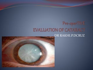
Pre operative analysis for cataract surgery
•Download as PPTX, PDF•
50 likes•13,962 views

Pre operative analysis for cataract surgery

Recommended
More Related Content
What's hot
What's hot (20)
Similar to Pre operative analysis for cataract surgery
Similar to Pre operative analysis for cataract surgery (20)
More from Dr Rakhi Dcruz
Recently uploaded
Recently uploaded (20)
Pre operative analysis for cataract surgery
- 2. PRE-OPERATIVE ASSESSMENT OCULAR HISTORY SYSTEMIC HISTORY OCULAR EXAMINATION OCULAR INVESTIGATIONS LABOROTARY INVESTIGATIONS
- 3. OCULAR HISTORY Ocular Symptoms Like Blurring Of Vision,coloured Halos,diplopia,glare Nuclear Cataract-distant> Near Posterior Subcapsular-near>distant Duration And Progression H/O Of Previous Intraocular Disease H/O Previous Cataract Surgery H/O Intraocular Injuries
- 4. SYSTEMIC HISTORY H/o of all systemic diseases,current medications patient is on.. Medications relevant to eye surgery Systemic alpha blockers(tamsulosin)-floppy iris syndrome Antiplatelets and anticoagulants Antihypertensives like diuretics-electrolyte imbalances Long term steroids-delayed healing Drug allergy to sulfonamides and other antibiotics
- 7. OCULAR EXAMINATION Visual acuity-distant and near vision Head posture & ocular posture-cover-uncover test Ocular movements Ocular adnexa- blepharitis,ectropion,entropion,lagophthalmos Lacrimal sac syrininging to r/o dacrocystitis Conjuctiva-congestion,scarring,symblepharon
- 8. CORNEA-stromal opacity and prominent arcus senilis Decreased endothelial count as in corneal guttata-post op decompensation Specular microscopy and pachymetry to assess the risk and to take precaution If abnormal or C- thickness > 600 µm is poor prognosis for corneal clarity. Endothelial deposits and keratic precipitates-uveitis or glaucoma First treat the intraocular diseases..symptm free period of 4-6 months for uvietis,steroids to prevent relapse
- 9. ANTERIOR CHAMBER-Shallow (intumescent of lens or forward displacement by posterior pathology) . Gonioscopy to rule out the angle abnormalities (synechia, neovascularization). PUPIL- Reacting promptly to light-both direst and consensual Presence of RAPD-Implies substantial additional pathology Readily dilating with mydriatics
- 10. lens Size of lens nucleus and grading of nuclear sclerosis- for planning size of incision and type of surgery Nuclear cataract are harder and need more power with phaco Black nuclear opacity-extremely dense-ECCE Postr polar cataract-prevent posterior capsular dehiscence and subsequent vitreous disturbances- avoid HYDRODISSECTION
- 11. ZONULAR APPARATUS –examine under mydriasis. Pseudoexfoliation weak zonule, fragile capsule & poor mydriasis SCLERA-prominent explant/encircling band for prior RD OR Eye is particularly large/sclera is thin-peri an retrobulbar local anesthesia should be avoided
- 12. FUNDUS EXAMINATION A thorough fundus examination is important. Retinal and optic nerve function must be assessed pre- op,Because if it is defective operation becomes valueless. Pathology such as ARMD,RETINAL DETACHMENT Can adversely affect visual outcome..hence a thorough fundus evaluation is important. In eyes with very dense opacity,when fundus cannot be seen 5 tests are of value
- 13. 1.PROJECTION OF LIGHT 2.2 POINT LIGHT DISCRIMINATION 3.MADDOX ROD 4.ENTOPIC VIEW OF RETINA 5.USG B SCAN-r/o vitreous haemorrage,retinal detachment,intraocular tumour & posterior staphyloma. Foveal ERG
- 14. INTRAOCULAR PRESSURE Can be raised due to swellin of lens in INCIPIENT STAGE/due to phacolytic glaucomain which case extraction is indicated. Primary glaucoma can be pre-excistent If galucoma glaucome medically controlled-lens extraction If NOT,perform a trabeculectomy followed by cataract extracion/combined procedure.
- 15. REFRACTIVE ERROR Its critical to obtain patients pre-operative refractive status in order to guide IOL implant selection. BIOMETRY facilitate calculation of lens power likely to result in desired post op refractory outcome. It involves 1.Keratometry 2.A SCAN AXIAL LENGTH-curvature of anterior corneal surface calculation by interferometry apparatus.
- 16. Use SRK formula (Sanders, Retlaff & Kraff) P = A – 2.5L – 0.9K P : Lens implant power for emmetropia (D) L : Axial length (mm) K : Average keratometric reading (D) A : Constant specific to the lens implant to be used That A = 113 for AC lenses & 119 for PC lenses. many other formulas like HAIGIS,HOFFER,HOLLADAY etc are also used.
- 17. CORNEAL PACHYMETRY * Ultrasonic pachymeters can accurately & reliably measure endothelial cell function. * If thickness > 600 µm maybe consistent with corneal edema & endothelium dysfunction that increase the likelihood postoperative clinical corneal edema.
- 18. SPECULAR MICROSCOPY: (endothelium cells) * A normal cell count > 2400 cells/mm2 * If a cell count fewer than 1000 cells/mm2 is risk of postoperative corneal decompensation.
- 19. After examination,we need to assess potential visual function after cataract removal. 1.GUYTON MINKOUSKI POTENTIAL ACUITY METER LOTMAR, RODENSTOCK TYPE LASER INTERFEROMETER(uses coherent white light/helium/neon laser generated interface stripes /fringes)
- 21. GLARE DISABILITY TESTING Brightness acuity tester or Miller nadler glare testing device Simple alternative is snellens chart kept indoor and outdoor in sunlight or a penlight shining obliquely towards pupil.
- 24. Contrast sensitivity Contrast sensitivity drops with cataract Wallmount charts for testing (PELLI ROBSON ,TERRY CHARTS) VEP(VISUALY EVOKED POTENTIAL) is more specific and it require intact macula and optic nerve besides cortical centre.
- 26. INFORMED WRITTEN CONSENT Patient should give full informed written consent before catarcat surgery. 1 in 1000-achieves very little or no sight 1 in 10000-lose eye completely Mild complications-periocular echymosis,raised IOP,mild iridocyclitis,wound leak. Moderate-posterior capsular rupture,zonular dehiscence,corneal decompensation,CME,RD(1%) SEVERE- ENDOPTHALMITIS(0.1%)SUPRACHOROIDAL HGE
- 27. LAB INVESTIGATIONS NORMAL-RBS,ECG,SCREENING,BP,XTD XRAY,URINE R/E,BRE,RFT,APTT,PT INR-in patients with individual risk factors or planned for general aneasthesia, Preop-antibiotic eye drops QID-3 DAYS PRIOR TO SX. ANTIANXIETY DRUGS if pt apprehensive Preparing eye-cutting lashes Asked to take a normal meal,normal sleep,normal bath,continue systemic medications .
- 28. Thank you...