Upper Extremity Trauma Shoulder
•Download as PPSX, PDF•
1 like•96 views

lecture done by Prof. Ken Schreibman, PhD/MD Professor of Radiology for more radiology lecture plez visit : https://radiologymadeeasy.com/

Recommended
Recommended
More Related Content
What's hot
What's hot (20)
Similar to Upper Extremity Trauma Shoulder
Similar to Upper Extremity Trauma Shoulder (20)
More from Radiology Made Easy
Recently uploaded
Recently uploaded (20)
Upper Extremity Trauma Shoulder
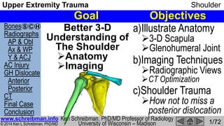
- 1. Bones Radiographs AP & Obl Ax & WP Y & ACJ AC Injury GH Dislocate Anterior Posterior CT Final Case Conclusion © 2014 Ken L Schreibman, PhD/MD www.schreibman.info Upper Extremity Trauma Shoulder 1/72 S C H Goal Objectives Better 3-D Understanding of The Shoulder Anatomy Imaging a)Illustrate Anatomy 3-D Scapula Glenohumeral Joint b)Imaging Techniques Radiographic Views CT Optimization c)Shoulder Trauma How not to miss a posterior dislocation www.schreibman.info Ken Schreibman, PhD/MD Professor of Radiology University of Wisconsin – Madison
- 2. Bones Radiographs AP & Obl Ax & WP Y & ACJ AC Injury GH Dislocate Anterior Posterior CT Final Case Conclusion © 2014 Ken L Schreibman, PhD/MD www.schreibman.info Upper Extremity Trauma Shoulder 2/72 S C H Shoulder: 3 Bones Scapula
- 3. Bones Radiographs AP & Obl Ax & WP Y & ACJ AC Injury GH Dislocate Anterior Posterior CT Final Case Conclusion © 2014 Ken L Schreibman, PhD/MD www.schreibman.info Upper Extremity Trauma Shoulder 3/72 S C H “Flat Bone” Scapula Skull Pelvis Sternum Ribs As opposed to “Long Bones” Humerus Clavicle … Scapula has complex 3-D anatomy Helps to look at it from multiple views Scapula: Anterior ViewScapula:
- 4. Bones Radiographs AP & Obl Ax & WP Y & ACJ AC Injury GH Dislocate Anterior Posterior CT Final Case Conclusion © 2014 Ken L Schreibman, PhD/MD www.schreibman.info Upper Extremity Trauma Shoulder 4/72 S C H Body of Scapula Parts: Glenoid Shallow Socket Dislocates… Body Triangular 3 Margins 2 Angles Scapula: AnteriorScapula: Anterior Medial View
- 5. Bones Radiographs AP & Obl Ax & WP Y & ACJ AC Injury GH Dislocate Anterior Posterior CT Final Case Conclusion © 2014 Ken L Schreibman, PhD/MD www.schreibman.info Upper Extremity Trauma Shoulder 5/72 S C H Scapula: Anterior ViewMedial View Parts: Body Razor Thin “Shoulder Blade” No articular surfaces Origin of all 4 Rotator Cuff (RC) Muscles Teres Scapula: Medial View Body of Scapula Anterior View
- 6. Bones Radiographs AP & Obl Ax & WP Y & ACJ AC Injury GH Dislocate Anterior Posterior CT Final Case Conclusion © 2014 Ken L Schreibman, PhD/MD www.schreibman.info Upper Extremity Trauma Shoulder 6/72 S C H Scapula: Medial View Parts: Body Spine ANTERIOR POSTERIOR
- 7. Bones Radiographs AP & Obl Ax & WP Y & ACJ AC Injury GH Dislocate Anterior Posterior CT Final Case Conclusion © 2014 Ken L Schreibman, PhD/MD www.schreibman.info Upper Extremity Trauma Shoulder 7/72 S C H Scapula: Posterior Medial View Parts: Body Spine Posterior Structure Off back of Body Defines RC muscles Supraspinatus Above the spine Infraspinatus Below the spine
- 8. Bones Radiographs AP & Obl Ax & WP Y & ACJ AC Injury GH Dislocate Anterior Posterior CT Final Case Conclusion © 2014 Ken L Schreibman, PhD/MD www.schreibman.info Upper Extremity Trauma Shoulder 8/72 S C H Parts: Body Spine Posterior Structure Off back of Body Origin of RC Muscles Supra- spinatus Infra- spinatus Scapula: Posterior Medial ViewScapula: Posterior View
- 9. Bones Radiographs AP & Obl Ax & WP Y & ACJ AC Injury GH Dislocate Anterior Posterior CT Final Case Conclusion © 2014 Ken L Schreibman, PhD/MD www.schreibman.info Upper Extremity Trauma Shoulder 9/72 S C H Scapula: Lateral View “Y-view” 3 Limbs: Body Inferior Spine Posterior Coracoid Anterior Glenoid Shallow Socket ANTPOST Coracoid not Coronoid Coronoid is in Elbow Coronoid from Elbow Imaging ASRT@RSNA 2013
- 10. Bones Radiographs AP & Obl Ax & WP Y & ACJ AC Injury GH Dislocate Anterior Posterior CT Final Case Conclusion © 2014 Ken L Schreibman, PhD/MD www.schreibman.info Upper Extremity Trauma Shoulder 10/72 S C H Scapula: Anterior Lateral View ANTPOSTLateral “Y” View Coracoid Most anterior part of the scapula Arises from anterior glenoid Acromion Arises from posterior spine Points anterior
- 11. Bones Radiographs AP & Obl Ax & WP Y & ACJ AC Injury GH Dislocate Anterior Posterior CT Final Case Conclusion © 2014 Ken L Schreibman, PhD/MD www.schreibman.info Upper Extremity Trauma Shoulder 11/72 S C H Scapula: Anterior ViewClavicle: A Cor US Bill of Rights “Long Bone”, but not a straight bone elongated-S elongated-f Integral Symbol fastener of the shoulder[L] “collarbone”, “key” etymonline.com
- 12. Bones Radiographs AP & Obl Ax & WP Y & ACJ AC Injury GH Dislocate Anterior Posterior CT Final Case Conclusion © 2014 Ken L Schreibman, PhD/MD www.schreibman.info Upper Extremity Trauma Shoulder 12/72 S C H Clavicle: 2 JointsClavicle: Acromial-Clavicular Joint (Lateral end) Sterno-Clavicular Joint (Medial end) 0 20 40 60 80 100 120 140 13-20 20-29 30-39 40-49 50-59 60-69 70-79 >80 per100,000p-y ‡Robinson. J Bone Joint Surg [Br] 1998;80-B:476-84 Female Male Clavicle Fracture Incidence Rate‡ Only bone connects scapula to the thorax Shoulder has wide ROM The most fractured bone? 3-10% of all fractures‡ 35% of shoulder injuries† Males<20yo †J Bone Joint Surg [Am] 2009;91:447-60 fastener of the shoulder
- 13. Bones Radiographs AP & Obl Ax & WP Y & ACJ AC Injury GH Dislocate Anterior Posterior CT Final Case Conclusion © 2014 Ken L Schreibman, PhD/MD www.schreibman.info Upper Extremity Trauma Shoulder 13/72 S C H Humerus:Humerus: 1 Head, 2 Necks Anatomic Neck Google Conjoined lambs Lateral view Surgical Neck Proximal Humerus Anterior view MOST common site for proximal humeral fractures This is the neck of interest to the Surgeons LEAST common site for proximal humeral fractures Articular Head Greater Tubercle Lesser Tubercle Hemi- sphere
- 14. Bones Radiographs AP & Obl Ax & WP Y & ACJ AC Injury GH Dislocate Anterior Posterior CT Final Case Conclusion © 2014 Ken L Schreibman, PhD/MD www.schreibman.info Upper Extremity Trauma Shoulder 14/72 S C H Neer Classification Proximal Humerus Fractures Neer CS. Displaced proximal humeral fractures. I. Classification and evaluation. J Bone Joint Surg Am. 1970;52 (6): 1077-89 F,T 56yoM Count Parts (displaced>1cm, angled>45°) not fragments Surgical Neck Proximal fragment Distal fragment D,P 52yoF 1-Part Fx Displaced<1cm Angled<45° Surgical Neck Greater Tubercle B,P 85yoF 2-Part Fx Angled > 45° Surgical Neck B,P 65yoM 3-Part Fx SN: Displaced > 1cm GT: Displaced > 1cm 1-Part Fx Displaced<1cm Angled<45° Surgical Neck GT1 2 3 1 2 3 ?
- 15. Bones Radiographs AP & Obl Ax & WP Y & ACJ AC Injury GH Dislocate Anterior Posterior CT Final Case Conclusion © 2014 Ken L Schreibman, PhD/MD www.schreibman.info Upper Extremity Trauma Shoulder 15/72 S C H Humerus: External vs Internal Rotation GT External Rotation Profiles GT LT GT Humerus Neutral Internal Rotation GT en face GTLT LT GT profiled GT en face Looks like Ice cream cone I = Ice cream I = Internal rotation Animated GIF S,D 32yoF
- 16. Bones Radiographs AP & Obl Ax & WP Y & ACJ AC Injury GH Dislocate Anterior Posterior CT Final Case Conclusion © 2014 Ken L Schreibman, PhD/MD www.schreibman.info Upper Extremity Trauma Shoulder 16/72 S C H Shoulder: 3 Bones We need to examine BOTH joints on ALL radiographic views Shoulder: 3 Bones & 2 Joints Acromial-Clavicular Joint AC Joint “Gliding Joint” Superior view Anterior view Gleno- Humeral Joint Inferior view Axillary view Anterior view Acro- mion A PGH Joint is ~40° oblique to AP GH Joint = Ball-in-Socket Humeral Head = ½ Ball Glenoid = Shallow Socket
- 17. Bones Radiographs AP & Obl Ax & WP Y & ACJ AC Injury GH Dislocate Anterior Posterior CT Final Case Conclusion © 2014 Ken L Schreibman, PhD/MD www.schreibman.info Upper Extremity Trauma Shoulder 17/72 S C H Radiographs: AP view Shows alignment of AC Joint Shows lateral view of Surgical Neck Does not profile GH Joint nor GT Marty 12yoM X-Rays AP Humerus Int. Rotated K,C 17yoM Anterior view AP view
- 18. Bones Radiographs AP & Obl Ax & WP Y & ACJ AC Injury GH Dislocate Anterior Posterior CT Final Case Conclusion © 2014 Ken L Schreibman, PhD/MD www.schreibman.info Upper Extremity Trauma Shoulder 18/72 S C H Radiographs: Oblique Shows alignment of AC Joint Shows AP view of Surgical Neck Does profile GH Joint and GT Humerus Ext. Rotated Grashey* viewAnterior Medial view K,C 17yoM *1905 Atlas typischer Röntgenbilder vom normalen Menschen p.38-39 [books.google.com] GHJ GT
- 19. Bones Radiographs AP & Obl Ax & WP Y & ACJ AC Injury GH Dislocate Anterior Posterior CT Final Case Conclusion © 2014 Ken L Schreibman, PhD/MD www.schreibman.info Upper Extremity Trauma Shoulder 19/72 S C H Radiographs: Technical Points Both AP & Oblique: 1)Shot Standing 2)Shield Genitals 3)Boomerang Filter Allows good exposure of GHJ without overexposure of ACJ Less stuff to penetrate here Than here
- 20. Bones Radiographs AP & Obl Ax & WP Y & ACJ AC Injury GH Dislocate Anterior Posterior CT Final Case Conclusion © 2014 Ken L Schreibman, PhD/MD www.schreibman.info Upper Extremity Trauma Shoulder 20/72 S C H Boomerang Filter C,L 42yoF A,T 30yoM Sometimes we can see the filter on radiographs Usually we see only the internal radiopaque tracer chain~$300 coneinstruments.com Well exposed ACJ Well exposed GHJ
- 21. Bones Radiographs AP & Obl Ax & WP Y & ACJ AC Injury GH Dislocate Anterior Posterior CT Final Case Conclusion © 2014 Ken L Schreibman, PhD/MD www.schreibman.info Upper Extremity Trauma Shoulder 21/72 S C H Boomerang Filter C,A 80yoF Technologist forget to use boomerang filter ACJ way over-exposed Repeated with boomerang filter in place ACJ now well-exposed
- 22. Bones Radiographs AP & Obl Ax & WP Y & ACJ AC Injury GH Dislocate Anterior Posterior CT Final Case Conclusion © 2014 Ken L Schreibman, PhD/MD www.schreibman.info Upper Extremity Trauma Shoulder 22/72 S C H Radiographs: Need Orthogonal Views ┼ AP/Obl = Orthogonal views of humerus Internal/External rotation of humerus AP/Obl ≠ Orthogonal views of GH joint AP doesn’t even profile glenohumeral joint The 3 Orthogonal views to the GHJ are: Oblique “Grashey” Axillary (supine) West Point (prone)
- 23. Bones Radiographs AP & Obl Ax & WP Y & ACJ AC Injury GH Dislocate Anterior Posterior CT Final Case Conclusion © 2014 Ken L Schreibman, PhD/MD www.schreibman.info Upper Extremity Trauma Shoulder 23/72 S C H Radiographs: Axillary view (supine) F,K 16yoF X-Rays Coracoid (anterior) ACJ Articular surface humeral head Articular surface glenoid Profiles glenohumeral joint Width Arthritis Alignment Dislocations
- 24. Bones Radiographs AP & Obl Ax & WP Y & ACJ AC Injury GH Dislocate Anterior Posterior CT Final Case Conclusion © 2014 Ken L Schreibman, PhD/MD www.schreibman.info Upper Extremity Trauma Shoulder 24/72 S C H F,K 16yoF Radiographs: West Point view (prone) Profiles glenohumeral joint Width Arthritis Alignment Dislocations X-Rays Coracoid (anterior) ACJ Articular surface humeral head Articular surface glenoid
- 25. Bones Radiographs AP & Obl Ax & WP Y & ACJ AC Injury GH Dislocate Anterior Posterior CT Final Case Conclusion © 2014 Ken L Schreibman, PhD/MD www.schreibman.info Upper Extremity Trauma Shoulder 25/72 S C H Radiographs: West Point vs Axillary S,T 39yoM recurrent anterior shoulder dislocations AP view (upright) QA: No filter Anterior Glenoid 3 metal Mitek suture anchors in Anterior Glenoid Axillary view (supine) Coracoid (anterior) West Point view (prone) Coracoid (anterior)Bankart Reconstruction WestPointview (prone) Coracoid (anterior) Both well show GHJ width/alignment Ax: Anterior glenoid overlaps clavicle WP: Well shows anterior glenoid Parallelism Parallelism
- 26. Bones Radiographs AP & Obl Ax & WP Y & ACJ AC Injury GH Dislocate Anterior Posterior CT Final Case Conclusion © 2014 Ken L Schreibman, PhD/MD www.schreibman.info Upper Extremity Trauma Shoulder 26/72 S C H Radiographs: UW 3-view series Standard (trauma, pain) 1)AP 2)Oblique 3)Axillary Instability (3-views Glenohumeral Joint) 1)Oblique 2)Axillary 3)West Point If need orthogonal views Scapula,ACJ 4)Lateral (“Scapular Y”, “Arch”/“Outlet”) 2-views of: ACJ Proximal Humerus 2-views of: Glenohumeral Joint
- 27. Bones Radiographs AP & Obl Ax & WP Y & ACJ AC Injury GH Dislocate Anterior Posterior CT Final Case Conclusion © 2014 Ken L Schreibman, PhD/MD www.schreibman.info Upper Extremity Trauma Shoulder 27/72 S C H Radiographs: Scapular Y view (PA) Orthog.view: Scapula Body Coracoid Spine Acromion AC Joint For discussion of Y vs Arch views: F,K 56yoM PA Merrill's Atlas of Radiographic Positioning and Procedures 12 Ed. Vol. 1 ©2012 [Print Replica] [Kindle Edition] Body Body Cor Cor ACJ ACJ
- 28. Bones Radiographs AP & Obl Ax & WP Y & ACJ AC Injury GH Dislocate Anterior Posterior CT Final Case Conclusion © 2014 Ken L Schreibman, PhD/MD www.schreibman.info Upper Extremity Trauma Shoulder 28/72 S C H Radiographs: AC Joints (Bilateral) Anatomic alignment of Acromia with lateral ends of Clavicles Marty 18yoM W,E 26yoF Without weights This is what we do at UW Merrill's Atlas of Radiographic Positioning and Procedures 12 Ed. Vol. 1 ©2012 [Print Replica] [Kindle Edition] p.209
- 29. Bones Radiographs AP & Obl Ax & WP Y & ACJ AC Injury GH Dislocate Anterior Posterior CT Final Case Conclusion © 2014 Ken L Schreibman, PhD/MD www.schreibman.info Upper Extremity Trauma Shoulder 29/72 S C H Radiographs: AC Joints (Bilateral) Merrill's Atlas of Radiographic Positioning and Procedures 12 Ed. Vol. 1 ©2012 [Print Replica] [Kindle Edition] Fig. 5-55 With weights We don’t use weights at UW “Weights should be attached to wrists as shown and not held in hands.”
- 30. Bones Radiographs AP & Obl Ax & WP Y & ACJ AC Injury GH Dislocate Anterior Posterior CT Final Case Conclusion © 2014 Ken L Schreibman, PhD/MD www.schreibman.info Upper Extremity Trauma Shoulder 30/72 S C H Shoulder: 3 BonesShoulder: 3 Bones & 2 Joints Scapula Gleno- Humeral Joint Acromial-Clavicular Joint Acromion Coracoid Rockwood & Green’s Fractures in Adults 8th Ed. ©2015 [Kindle Edition] Chapter 41 2 Ligaments attach clavicle to scapula: AC Lig Acromio-Clavicular CC Lig Coraco-Clavicular AC injury types based on: Which ligaments are torn Degree/direction of A-C displacement
- 31. Bones Radiographs AP & Obl Ax & WP Y & ACJ AC Injury GH Dislocate Anterior Posterior CT Final Case Conclusion © 2014 Ken L Schreibman, PhD/MD www.schreibman.info Upper Extremity Trauma Shoulder 31/72 S C H Acromioclavicular Injury: Type 1 AC Lig: Sprain (intact) CC Lig: Intact ACJ: Aligned Radiographs: Normal Rockwood & Green’s Fractures in Adults 8th Ed. ©2015 [Kindle Edition] Fig 41-2 AC injuries most commonly occur in males <30 related to contact sports Galen (120-199AD) diagnosed his own AC dislocation received from wrestling. Treated himself with tight bandages to hold clavicle down, keeping arm elevated. He abandoned the treatment after only a few days as it was so uncomfortable. (Low compliance rate of shoulder bracing)
- 32. Bones Radiographs AP & Obl Ax & WP Y & ACJ AC Injury GH Dislocate Anterior Posterior CT Final Case Conclusion © 2014 Ken L Schreibman, PhD/MD www.schreibman.info Upper Extremity Trauma Shoulder 32/72 S C H Acromioclavicular Injury: Type 2 AC Lig: Torn CC Lig: Intact ACJ: Subluxated While clavicle appears displaced up, it’s the scapula that’s displaced down D,V 21yoM Rockwood & Green’s Fractures in Adults 8th Ed. ©2015 [Kindle Edition] Loc 60286 AsymptomaticSymptomatic
- 33. Bones Radiographs AP & Obl Ax & WP Y & ACJ AC Injury GH Dislocate Anterior Posterior CT Final Case Conclusion © 2014 Ken L Schreibman, PhD/MD www.schreibman.info Upper Extremity Trauma Shoulder 33/72 S C H Acromioclavicular Injury: Type 3 AC Lig: Torn CC Lig: Torn ACJ: Dislocated C↔C distance is increased less than twice the normal distance H,C 30yoF AsymptomaticSymptomatic 10mm18mm
- 34. Bones Radiographs AP & Obl Ax & WP Y & ACJ AC Injury GH Dislocate Anterior Posterior CT Final Case Conclusion © 2014 Ken L Schreibman, PhD/MD www.schreibman.info Upper Extremity Trauma Shoulder 34/72 S C H Acromioclavicular Injury: Type 5 AC Lig: Torn CC Lig: Torn ACJ: Very Dislocated C↔C distance is increased more than twice the normal distance L,L 42yoM AsymptomaticSymptomatic Deltoid/Trapezius muscles are torn from clavicle 14mm32mm
- 35. Bones Radiographs AP & Obl Ax & WP Y & ACJ AC Injury GH Dislocate Anterior Posterior CT Final Case Conclusion © 2014 Ken L Schreibman, PhD/MD www.schreibman.info Upper Extremity Trauma Shoulder 35/72 S C H Acromioclavicular Injury: Type 4 AC Lig: Torn CC Lig: Torn Clavicle Posterior to Acromion Hard to see on AP view Need to look at Axillary view AP view W,N 29yoF Axillary view Courtesy of Michael Tuite, MD Vice Chair, UW Radiology Dept. This type is very rare Subtle overlap of Acromion on Clavicle Cor (Ant) C A Clavicle POSTERIOR to Acromion
- 36. Bones Radiographs AP & Obl Ax & WP Y & ACJ AC Injury GH Dislocate Anterior Posterior CT Final Case Conclusion © 2014 Ken L Schreibman, PhD/MD www.schreibman.info Upper Extremity Trauma Shoulder 36/72 S C H Acromioclavicular Injury: Type 6 Rockwood & Green’s Fractures in Adults 8th Ed. ©2015 [Kindle Edition] Fig 41-14 This type is so rare I’ve never seen one
- 37. Bones Radiographs AP & Obl Ax & WP Y & ACJ AC Injury GH Dislocate Anterior Posterior CT Final Case Conclusion © 2014 Ken L Schreibman, PhD/MD www.schreibman.info Upper Extremity Trauma Shoulder 37/72 S C H Shoulder: 3 Bones Acromio-Clavicular Joint Scapula Gleno- Humeral Joint GHJ: Shallow Socket Glenoid covers only ~25% humeral head1 360° range of motion Most dislocated joint 45% of ALL dislocations2 ~70,000/year in US3 Rockwood & Green [Kindle Edition] 1Loc 57671 2Loc57623 Shoulder: 3 Bones & 2 Joints 0 20 40 60 80 100 0-9 10-19 20-29 30-39 40-49 50-59 60-69 70-79 80-89 >90 per100,000p-y Male Female Bimodal distribution ♂<30: Sports ♀>80: Falls at home Shoulder Dislocation Incidence Rate 3Zacchilli: J Bone Joint Surg Am, 2010;92(3):542-549
- 38. Bones Radiographs AP & Obl Ax & WP Y & ACJ AC Injury GH Dislocate Anterior Posterior CT Final Case Conclusion © 2014 Ken L Schreibman, PhD/MD www.schreibman.info Upper Extremity Trauma Shoulder 38/72 S C H Shoulder Range of Motion Nature v498 483-6 27June2013
- 39. Bones Radiographs AP & Obl Ax & WP Y & ACJ AC Injury GH Dislocate Anterior Posterior CT Final Case Conclusion © 2014 Ken L Schreibman, PhD/MD www.schreibman.info Upper Extremity Trauma Shoulder 39/72 S C H Scapula: Lateral View (Y-view)Gleno-Humeral Dislocations ANTPOST Glenoid Shallow Socket 3 Limbs: Body Inferior Coracoid Anterior Spine Posterior Posterior Dislocations Straight Posterior Hard to see! 3% of dislocations Missed 50%! Inferior Dislocation < ½% of dislocations “Luxatio Erecta” Clinically obvious Anterior Dislocations Anterior-Inferior Easy to see! 97% of dislocations Krøner. The epidemiology of shoulder dislocations. Arch Orthop Trauma Surg. 1989;108:288-90.
- 40. Bones Radiographs AP & Obl Ax & WP Y & ACJ AC Injury GH Dislocate Anterior Posterior CT Final Case Conclusion © 2014 Ken L Schreibman, PhD/MD www.schreibman.info Upper Extremity Trauma Shoulder 40/72 S C H GH Dislocations: Inferior B,T 40yoM 1 hour later after reduction in ED Luxatio Erecta [L] “Dislocation” “Upright” Humerus stuck in abduction “Hands up dislocation”
- 41. Bones Radiographs AP & Obl Ax & WP Y & ACJ AC Injury GH Dislocate Anterior Posterior CT Final Case Conclusion © 2014 Ken L Schreibman, PhD/MD www.schreibman.info Upper Extremity Trauma Shoulder 41/72 S C H GH Dislocations: Anterior S,T 39yoM recurrent anterior shoulder dislocations AP view Axillary viewOblique view Y view AP view Axillary viewOblique view Y view Normal Overlap Normal Overlap Parallelism Parallelism Abnormal Overlap Lack of Parallelism Lack of Parallelism Humerus Anterior to Glenoid Humerus Anterior to Glenoid H G L O C A T E D D I S L O C A T E D
- 42. Bones Radiographs AP & Obl Ax & WP Y & ACJ AC Injury GH Dislocate Anterior Posterior CT Final Case Conclusion © 2014 Ken L Schreibman, PhD/MD www.schreibman.info Upper Extremity Trauma Shoulder 42/72 S C H GH Dislocations: AnteriorGH Dislocations: Subcoracoid Humerus Anterior-Inferior to Glenoid Easy to see on all radiographic views ⅔ Anterior Dislocations are subcoracoid M,D 37yoM H Cor AP view Oblique view Y view H Cor H Cor G
- 43. Bones Radiographs AP & Obl Ax & WP Y & ACJ AC Injury GH Dislocate Anterior Posterior CT Final Case Conclusion © 2014 Ken L Schreibman, PhD/MD www.schreibman.info Upper Extremity Trauma Shoulder 43/72 S C H GH Dislocations: Subglenoid Humerus Anterior-Inferior to Glenoid Easy to see on all radiographic views ⅓ Anterior Dislocations are subglenoid Y,C 90yoF H AP view Axillary view G Oblique view H G H G
- 44. Bones Radiographs AP & Obl Ax & WP Y & ACJ AC Injury GH Dislocate Anterior Posterior CT Final Case Conclusion © 2014 Ken L Schreibman, PhD/MD www.schreibman.info Upper Extremity Trauma Shoulder 44/72 S C H GH Dislocations: Anterior 2 Very rare types (I’ve never seen either…) Subclavicular www.radiologyassistant.nl Intrathoracic Courtesy of Michael Tuite, MD Vice Chair, UW Radiology Dept.
- 45. Bones Radiographs AP & Obl Ax & WP Y & ACJ AC Injury GH Dislocate Anterior Posterior CT Final Case Conclusion © 2014 Ken L Schreibman, PhD/MD www.schreibman.info Upper Extremity Trauma Shoulder 45/72 S C H GH Dislocations: Anterior Humerus Anterior-Inferior to Glenoid Easy to see on all radiographic views AP: Abnormal G-H overlap Obl: Lack of G-H parallelism Ax: Humeral Head Ant. to Glenoid Subcoracoid=⅔, Subglenoid=⅓ Subclavicular, Intrathoracic, Luxatio Erecta = all rare Can have characteristic fractures: Humeral Head (Hill-Sachs) 40-90% single disloc. ~100% recurrent dislocations Anterior Glenoid (Bony Bankart) Rockwood & Green [Kindle Edition] Loc 57895
- 46. Bones Radiographs AP & Obl Ax & WP Y & ACJ AC Injury GH Dislocate Anterior Posterior CT Final Case Conclusion © 2014 Ken L Schreibman, PhD/MD www.schreibman.info Upper Extremity Trauma Shoulder 46/72 S C H GH Dislocations: AnteriorGH Dislocations: Hill-Sachs Posterior Humeral Head impacted on Anterior Glenoid Creating wedged fracture in the postero-supero- lateral humeral head Hill-Sachs Defect Hill, Sachs: The Grooved Defect of the Humeral Head. Radiology 1940; 35:690-700 P,D 24yoF AP view Axillary viewOblique view AP view Axillary viewOblique view G H H-S H-S Normal Overlap Parallelism Abnormal Overlap Lack of Parallelism Humerus Anterior to Glenoid Humerus aligned with Glenoid H-S R E L O C A T E D D I S L O C A T E D
- 47. Bones Radiographs AP & Obl Ax & WP Y & ACJ AC Injury GH Dislocate Anterior Posterior CT Final Case Conclusion © 2014 Ken L Schreibman, PhD/MD www.schreibman.info Upper Extremity Trauma Shoulder 47/72 S C H Mechanism Humeral Head Axillary view Humeral Head Anterior humerus dislocates, postero-superior head impacts into anterior-inferior glenoid (creating Hill-Sachs fracture) ± fracture antero- inferior glenoid Bankart fracture “Bony Bankart” The defect Bankart described was not of the bone, but of the cartilaginous labrum H-S Bankart: The Pathology and Treatment of Recurrent Dislocation of the Shoulder. JBJS 1938 v26 p23-9 Bankart Fx
- 48. Bones Radiographs AP & Obl Ax & WP Y & ACJ AC Injury GH Dislocate Anterior Posterior CT Final Case Conclusion © 2014 Ken L Schreibman, PhD/MD www.schreibman.info Upper Extremity Trauma Shoulder 48/72 S C H GH Dislocations: Bankhart Fx H,D 35yoM P,C 32yoM
- 49. Bones Radiographs AP & Obl Ax & WP Y & ACJ AC Injury GH Dislocate Anterior Posterior CT Final Case Conclusion © 2014 Ken L Schreibman, PhD/MD www.schreibman.info Upper Extremity Trauma Shoulder 49/72 S C H Bankart: Often best seen on WP Oblique view Axillary view West Point view ? ? No fracture seen ! ! Axillary view: Clavicle overlaps anterior glenoid West Point view: Well shows anterior glenoid E,H 20yoM
- 50. Bones Radiographs AP & Obl Ax & WP Y & ACJ AC Injury GH Dislocate Anterior Posterior CT Final Case Conclusion © 2014 Ken L Schreibman, PhD/MD www.schreibman.info Upper Extremity Trauma Shoulder 50/72 S C H Dislocations: Anterior v Posterior Anterior Dislocations (97%) Goes Anterior & Inferior Easy to see Indirect trauma Rarely from direct blow 48% fall at home. 35% during sports. Posterior Dislocations (3%) Goes Straight Posterior Harder to see 67% Trauma (Falls > MVA > Sports) 31% Seizure 2% Electrocution W,F 19yoM Recurrent S,C 58yoF Bike v Car Robinson JBJS Am 2011;93(17)1605-1613 Internal Rotators overwhelm weaker External Rotators Zacchilli JBJS Am 2010;92(3):542-549
- 51. Bones Radiographs AP & Obl Ax & WP Y & ACJ AC Injury GH Dislocate Anterior Posterior CT Final Case Conclusion © 2014 Ken L Schreibman, PhD/MD www.schreibman.info Upper Extremity Trauma Shoulder 51/72 S C H Posterior Dislocation Clues: 1 Humerus Stuck in Internal Rotation S,C 58yoF Bike v Car GT en face Humeral Head is in Internal Rotation Humeral Head still is in Internal Rotation GT en face
- 52. Bones Radiographs AP & Obl Ax & WP Y & ACJ AC Injury GH Dislocate Anterior Posterior CT Final Case Conclusion © 2014 Ken L Schreibman, PhD/MD www.schreibman.info Upper Extremity Trauma Shoulder 52/72 S C H Posterior Dislocation Clues: 2 Lack of Parallelism on Oblique View S,C 58yoF Bike v Car Dislocated Relocated Articular surface humeral head Articular surface glenoid Parallelism Lack of Parallelism
- 53. Bones Radiographs AP & Obl Ax & WP Y & ACJ AC Injury GH Dislocate Anterior Posterior CT Final Case Conclusion © 2014 Ken L Schreibman, PhD/MD www.schreibman.info Upper Extremity Trauma Shoulder 53/72 S C H Posterior Dislocation Clues: 3! Look at the Axillary/WP View! Oblique AP Axillary? Coracoid (anterior) Lack of Parallelism Humeral Head stuck in Internal Rotation West Point! Humeral Head Posterior to Glenoid! G H
- 54. Bones Radiographs AP & Obl Ax & WP Y & ACJ AC Injury GH Dislocate Anterior Posterior CT Final Case Conclusion © 2014 Ken L Schreibman, PhD/MD www.schreibman.info Upper Extremity Trauma Shoulder 54/72 S C H Posterior Dislocation Clues: Bonus Trough Line Sign (Reverse Hill-Sachs) Anterior Dislocation: Anterior Glenoid impacts into Posterior Humerus “Hill-Sachs” Posterior Dislocation: Posterior Glenoid impacts into Anterior Humerus Trough Line Sign AP West Point TLS TLS Axial CT (after relocation) GT LT Biceps TLS AJR 1978; 130: 945-950
- 55. Bones Radiographs AP & Obl Ax & WP Y & ACJ AC Injury GH Dislocate Anterior Posterior CT Final Case Conclusion © 2014 Ken L Schreibman, PhD/MD www.schreibman.info Upper Extremity Trauma Shoulder 55/72 S C H Shoulder Exams at UW DOR Searched UW PACS (10/1/13 – 9/30/14) Body Region: Shoulder. Sorted by Modality Recorded total number of exams each month Counted number of MR and CT exams Calculated number of radiographic exams (RG) Doesn’t include fluoroscopic injections (not listed by joint) »We do daily shoulder injections for pain (steroids), MR-Arthro (Gd) »Train all UW Residents Doesn’t include Ultrasound (not listed by joint) »UW MSK US Clinic »Dx: Rotator Cuff »Rx: Steroids. Lavage Ca++ Tendonitis This is actual research I did for this talk 1128 200 13 0 200 400 600 800 1000 1200 RG MR CT >37/day ~10/day <1/day Ave number shoulder exams per month
- 56. Bones Radiographs AP & Obl Ax & WP Y & ACJ AC Injury GH Dislocate Anterior Posterior CT Final Case Conclusion © 2014 Ken L Schreibman, PhD/MD www.schreibman.info Upper Extremity Trauma Shoulder 56/72 S C H Shoulder: What to Order When Radiographs (Obl + Ax + AP/WP ± Y) Hx: Trauma (Fractures, Dislocations) Hx: Pain (Arthritis, Calcific Tendonitis, …) MR Rotator Cuff tears (without contrast) Labral tears (with intra-articular contrast) US Dx: Rotator Cuff tears (sizeable tears) Rx: Calcific Tendonitis Lavage* CT: Mostly for surgical planning *Lee & Rosas AJR online 2010 v195 n3 Video Article UWMF Charges 2014 $207 $2,946 $1,473 $665 $1,519 $4,325
- 57. Bones Radiographs AP & Obl Ax & WP Y & ACJ AC Injury GH Dislocate Anterior Posterior CT Final Case Conclusion © 2014 Ken L Schreibman, PhD/MD www.schreibman.info Upper Extremity Trauma Shoulder 57/72 S C H Optimizing Bone CT: General There are always 3 things technologists can do to optimize Bone CT 1) Optimize Patient Positioning Try to center the bone Get other bones/metal out of scanning FOV 2) Optimize Scanning Technique Thin slices, 50% overlap Use small focal spot, small display FOV 3) Optimize Reformats 2D: Angle slices relative to ANATOMY 3D: Rotate & Segment
- 58. Bones Radiographs AP & Obl Ax & WP Y & ACJ AC Injury GH Dislocate Anterior Posterior CT Final Case Conclusion © 2014 Ken L Schreibman, PhD/MD www.schreibman.info Upper Extremity Trauma Shoulder 58/72 S C H Optimizing Bone CT: Shoulder 1) Optimize Patient Positioning Try to center the bone Get other bones out of scanning FOV This depends on body habitus This does not S,A 66yoMCT: AP Scout Shrug UP ipsilateral Scooch patient over Shrug DOWN contra- lateral “Schreibman Shrug” Gets contralateral shoulder out of scan FOV, minimizing streak artifacts from that side
- 59. Bones Radiographs AP & Obl Ax & WP Y & ACJ AC Injury GH Dislocate Anterior Posterior CT Final Case Conclusion © 2014 Ken L Schreibman, PhD/MD www.schreibman.info Upper Extremity Trauma Shoulder 59/72 S C H Optimizing Bone CT: Shoulder 1) Optimize Patient Positioning Try to center the bone Get other bones out of scanning FOV GET METAL OUT OF SCANNING FOV! This depends on body habitus This does not C,B 83yoF“Schreibman Shrug” Gets metal contralateral shoulder out of scan FOV ABER keeps metal contralateral shoulder within the scan FOV CT: AP ScoutCT: AP Scout
- 60. Bones Radiographs AP & Obl Ax & WP Y & ACJ AC Injury GH Dislocate Anterior Posterior CT Final Case Conclusion © 2014 Ken L Schreibman, PhD/MD www.schreibman.info Upper Extremity Trauma Shoulder 60/72 S C H Optimizing Bone CT: General 2) Optimize Scanning Technique (This is what my physicist tells me..) a) Use Small Focal Spot Cannot manually select small focal spot Small focal spot comes on automatically if the mA<particular value, based upon the kV Ask your Application person for your CT scanner Can use Automatic Exposure Control (AEC) Set the Max mA value to be less than the maximum allowed mA for the small focal spot
- 61. Bones Radiographs AP & Obl Ax & WP Y & ACJ AC Injury GH Dislocate Anterior Posterior CT Final Case Conclusion © 2014 Ken L Schreibman, PhD/MD www.schreibman.info Upper Extremity Trauma Shoulder 61/72 S C H GE CT Scanner mA Limits What kV to use? Adults: At least 120 Large Adults: Use 140 Small child: Use 100 Scanner Name Scan FOV 140 kV 120 kV 100 kV 80 kV Discovery CT750HD Normal mode: Large Focal Spot 715 835 800 700 Hi Res mode: Large Focal Spot 540 625 750 700 Normal mode: Small Focal Spot 10 - 490 10 - 570 10 - 680 10 - 620 Hi Res mode: Small Focal Spot 10 - 360 10 - 420 10 - 500 10 - 620 LightSpeed VCT 64, LightSpeed 16 Pro, & Optima CT 580 Large Focal Spot 715 800 770 675 Small Focal Spot 10 - 335 10 - 335 10 - 310 10 - 300 Revolution Evo & Optima CT660 Large Focal Spot 515 560 480 400 Small Focal Spot 10 - 170 10 - 200 10 - 240 10 - 300 LightSpeed 16, & LightSpeed 8 Large Focal Spot 380 440 420 400 Small Focal Spot 10 - 170 10 - 200 10 - 240 10 - 300 Courtesy of Frank Ranallo, PhD, DABR Physicist- UW Radiology Department
- 62. Bones Radiographs AP & Obl Ax & WP Y & ACJ AC Injury GH Dislocate Anterior Posterior CT Final Case Conclusion © 2014 Ken L Schreibman, PhD/MD www.schreibman.info Upper Extremity Trauma Shoulder 62/72 S C H Optimizing Bone CT: General 2) Optimize Scanning Technique (This is what my physicist tells me..) b) Thin slices with 50% overlap Shoulder: Thin but not too thin (1-1.5mm) <1mm slices may be too noisy (We use 1.25mm) 50% overlap yields better reformats Adds information to the stack of axial images Pitch close to 0.5 Reduces helical artifacts Uses less mA, hence use small focal spot
- 63. Bones Radiographs AP & Obl Ax & WP Y & ACJ AC Injury GH Dislocate Anterior Posterior CT Final Case Conclusion © 2014 Ken L Schreibman, PhD/MD www.schreibman.info Upper Extremity Trauma Shoulder 63/72 S C H Optimizing Bone CT: General 2) Optimize Scanning Technique (This is what my physicist tells me..) c) Use smallest possible display FOV to maximize resolution Display FOV always = 512 pixels Display FOV smaller pixel size Smaller pixel size higher resolution Just a little math… 50cm display FOV / 512 pixels pixel size ≈ 1 mm 25cm display FOV / 512 pixels pixel size ≈ ½mm
- 64. Bones Radiographs AP & Obl Ax & WP Y & ACJ AC Injury GH Dislocate Anterior Posterior CT Final Case Conclusion © 2014 Ken L Schreibman, PhD/MD www.schreibman.info Upper Extremity Trauma Shoulder 64/72 S C H Optimizing Bone CT: Shoulder 2) Optimize Scanning Technique (This is what my physicist tells me..) d) Use “Ultra High Resolution” (UHR)… …if available on your CT scanner On any CT scanner, resolution degrades dramatically as you move away from center This will always be an issue with shoulders Hi Res uses fluctuating focal spot position Minimizes off-center sharpness degradation Particularly useful for shoulders
- 65. Bones Radiographs AP & Obl Ax & WP Y & ACJ AC Injury GH Dislocate Anterior Posterior CT Final Case Conclusion © 2014 Ken L Schreibman, PhD/MD www.schreibman.info Upper Extremity Trauma Shoulder 65/72 S C H Optimizing Bone CT: Shoulder 3) Optimize Reformats Angle slices relative to ANATOMY Not relative to table Slices should not be coronal to the table Also, all these annotations should be turned off CT: Axial image through GHJ Coronal slices angled perpendicular to GHJ CT: AP Scout Overly aggressive shrugs: Angle axial reformats Sagittal slices angled parallel to GHJ
- 66. Bones Radiographs AP & Obl Ax & WP Y & ACJ AC Injury GH Dislocate Anterior Posterior CT Final Case Conclusion © 2014 Ken L Schreibman, PhD/MD www.schreibman.info Upper Extremity Trauma Shoulder 66/72 S C H Optimizing Bone CT: Shoulder 3b)Optimize 3-D Reformats Series of 36 rotating images, 10° intervals Rotate around both vertical and horizontal axes Disarticulate humerus/scapula
- 67. Bones Radiographs AP & Obl Ax & WP Y & ACJ AC Injury GH Dislocate Anterior Posterior CT Final Case Conclusion © 2014 Ken L Schreibman, PhD/MD www.schreibman.info Upper Extremity Trauma Shoulder 67/72 S C H One final case… R,D 58yoM: Cleaning gutters, fell from 6ft ladder. Fell on elbow, shoulder pain Direct trauma was to elbow Indirect trauma was to shoulder Elbow Laceration Surgical Neck Fracture Angulated > 45° 2-Part Fracture (at least) Anterior Dislocation Subcoracoid GT FxTriceps Spur
- 68. Bones Radiographs AP & Obl Ax & WP Y & ACJ AC Injury GH Dislocate Anterior Posterior CT Final Case Conclusion © 2014 Ken L Schreibman, PhD/MD www.schreibman.info Upper Extremity Trauma Shoulder 68/72 S C H Multiple Views… R,D 58yoM: Cleaning gutters, fell from 6ft ladder. Fell on elbow, shoulder pain Cor(Ant) Axillary: Dislocated AP: Dislocated Axillary: Relocated AP: Relocated
- 69. Bones Radiographs AP & Obl Ax & WP Y & ACJ AC Injury GH Dislocate Anterior Posterior CT Final Case Conclusion © 2014 Ken L Schreibman, PhD/MD www.schreibman.info Upper Extremity Trauma Shoulder 69/72 S C H 2D Reformats R,D 58yoM: Cleaning gutters, fell from 6ft ladder. Fell on elbow, shoulder pain AP: Relocated 16:05 AP: CT scout 17:39 CT: Axial slice through GHJ CT: Coronal Reformat (Perpendicular to GHJ) CT: Sagittal Reformat (Parallel to GHJ) Bankart Fx Bankart Fx Coracoid Fx CT:
- 70. Bones Radiographs AP & Obl Ax & WP Y & ACJ AC Injury GH Dislocate Anterior Posterior CT Final Case Conclusion © 2014 Ken L Schreibman, PhD/MD www.schreibman.info Upper Extremity Trauma Shoulder 70/72 S C H 3D Reformats R,D 58yoM: Cleaning gutters, fell from 6ft ladder. Fell on elbow, shoulder pain 36 Images: Vertical Rotation Axis36 Images: Horizontal Rotation AxisRibs RemovedRibs & Clavicle RemovedReoriented to better show GH JointScapula only Humerus only Bankart Fx Coracoid Fx Sagittal Reformat (Parallel to GHJ) Coronal Reformat (Perpendicular to GHJ) Post ORIF CT:
- 71. Bones Radiographs AP & Obl Ax & WP Y & ACJ AC Injury GH Dislocate Anterior Posterior CT Final Case Conclusion © 2014 Ken L Schreibman, PhD/MD www.schreibman.info Upper Extremity Trauma Shoulder 71/72 S C H Goal: Better 3-D Understanding of Shoulder Objectives: a)Illustrate Anatomy: 3-D Scapula, Glenohumeral Joint b)Imaging Techniques: Radiographs, CT Optimization c)Shoulder Trauma: How not to miss posterior dislocation Three Easily Missed Dislocations: 1) Every Elbow, look for Radial Head dislocation 2) Every Shoulder, look for Posterior dislocation 3) Every Foot, look for Lisfranc dislocation Can download this and all of my lectures in various formats
- 72. Bones Radiographs AP & Obl Ax & WP Y & ACJ AC Injury GH Dislocate Anterior Posterior CT Final Case Conclusion © 2014 Ken L Schreibman, PhD/MD www.schreibman.info Upper Extremity Trauma Shoulder 72/72 S C H Questions?