SlideShare a Scribd company logo
1 of 5
Download to read offline
Radiography of the acutely injured shoulder
M.J. Neep a,*, A. Aziz b
a
Department of Medical Imaging, University College London Hospital, 235 Euston Road, London NW1 2BU, United Kingdom
b
School of Dentistry and Health Sciences, Charles Sturt University, Wagga Wagga, New South Wales 2650, Australia
a r t i c l e i n f o
Article history:
Received 19 August 2010
Received in revised form
14 January 2011
Accepted 26 January 2011
Available online 22 February 2011
Keywords:
Modified axial
Trauma
Examination
X-ray
Lateral scapula
Chi-squared
a b s t r a c t
Routine radiological examination of the acute shoulder has been unchanged in radiology departments for
many years. At UCLH (University College London Hospitals, UK) this examination consists of two
projections, an AP (antero-posterior) and an LS (lateral scapula). Following a review of the related
literature and the possible advantages of an axial style projection, a study was performed to evaluate
whether a new projection named modified trauma axial (MTA) shoulder projection could replace the
existing LS projection in the routine examination of the acute shoulder.
A retrospective analysis of 244 acute shoulder examinations over a 5-month period was performed. AP,
LS and MTA projections were taken with paired AP and LS, and AP and MTA radiographs were reported
separately. 97 traumatic abnormalities were reported using AP and MTA whilst only 64 abnormalities
were reported using AP and LS views. The MTA projection demonstrated it was significant for evaluating
articular surfaces of the humeral head and glenoid, defects in the humeral head, greater tuberosity
fractures, glenoid fractures and fractures of the acromion.
It was established that if the LS projection was replaced with the MTA view no traumatic pathologies
would have been overlooked and in fact there was a 52% increase in traumatic abnormalities detected.
Use of a chi-squared test demonstrated a highly significant difference in the number of traumatic
abnormalities detected between the two pairs of projection combinations (p ¼ 0.0004).
Based on this study and the examined literature the routine examination of the acutely injured
shoulder is recommended to include the AP and MTA projections only.
Ó 2011 The College of Radiographers. Published by Elsevier Ltd. All rights reserved.
Introduction
Shoulder trauma is both frequent and sometimes difficult to
diagnose. This is due to the complex anatomy of the shoulder girdle
and the difficulty in seeing abnormalities with plain film radiog-
raphy; which is almost always the first imaging modality used on
patients presenting to Accident and Emergency.1
These factors have
led to the development of numerous radiographic projections, each
one designed to optimise interpretation of specific anatomical parts
of the shoulder girdle.
To image shoulder injuries what is needed are good quality
radiographs of the glenohumeral region in two planes. Routine
radiographic projections for imaging of the acute shoulder include
the AP with an additional projection; either a lateral of the scapula
and proximal humerus (LS) or an axial projection of the shoulder.2e4
The AP projection is a keycomponent of the routine trauma shoulder
series. It provides an overall impression of the glenohumeral joint,
acromioclavicular joint, proximal humerus and clavicle which are
vital in the initial radiographic evaluation of shoulder injuries. At
UCLH (UK), the AP projection is paired with an LS projection as the
routine examination of the acutely injured shoulder.
Despite numerous papers published over the last century on the
benefits of utilisation of either a lateral or axial style projection in
addition to the AP projection, opinion still remains divided. The LS
projection (also called the ‘Y’ view or trans-scapular) was originally
presented by McLaughlin5
in 1952,and laterby Neer,6
Rubin, Grayand
Green7
and DeSmet.8
Advocates of the LS projection felt that the
routine axial projection was too difficult to obtain technically and
painful for the patient. In their opinion, the LS view provided a tech-
nique that was easy to perform in a trauma examination. Others,
however, believe that the LS projection is difficult to obtain in young
thin patients and almost impossible in obese and elderly kyphotic
patients.9e11
Furthermore, the LS projection is considered difficult to
interpret due to overlying structures on the resultant radiograph.12
The earliest technique for the axial projection was described by
Pfister13
in 1910 and many modifications have since been
reported.10,14e26
Advocates of the axial style projection felt it was
* Corresponding author.
E-mail address: emjahuk@yahoo.co.uk (M.J. Neep).
Contents lists available at ScienceDirect
Radiography
journal homepage: www.elsevier.com/locate/radi
1078-8174/$ e see front matter Ó 2011 The College of Radiographers. Published by Elsevier Ltd. All rights reserved.
doi:10.1016/j.radi.2011.01.006
Radiography 17 (2011) 188e192
the most valuable for evaluating the articular surfaces of the
humeral head and the glenoid, and provided an excellent view of
the relationship of the glenohumeral joint. The common disad-
vantages associated with the axial (and most of the modified axial)
projections were the need of abduction of the arm or necessity of an
erect body position.27,28
Thus in a trauma examination, radiography
of the acute shoulder is commonly restricted to the AP and LS
projections. The basis for implementation of an MTA projection is
that it can be performed with ease, irrespective of a patient’s
positioning (erect or supine), size or body habitus.
The purpose behind this study is to evaluate whether the
implementation of an MTA shoulder projection could replace the
existing LS projection from the routine examination of the acute
shoulder by gaining an increase in diagnostic information, with
no other detrimental consequences. As with all radiographic
imaging the DRL (radiation dose) is important and as such we
planned to calculate and compare this for the projections we are
investigating.
Materials and methods
All patients presenting to UCLH Accident and Emergency
department with acute shoulder trauma during a 5-month period
were assessed by a casualty Doctor. If radiological examination was
considered appropriate, three radiographic projections were taken;
an AP (with neutral rotation), an LS and an MTA. Examination was
purely based on clinical need for radiographic imaging of the
shoulder; with no exclusion on age, pregnancy status or extent of
trauma. As clinical examination of the shoulder in trauma alone is
unreliable and radiographic examination a cornerstone of diagnosis
the experience of the casualty doctor was not documented.
Ethical approval for this study was obtained from the UCLH
ethics committee prior to introduction of the MTA projection. A 5-
month period was used as during that time a sufficient amount of
shoulder trauma and hence projections would have taken place for
statistical analysis to show significance. Due to the retrospective
design it was felt that a 5-month period would be sufficient (with
on average 1 to 2 examinations completed per day).
The erect AP and LS radiographs were obtained according to
Clark’s3
guide to radiographic positioning. The MTA was obtained
by placing the patient in the seated position with their back to the
X-ray table or horizontal bucky. The arm was positioned with
external rotation as was physically possible, to allow for greater
visualisation of the greater tuberosity. The thorax remained parallel
to the edge of the table (Fig. 1). The detector was placed horizon-
tally behind the humerus and in contact with the arm. The X-ray
tube was angled 45 caudally from the vertical (Fig. 2), so the
central ray passed through the glenohumeral joint towards the
Figure 1. Patient’s thorax remains parallel to X-ray table or horizontal bucky for the
MTA projection. Figure 2. Positioning for the MTA shoulder projection with the patient erect.
M.J. Neep, A. Aziz / Radiography 17 (2011) 188e192 189
centre of the detector. The resultant radiograph produced a macro
appearance due to the large object to detector distance, while still
accurately demonstrating the relationship of the humeral head to
the glenoid fossa. In addition, this projection provided effective
visualisation of the distal clavicle, coracoid process and acromion
process (Fig. 3). This projection was also utilised in the supine
position, whereby the only change in technique was that the
detector was placed parallel to the patient’s thorax (Fig. 4). The
source to skin distance remained constant for both positions. The
only difference in radiographic appearance between the erect and
supine resultant images was a minimal reduction in the magnifi-
cation regarding the latter. This lack of magnification did not
significantly effect the detection of abnormal appearances in this
study.
A retrospective review of the examinations was obtained by
utilising the Picture Archiving and Communication Systems (PACS)
database. The three projections were divided into two folders that
were saved onto the PACS network. The first folder contained only
the AP and LS projections and the second folder contained only the
AP and MTA projections.
Two assessors, qualified to interpret musculoskeletal radio-
graphs, were asked to evaluate each examination for acute trau-
matic pathology and quality of resultant projection in each of the
two folders. With regard to traumatic pathology they were asked to
classify whether the examination was normal or abnormal and
then to give a brief interpretation of their findings in free text.
Robinson, Wilson, Coral, Murphy and Verow,29
outlined a few
guidelines for categorisation of examinations, which this study
utilised. They stated a normal finding shall include anatomical
variants, non-traumatic pathology, old fractures and evidence of
previous surgery unless specifically related to the presentation. In
addition, they stated an abnormal finding included joint effusions,
fractures, dislocations, subluxations and soft tissue swelling.
The assessors also recorded the quality of the resultant projec-
tion, with regards to radiographic positioning, by scoring the
quality as optimal, sub-optimal or non-diagnostic. Each of these
categories were allocated a score, optimal being 3, sub-optimal 2
and non-diagnostic 1.
One month after the assessors interpreted the first folder, they
were each presented with the second folder. This one-month delay
was designed to limit bias due to case recall. Once all the infor-
mation was obtained by the investigator, the overall results were
compared. Had there been any discrepancies between the two
assessors, a third assessor would have been utilised, however, this
was not necessary.
Finally, this study determined a local DRL for the MTA projec-
tion. Erect and supine data was averaged for the MTA projection.
The DRL was calculated by a medical physicist at UCLH. This
allowed comparison with the already existing local DRL for the LS
radiograph in order to investigate which of these two projections
generates a lower dose level for patients.
Results
Over the 5-month period from 22 November 2008 until 22 April
2009, 244 examinations of acute shoulder trauma were performed.
All 244 examinations had three projections taken during their
radiological examination, no examinations were excluded from
analysis (n ¼ 244). The radiographic findings and the frequency
with which they were interpreted for each of the pairs of projec-
tions are shown in Table 1. 64 traumatic abnormalities were diag-
nosed with the AP and LS views. 97 traumatic abnormalities were
diagnosed with the AP and MTA projections.
Only 2 Hill-Sachs lesions were demonstrated in the AP and LS
combination whilst 19 were identified with the AP and MTA
projections. Similarly only 2 glenoid rim fractures (including
Bankart lesions) were visible on the AP and LS views whilst 8 were
demonstrated on AP and MTA. No fractures of the acromion were
seen and only 3 greater tuberosity avulsions were identified with
Figure 3. Resultant MTA image.
Figure 4. Positioning for the MTA shoulder projection with the supine patient.
Table 1
Traumatic pathologies detected with the different paired projections.
Traumatic Pathology AP  LS AP  MTA
Acromioclavicular dislocation 3 3
Acromioclavicular subluxation 15 15
Fracture of acromion 0 2
Fracture of clavicle 5 5
Fracture of humeral head 1 1
Fracture of humeral neck 4 4
Fracture of humeral head and neck 11 11
Avulsion of greater tuberosity 3 8
Hill-Sachs lesion 2 19
Fracture of scapula body 2 2
Fracture of glenoid rim (inc. Bankart lesion) 2 8
Anterior glenohumeral dislocation 13 13
Anterior subluxation of humeral head 0 1
Inferior subluxation of humeral head 1 1
Posterior glenohumeral dislocation 0 1
Sub-acrominal effusion 2 3
Total 64 97
M.J. Neep, A. Aziz / Radiography 17 (2011) 188e192190
AP and LS radiographs compared with 2 and 8 respectively for the
AP and MTA radiographs.
Of the 244 examinations, the image quality was optimal (with
a score of 3) in all cases other than 4 non-diagnostic LS projections
and 3 MTA projections.
The local DRL for the MTA projection was calculated to equal
261 mGycm2
and was compared with the existing local DRL for the
LS view which was 352 mGycm2
.
Statistical chi-squared test was performed on the AP and LS vs
AP and MTA data and a p value was found to be 0.0004 as shown in
Table 2. This was calculated using an expected frequency of 30% but
was also highly significant with both higher and lower expected
frequencies. 30% frequency was chosen as this was the approximate
percentage of traumatic abnormalities detected using the AP and LS
projection combination.
Discussion
The AP projection paired with the MTA projection was the most
effective radiograph combination for demonstrating traumatic
abnormalities of the shoulder. The AP and MTA views identified 33
more traumatic pathologies than the AP and LS. This finding was in
agreement with multiple previous authors who advocated the
importance of including an axial projection in the routine exami-
nation of the acute shoulder.11,12,28,30,31
The AP and MTA were the most successful in demonstrating
Hill-Sachs lesions, glenoid rim fractures, acromion fractures and
fractures of the greater tuberosity. This radiographic combination
also detected all of the dislocations and subluxations of the gle-
nohumeral joint and acromioclavicular joint. Previous studies
support this opinion by acknowledging that the modified axial
projection demonstrates specific traumatic pathologies that are not
easily detected on an LS projection or the AP alone.11,12,27,31
The AP and LS radiograph combination was far inferior in
diagnosing traumatic pathologies of the acute shoulder. If the AP
and LS projections alone were used to diagnose the 244 patients in
this study, 23 examinations would have been incorrectly reported
as normal. Consequently a majority of these patients would have
been discharged without correct clinical management leading to
possible serious implications. The LS projection did not identify any
abnormality that was not detected by the MTA projection, so
replacing the LS with the MTA projection would have only led to an
increase in diagnostic accuracy of traumatic abnormalities.
Numerous earlier studies acknowledged that previous modified
axial projections were difficult to obtain in the trauma
setting.11,12,27,28,30,31
This was not demonstrated to be true in this
study. All 244 examinations obtained the three projections
required and informal feedback from the radiographers reported
ease in acquiring the MTA projection. This study also demonstrated
that the quality of the MTA resultant images, with regards to
radiographic positioning, was effectively the same as the LS pro-
jection (under 0.5% difference using the above scoring system).
Not only is the MTA projection easier to perform, painless for the
patient and gains more diagnostic information, it also yields a lower
radiation dose level by 35% when compared with the dose level of
the LS projection.
The chi-squared test demonstrated that the difference between
the number of traumatic abnormalities detected between the two
different projection combinations was highly significant
(p ¼ 0.0004). For this study’s statistical calculation, an expected
frequency of 30% was utilised. Just as easily a higher or lower
expected frequency would have elicited significant results. The
result of the statistical analysis supports this study’s clinical results,
that the AP and MTA are the most successful projection combina-
tion in detecting traumatic abnormalities of the shoulder.
As this study has shown, for traumatic imaging of the shoulder
joint the accepted views that we have used for many years can be
improved by the MTA projection, with increased diagnostic effec-
tiveness. A possible interesting and useful study could be done
looking at non-traumatic pathology and the MTA view. It is possibly
as good as the axial view and may be the radiograph of choice in
patients who are difficult to position due to body habitus, arthritis
and limited range of movement.
Conclusion
Based on this study, the MTA projection is easy to obtain,
painless for the patient and provides an increase in diagnostic
information in comparison to the LS projection. The MTA is perti-
nent for evaluating the articular surfaces of the humeral head and
the glenoid. In addition, defects in the humeral head, greater
tuberosity fractures, glenoid fractures and fractures of the acromion
are also clearly visualised.
This study demonstrated that if the LS projection was replaced
by the MTA view in the routine examination of the acutely injured
shoulder, no traumatic pathologies would have been overlooked. In
fact, without the MTA projection, 33 traumatic abnormalities
would have gone undetected. Statistical analysis showed high
significance (p ¼ 0.0004) in the number of traumatic abnormalities
detected between the two pairs of projection combinations.
Therefore, it is recommended standard practice for radiographic
examinations of traumatic shoulder injuries to include the AP and
MTA projections only.
References
1. Inman JB, Saunders DM, Abbott LC. Observations on the function of the
shoulder joint. Journal of Bone and Joint Surgery 1944;26:1e30.
2. Bontrager KL, Lampignamo JP. Textbook of radiographic positioning and related
anatomy. 6th ed. St. Louis, Mo: Mosby; 2005. p. 193e201.
3. Clark KC. Positioning in radiography. 10th ed. London: Heineman Medical
Books; 1979.
4. Merrill V. Shoulder girdle. In: Ballinger PW, editor. Merrill’s atlas of
radiographic positions and radiographic procedures. 6th ed. St Louis, Mo: Mosby;
1986. p. 101e50.
5. McLaughlin HL. Posterior dislocation of the shoulder. Journal of Bone and Joint
Surgery 1952;34a:584e90.
6. Neer CS. Displaced proximal humerus fracture, parts I and II. Journal of Bone and
Joint Surgery 1970;52a:1077e103.
7. Rubin SA, Gray RL, Green WR. The shoulder ‘Y’ view: diagnostic aid in shoulder
trauma. Radiology 1976;110:725e6.
8. DeSmet A. Anterior oblique projection in radiography of the traumatized
shoulder. American Journal of Radiology 1980;34:515e8.
9. Horsfield D, Renton P. The ‘other view’ in the radiography of shoulder trauma.
Radiography 1980;4:213e4.
10. Wallace WA, Hellier M. Improving radiographs of the injured shoulder. Radi-
ography 1983;49(589):229e33.
11. Brems-Dalgaard E, Davidson E, Sloth C. Radiographic examination of the acute
shoulder. European Journal of Radiology 1990;11:10e4.
Table 2
Results of Chi-squared analysis.
No abnormality Abnormality Totals
Observed data
AP þ LS 180 64 244
AP þ MTA 147 97 244
Totals 327 161
n ¼ 244
Expected data 30% frequency of abnormality
AP þ LS 170.8 73.2 244
AP þ MTA 170.8 73.2 244
Totals 341.6 146.4
n ¼ 244
p ¼ 0.0004
M.J. Neep, A. Aziz / Radiography 17 (2011) 188e192 191
12. Silfverskiold JP, Straehley DJ, Jones WW. Roentgenographic evaluation of sus-
pected shoulder dislocation: a prospective study comparing the axillary view
and the scapular ‘Y’ view. Orthopedics 1990;13:63e9.
13. Pfister A. Diagnosis of shoulder injury by X-ray. Clinical Medicine 1910;6:
178e9.
14. Lawrence WS. A new position in radiographing the shoulder joint. Roentgen-
ology 1915;2:728e30.
15. Jordan H. New technique for the roentgen examination of the shoulder joint.
Radiology 1935;25:480e4.
16. Cleaves EN. A new film holder for roentgen examination of the shoulder.
American Journal of Radiology 1941;45:288e90.
17. Knuttsson F. On axial projection of the shoulder-joint. Acta Radiologica
1948;30:214e6.
18. Warrick CK. Posterior dislocation of the shoulder joint. British Journal of Radi-
ology 1965;38:758e61.
19. Bloom MH, Obata WG. Diagnosis of posterior dislocation of the shoulder with
use of the velpeau axillary and angle up roentgenographic view. Journal of Bone
and Joint Surgery 1967;49a:943e9.
20. Rokous R, Feagin JA, Abbot HG. Modified axillary roentgenograph: a useful
adjunct in the diagnosis of recurrent instability of the shoulder. Clinical
Orthopaedics 1972;82:84e6.
21. Teigte RA, Ciullo JV. CAM axillary X-ray. Exhibit to the annual meeting of
the American academy of orthopedic surgeons. Orthopaedic Transactions
1982;6:451.
22. Garth WP, Slappery LE, Ochs CW. Roentgenographic demonstration of insta-
bility of the shoulder: the apical oblique projection. Journal of Bone and Joint
Surgery 1984;66a:1450e3.
23. Oppenhiem WL, Dawson EG, Quinlan C, Graham SA. The cephaloscapular
projection. Clinical Orthopaedics 1985;195:191e5.
24. Hobbs D. Alternate axial shoulder projections. Radiologic Technology
2005;76:434e5.
25. Geusens E, Pan S, Verhulst D, Brys P. The modified axillary view of the shoulder,
a painless alternative. Emergency Radiology 2006;12:227e30.
26. Wilkinson K. Alternate trauma shoulder projection. Radiologic Technology
2006;78:11e2.
27. Flinn RM, MacMillan CL, Campbell DR, Fraser DB. Optimal radiography of the
acutely injured shoulder. Journal of Association of Canadian Radiologists
1983;34:128e32.
28. Putkonen M, Lahde S, Puranen J, Palvansale M. The value of axial view in the
radiography of shoulder girdle e experiences with a new modification of
positioning. Rontgen-blatter 1988;41:158e62.
29. Robinson PJA, Wilson D, Coral A, Murphy A, Verow P. Variation between
experienced observers in the interpretation of accident and emergency
radiographs. British Journal of Radiology 1999;72:323e30.
30. Kornuth PJ, Salazar AM. The apical oblique view of the shoulder: its usefulness
in acute trauma. American Journal of Radiology 1987;149:113e6.
31. Sloth C, Just L. The apical oblique radiograph in examination of acute shoulder
trauma. European Journal of Radiology 1989;9:147e51.
M.J. Neep, A. Aziz / Radiography 17 (2011) 188e192192

More Related Content

What's hot

Spina bifida alternative approaches and treatment, based on evidence throug...
Spina bifida   alternative approaches and treatment, based on evidence throug...Spina bifida   alternative approaches and treatment, based on evidence throug...
Spina bifida alternative approaches and treatment, based on evidence throug...Clinical Surgery Research Communications
 
4a5a4e78 05c2-48ff-a17e-6952a767c6b7
4a5a4e78 05c2-48ff-a17e-6952a767c6b74a5a4e78 05c2-48ff-a17e-6952a767c6b7
4a5a4e78 05c2-48ff-a17e-6952a767c6b7ssuser6863bd
 
Spine surgery in india
Spine surgery in indiaSpine surgery in india
Spine surgery in indiaSwarali Mishra
 
Comparison of 3 d shoulder complex kinematic part 1
Comparison of 3 d shoulder complex kinematic part 1Comparison of 3 d shoulder complex kinematic part 1
Comparison of 3 d shoulder complex kinematic part 1Satoshi Kajiyama
 
ΑΝΑΘΕΩΡΗΣΗ ΣΥΝΔΕΣΜΟΠΛΑΣΤΙΚΗΣ ΠΡΟΣΘΙΟΥ ΧΙΑΣΤΟΥ ΣΥΝΔΕΣΜΟΥ ΓΟΝΑΤΟΣ ΜΕ ΑΝΑΤΟΜΙΚΗ ...
ΑΝΑΘΕΩΡΗΣΗ ΣΥΝΔΕΣΜΟΠΛΑΣΤΙΚΗΣ ΠΡΟΣΘΙΟΥ ΧΙΑΣΤΟΥ ΣΥΝΔΕΣΜΟΥ ΓΟΝΑΤΟΣ ΜΕ ΑΝΑΤΟΜΙΚΗ ...ΑΝΑΘΕΩΡΗΣΗ ΣΥΝΔΕΣΜΟΠΛΑΣΤΙΚΗΣ ΠΡΟΣΘΙΟΥ ΧΙΑΣΤΟΥ ΣΥΝΔΕΣΜΟΥ ΓΟΝΑΤΟΣ ΜΕ ΑΝΑΤΟΜΙΚΗ ...
ΑΝΑΘΕΩΡΗΣΗ ΣΥΝΔΕΣΜΟΠΛΑΣΤΙΚΗΣ ΠΡΟΣΘΙΟΥ ΧΙΑΣΤΟΥ ΣΥΝΔΕΣΜΟΥ ΓΟΝΑΤΟΣ ΜΕ ΑΝΑΤΟΜΙΚΗ ...STAVROS ALEVROGIANNIS
 
Radiological parameters in patients with patellofemoral pathology
Radiological parameters in patients with patellofemoral pathologyRadiological parameters in patients with patellofemoral pathology
Radiological parameters in patients with patellofemoral pathologyProfessor M. A. Imam
 
Spine Motion Lab MANS 2013 Azam Basheer MD
Spine Motion Lab MANS 2013 Azam Basheer MDSpine Motion Lab MANS 2013 Azam Basheer MD
Spine Motion Lab MANS 2013 Azam Basheer MDAzam Basheer
 
Arthoplasty vs ACDF Azam Basheer MD CNS AANS 2013
Arthoplasty vs ACDF Azam Basheer MD CNS AANS 2013Arthoplasty vs ACDF Azam Basheer MD CNS AANS 2013
Arthoplasty vs ACDF Azam Basheer MD CNS AANS 2013Azam Basheer
 
Minimally invasive surgery in orthopedics
Minimally invasive surgery in orthopedicsMinimally invasive surgery in orthopedics
Minimally invasive surgery in orthopedicsSpringer
 
Hip involvement negatively impact the postoperative radiographic outcomes aft...
Hip involvement negatively impact the postoperative radiographic outcomes aft...Hip involvement negatively impact the postoperative radiographic outcomes aft...
Hip involvement negatively impact the postoperative radiographic outcomes aft...Clinical Surgery Research Communications
 
Reliable method of radial and ulnar nerve identification during the posterior...
Reliable method of radial and ulnar nerve identification during the posterior...Reliable method of radial and ulnar nerve identification during the posterior...
Reliable method of radial and ulnar nerve identification during the posterior...BipulBorthakur
 
Prediction of Plantar Plate Injury using MRI
Prediction of Plantar Plate Injury using MRIPrediction of Plantar Plate Injury using MRI
Prediction of Plantar Plate Injury using MRIWenjay Sung
 
Lecture cervical adjacent level disease
Lecture cervical adjacent level diseaseLecture cervical adjacent level disease
Lecture cervical adjacent level diseaseSpiro Antoniades
 
Study of functional outcome following arthroscopic anatomical ACL reconstruct...
Study of functional outcome following arthroscopic anatomical ACL reconstruct...Study of functional outcome following arthroscopic anatomical ACL reconstruct...
Study of functional outcome following arthroscopic anatomical ACL reconstruct...Dr.Avinash Rao Gundavarapu
 
Total elbow arthroplasty for distal humerus fractures provided similar outcom...
Total elbow arthroplasty for distal humerus fractures provided similar outcom...Total elbow arthroplasty for distal humerus fractures provided similar outcom...
Total elbow arthroplasty for distal humerus fractures provided similar outcom...BipulBorthakur
 

What's hot (20)

Spina bifida alternative approaches and treatment, based on evidence throug...
Spina bifida   alternative approaches and treatment, based on evidence throug...Spina bifida   alternative approaches and treatment, based on evidence throug...
Spina bifida alternative approaches and treatment, based on evidence throug...
 
O -arm in spine surgery
O -arm in spine surgeryO -arm in spine surgery
O -arm in spine surgery
 
4a5a4e78 05c2-48ff-a17e-6952a767c6b7
4a5a4e78 05c2-48ff-a17e-6952a767c6b74a5a4e78 05c2-48ff-a17e-6952a767c6b7
4a5a4e78 05c2-48ff-a17e-6952a767c6b7
 
Spine surgery in india
Spine surgery in indiaSpine surgery in india
Spine surgery in india
 
Comparison of 3 d shoulder complex kinematic part 1
Comparison of 3 d shoulder complex kinematic part 1Comparison of 3 d shoulder complex kinematic part 1
Comparison of 3 d shoulder complex kinematic part 1
 
ΑΝΑΘΕΩΡΗΣΗ ΣΥΝΔΕΣΜΟΠΛΑΣΤΙΚΗΣ ΠΡΟΣΘΙΟΥ ΧΙΑΣΤΟΥ ΣΥΝΔΕΣΜΟΥ ΓΟΝΑΤΟΣ ΜΕ ΑΝΑΤΟΜΙΚΗ ...
ΑΝΑΘΕΩΡΗΣΗ ΣΥΝΔΕΣΜΟΠΛΑΣΤΙΚΗΣ ΠΡΟΣΘΙΟΥ ΧΙΑΣΤΟΥ ΣΥΝΔΕΣΜΟΥ ΓΟΝΑΤΟΣ ΜΕ ΑΝΑΤΟΜΙΚΗ ...ΑΝΑΘΕΩΡΗΣΗ ΣΥΝΔΕΣΜΟΠΛΑΣΤΙΚΗΣ ΠΡΟΣΘΙΟΥ ΧΙΑΣΤΟΥ ΣΥΝΔΕΣΜΟΥ ΓΟΝΑΤΟΣ ΜΕ ΑΝΑΤΟΜΙΚΗ ...
ΑΝΑΘΕΩΡΗΣΗ ΣΥΝΔΕΣΜΟΠΛΑΣΤΙΚΗΣ ΠΡΟΣΘΙΟΥ ΧΙΑΣΤΟΥ ΣΥΝΔΕΣΜΟΥ ΓΟΝΑΤΟΣ ΜΕ ΑΝΑΤΟΜΙΚΗ ...
 
Radiological parameters in patients with patellofemoral pathology
Radiological parameters in patients with patellofemoral pathologyRadiological parameters in patients with patellofemoral pathology
Radiological parameters in patients with patellofemoral pathology
 
Spine Motion Lab MANS 2013 Azam Basheer MD
Spine Motion Lab MANS 2013 Azam Basheer MDSpine Motion Lab MANS 2013 Azam Basheer MD
Spine Motion Lab MANS 2013 Azam Basheer MD
 
Arthoplasty vs ACDF Azam Basheer MD CNS AANS 2013
Arthoplasty vs ACDF Azam Basheer MD CNS AANS 2013Arthoplasty vs ACDF Azam Basheer MD CNS AANS 2013
Arthoplasty vs ACDF Azam Basheer MD CNS AANS 2013
 
Minimally invasive surgery in orthopedics
Minimally invasive surgery in orthopedicsMinimally invasive surgery in orthopedics
Minimally invasive surgery in orthopedics
 
Hip involvement negatively impact the postoperative radiographic outcomes aft...
Hip involvement negatively impact the postoperative radiographic outcomes aft...Hip involvement negatively impact the postoperative radiographic outcomes aft...
Hip involvement negatively impact the postoperative radiographic outcomes aft...
 
Reliable method of radial and ulnar nerve identification during the posterior...
Reliable method of radial and ulnar nerve identification during the posterior...Reliable method of radial and ulnar nerve identification during the posterior...
Reliable method of radial and ulnar nerve identification during the posterior...
 
Prediction of Plantar Plate Injury using MRI
Prediction of Plantar Plate Injury using MRIPrediction of Plantar Plate Injury using MRI
Prediction of Plantar Plate Injury using MRI
 
ACL disorders
ACL disordersACL disorders
ACL disorders
 
Lecture cervical adjacent level disease
Lecture cervical adjacent level diseaseLecture cervical adjacent level disease
Lecture cervical adjacent level disease
 
Ulnar dimelia – a rare and neglected anomaly of upper extremity
Ulnar dimelia – a rare and neglected anomaly of upper extremityUlnar dimelia – a rare and neglected anomaly of upper extremity
Ulnar dimelia – a rare and neglected anomaly of upper extremity
 
Groin us
Groin usGroin us
Groin us
 
Study of functional outcome following arthroscopic anatomical ACL reconstruct...
Study of functional outcome following arthroscopic anatomical ACL reconstruct...Study of functional outcome following arthroscopic anatomical ACL reconstruct...
Study of functional outcome following arthroscopic anatomical ACL reconstruct...
 
Total elbow arthroplasty for distal humerus fractures provided similar outcom...
Total elbow arthroplasty for distal humerus fractures provided similar outcom...Total elbow arthroplasty for distal humerus fractures provided similar outcom...
Total elbow arthroplasty for distal humerus fractures provided similar outcom...
 
Long bone measurement
Long bone measurementLong bone measurement
Long bone measurement
 

Viewers also liked

Pre hospital care of acutely injured patient by mohd taofiq et al.
Pre hospital care of acutely injured patient by mohd taofiq et al.Pre hospital care of acutely injured patient by mohd taofiq et al.
Pre hospital care of acutely injured patient by mohd taofiq et al.taofiq yinka
 
Initial Assessment And Management
Initial Assessment And ManagementInitial Assessment And Management
Initial Assessment And Managementkk 555888
 
Structured Approach to Critically Ill and Injured Patient
Structured Approach to Critically Ill and Injured PatientStructured Approach to Critically Ill and Injured Patient
Structured Approach to Critically Ill and Injured Patientmetriccertain
 
Triage In Emergency Department
Triage In Emergency DepartmentTriage In Emergency Department
Triage In Emergency DepartmentFrank Smith
 
Care of patient in acute biologic crisis
Care of patient in acute biologic crisisCare of patient in acute biologic crisis
Care of patient in acute biologic crisisTosca Torres
 

Viewers also liked (7)

Pre hospital care of acutely injured patient by mohd taofiq et al.
Pre hospital care of acutely injured patient by mohd taofiq et al.Pre hospital care of acutely injured patient by mohd taofiq et al.
Pre hospital care of acutely injured patient by mohd taofiq et al.
 
Introduction
IntroductionIntroduction
Introduction
 
Golden hour
Golden hourGolden hour
Golden hour
 
Initial Assessment And Management
Initial Assessment And ManagementInitial Assessment And Management
Initial Assessment And Management
 
Structured Approach to Critically Ill and Injured Patient
Structured Approach to Critically Ill and Injured PatientStructured Approach to Critically Ill and Injured Patient
Structured Approach to Critically Ill and Injured Patient
 
Triage In Emergency Department
Triage In Emergency DepartmentTriage In Emergency Department
Triage In Emergency Department
 
Care of patient in acute biologic crisis
Care of patient in acute biologic crisisCare of patient in acute biologic crisis
Care of patient in acute biologic crisis
 

Similar to Detecting Shoulder Injuries with Modified Axial X-rays

Open debridement and radiocapitellar replacement in primary and post-traumati...
Open debridement and radiocapitellar replacement in primary and post-traumati...Open debridement and radiocapitellar replacement in primary and post-traumati...
Open debridement and radiocapitellar replacement in primary and post-traumati...Alberto Mantovani
 
5. PCL repair
5. PCL repair5. PCL repair
5. PCL repairdrajun
 
Generic preoperative planning for proximal femoral osteotomy in the treatment...
Generic preoperative planning for proximal femoral osteotomy in the treatment...Generic preoperative planning for proximal femoral osteotomy in the treatment...
Generic preoperative planning for proximal femoral osteotomy in the treatment...Ciro Madrid Flores
 
Percutaneous fixation of bilateral anterior column acetabular fractures
Percutaneous fixation of bilateral anterior column acetabular fracturesPercutaneous fixation of bilateral anterior column acetabular fractures
Percutaneous fixation of bilateral anterior column acetabular fracturesApollo Hospitals
 
The role of radiation diagnostic methods in pathological changes of the hip j...
The role of radiation diagnostic methods in pathological changes of the hip j...The role of radiation diagnostic methods in pathological changes of the hip j...
The role of radiation diagnostic methods in pathological changes of the hip j...SubmissionResearchpa
 
Codo 1. elbow us anatomy, variants, and scanning technique
Codo 1. elbow us anatomy, variants, and scanning techniqueCodo 1. elbow us anatomy, variants, and scanning technique
Codo 1. elbow us anatomy, variants, and scanning techniquedaniel perez
 
The value of dynamic MRI in cervical spondylotic myelopathy: About 24 cases.T...
The value of dynamic MRI in cervical spondylotic myelopathy: About 24 cases.T...The value of dynamic MRI in cervical spondylotic myelopathy: About 24 cases.T...
The value of dynamic MRI in cervical spondylotic myelopathy: About 24 cases.T...Michel Triffaux
 
Post therapeutic i-131 whole body scan in
Post therapeutic i-131 whole body scan inPost therapeutic i-131 whole body scan in
Post therapeutic i-131 whole body scan infatmahoceny
 
Quantitative Analysis of Patellar Tendon Abnormality in Asymptomatic Professi...
Quantitative Analysis of Patellar Tendon Abnormality in Asymptomatic Professi...Quantitative Analysis of Patellar Tendon Abnormality in Asymptomatic Professi...
Quantitative Analysis of Patellar Tendon Abnormality in Asymptomatic Professi...Medical_Lab
 
Percutaneous fixation of bilateral anterior column acetabular fractures: A ca...
Percutaneous fixation of bilateral anterior column acetabular fractures: A ca...Percutaneous fixation of bilateral anterior column acetabular fractures: A ca...
Percutaneous fixation of bilateral anterior column acetabular fractures: A ca...Apollo Hospitals
 
Cervical spine clearance in polytrauma
Cervical spine clearance in polytraumaCervical spine clearance in polytrauma
Cervical spine clearance in polytraumaPonnilavan Ponz
 
N0141116569
N0141116569N0141116569
N0141116569iosrjce
 
Lateral condyle of humerus fracture in children
Lateral condyle of humerus fracture in childrenLateral condyle of humerus fracture in children
Lateral condyle of humerus fracture in childrenAnilKC5
 

Similar to Detecting Shoulder Injuries with Modified Axial X-rays (18)

Open debridement and radiocapitellar replacement in primary and post-traumati...
Open debridement and radiocapitellar replacement in primary and post-traumati...Open debridement and radiocapitellar replacement in primary and post-traumati...
Open debridement and radiocapitellar replacement in primary and post-traumati...
 
5. PCL repair
5. PCL repair5. PCL repair
5. PCL repair
 
Generic preoperative planning for proximal femoral osteotomy in the treatment...
Generic preoperative planning for proximal femoral osteotomy in the treatment...Generic preoperative planning for proximal femoral osteotomy in the treatment...
Generic preoperative planning for proximal femoral osteotomy in the treatment...
 
Percutaneous fixation of bilateral anterior column acetabular fractures
Percutaneous fixation of bilateral anterior column acetabular fracturesPercutaneous fixation of bilateral anterior column acetabular fractures
Percutaneous fixation of bilateral anterior column acetabular fractures
 
The role of radiation diagnostic methods in pathological changes of the hip j...
The role of radiation diagnostic methods in pathological changes of the hip j...The role of radiation diagnostic methods in pathological changes of the hip j...
The role of radiation diagnostic methods in pathological changes of the hip j...
 
Codo 1. elbow us anatomy, variants, and scanning technique
Codo 1. elbow us anatomy, variants, and scanning techniqueCodo 1. elbow us anatomy, variants, and scanning technique
Codo 1. elbow us anatomy, variants, and scanning technique
 
Imaging in sports injury
Imaging in sports injuryImaging in sports injury
Imaging in sports injury
 
The value of dynamic MRI in cervical spondylotic myelopathy: About 24 cases.T...
The value of dynamic MRI in cervical spondylotic myelopathy: About 24 cases.T...The value of dynamic MRI in cervical spondylotic myelopathy: About 24 cases.T...
The value of dynamic MRI in cervical spondylotic myelopathy: About 24 cases.T...
 
NM
NMNM
NM
 
Groin us
Groin usGroin us
Groin us
 
Post therapeutic i-131 whole body scan in
Post therapeutic i-131 whole body scan inPost therapeutic i-131 whole body scan in
Post therapeutic i-131 whole body scan in
 
Quantitative Analysis of Patellar Tendon Abnormality in Asymptomatic Professi...
Quantitative Analysis of Patellar Tendon Abnormality in Asymptomatic Professi...Quantitative Analysis of Patellar Tendon Abnormality in Asymptomatic Professi...
Quantitative Analysis of Patellar Tendon Abnormality in Asymptomatic Professi...
 
Percutaneous fixation of bilateral anterior column acetabular fractures: A ca...
Percutaneous fixation of bilateral anterior column acetabular fractures: A ca...Percutaneous fixation of bilateral anterior column acetabular fractures: A ca...
Percutaneous fixation of bilateral anterior column acetabular fractures: A ca...
 
Cervical spine clearance in polytrauma
Cervical spine clearance in polytraumaCervical spine clearance in polytrauma
Cervical spine clearance in polytrauma
 
Subperiosteal resection of mid-clavicle in sprengel's.pdf
Subperiosteal resection of mid-clavicle in sprengel's.pdfSubperiosteal resection of mid-clavicle in sprengel's.pdf
Subperiosteal resection of mid-clavicle in sprengel's.pdf
 
N0141116569
N0141116569N0141116569
N0141116569
 
ARTÍCULO sem 1 (2).pdf
ARTÍCULO sem 1 (2).pdfARTÍCULO sem 1 (2).pdf
ARTÍCULO sem 1 (2).pdf
 
Lateral condyle of humerus fracture in children
Lateral condyle of humerus fracture in childrenLateral condyle of humerus fracture in children
Lateral condyle of humerus fracture in children
 

More from Michael Neep

Neep-2013-Journal_of_Medical_Imaging_and_Radiation_Oncology
Neep-2013-Journal_of_Medical_Imaging_and_Radiation_OncologyNeep-2013-Journal_of_Medical_Imaging_and_Radiation_Oncology
Neep-2013-Journal_of_Medical_Imaging_and_Radiation_OncologyMichael Neep
 
Neep_et_al-2014-Journal_of_Medical_Imaging_and_Radiation_Oncology
Neep_et_al-2014-Journal_of_Medical_Imaging_and_Radiation_OncologyNeep_et_al-2014-Journal_of_Medical_Imaging_and_Radiation_Oncology
Neep_et_al-2014-Journal_of_Medical_Imaging_and_Radiation_OncologyMichael Neep
 
Workshop overview- SYDNEY
Workshop overview- SYDNEYWorkshop overview- SYDNEY
Workshop overview- SYDNEYMichael Neep
 

More from Michael Neep (6)

Neep-2013-Journal_of_Medical_Imaging_and_Radiation_Oncology
Neep-2013-Journal_of_Medical_Imaging_and_Radiation_OncologyNeep-2013-Journal_of_Medical_Imaging_and_Radiation_Oncology
Neep-2013-Journal_of_Medical_Imaging_and_Radiation_Oncology
 
Neep_et_al-2014-Journal_of_Medical_Imaging_and_Radiation_Oncology
Neep_et_al-2014-Journal_of_Medical_Imaging_and_Radiation_OncologyNeep_et_al-2014-Journal_of_Medical_Imaging_and_Radiation_Oncology
Neep_et_al-2014-Journal_of_Medical_Imaging_and_Radiation_Oncology
 
jmrs0061-0069
jmrs0061-0069jmrs0061-0069
jmrs0061-0069
 
International_Oct
International_OctInternational_Oct
International_Oct
 
International_Nov
International_NovInternational_Nov
International_Nov
 
Workshop overview- SYDNEY
Workshop overview- SYDNEYWorkshop overview- SYDNEY
Workshop overview- SYDNEY
 

Detecting Shoulder Injuries with Modified Axial X-rays

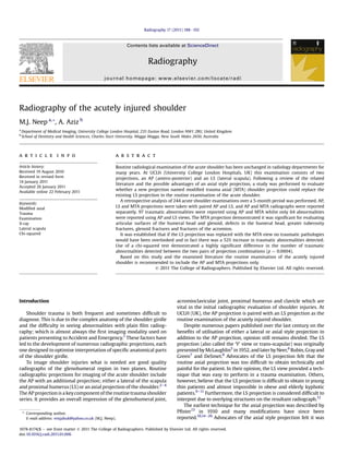
  • 1. Radiography of the acutely injured shoulder M.J. Neep a,*, A. Aziz b a Department of Medical Imaging, University College London Hospital, 235 Euston Road, London NW1 2BU, United Kingdom b School of Dentistry and Health Sciences, Charles Sturt University, Wagga Wagga, New South Wales 2650, Australia a r t i c l e i n f o Article history: Received 19 August 2010 Received in revised form 14 January 2011 Accepted 26 January 2011 Available online 22 February 2011 Keywords: Modified axial Trauma Examination X-ray Lateral scapula Chi-squared a b s t r a c t Routine radiological examination of the acute shoulder has been unchanged in radiology departments for many years. At UCLH (University College London Hospitals, UK) this examination consists of two projections, an AP (antero-posterior) and an LS (lateral scapula). Following a review of the related literature and the possible advantages of an axial style projection, a study was performed to evaluate whether a new projection named modified trauma axial (MTA) shoulder projection could replace the existing LS projection in the routine examination of the acute shoulder. A retrospective analysis of 244 acute shoulder examinations over a 5-month period was performed. AP, LS and MTA projections were taken with paired AP and LS, and AP and MTA radiographs were reported separately. 97 traumatic abnormalities were reported using AP and MTA whilst only 64 abnormalities were reported using AP and LS views. The MTA projection demonstrated it was significant for evaluating articular surfaces of the humeral head and glenoid, defects in the humeral head, greater tuberosity fractures, glenoid fractures and fractures of the acromion. It was established that if the LS projection was replaced with the MTA view no traumatic pathologies would have been overlooked and in fact there was a 52% increase in traumatic abnormalities detected. Use of a chi-squared test demonstrated a highly significant difference in the number of traumatic abnormalities detected between the two pairs of projection combinations (p ¼ 0.0004). Based on this study and the examined literature the routine examination of the acutely injured shoulder is recommended to include the AP and MTA projections only. Ó 2011 The College of Radiographers. Published by Elsevier Ltd. All rights reserved. Introduction Shoulder trauma is both frequent and sometimes difficult to diagnose. This is due to the complex anatomy of the shoulder girdle and the difficulty in seeing abnormalities with plain film radiog- raphy; which is almost always the first imaging modality used on patients presenting to Accident and Emergency.1 These factors have led to the development of numerous radiographic projections, each one designed to optimise interpretation of specific anatomical parts of the shoulder girdle. To image shoulder injuries what is needed are good quality radiographs of the glenohumeral region in two planes. Routine radiographic projections for imaging of the acute shoulder include the AP with an additional projection; either a lateral of the scapula and proximal humerus (LS) or an axial projection of the shoulder.2e4 The AP projection is a keycomponent of the routine trauma shoulder series. It provides an overall impression of the glenohumeral joint, acromioclavicular joint, proximal humerus and clavicle which are vital in the initial radiographic evaluation of shoulder injuries. At UCLH (UK), the AP projection is paired with an LS projection as the routine examination of the acutely injured shoulder. Despite numerous papers published over the last century on the benefits of utilisation of either a lateral or axial style projection in addition to the AP projection, opinion still remains divided. The LS projection (also called the ‘Y’ view or trans-scapular) was originally presented by McLaughlin5 in 1952,and laterby Neer,6 Rubin, Grayand Green7 and DeSmet.8 Advocates of the LS projection felt that the routine axial projection was too difficult to obtain technically and painful for the patient. In their opinion, the LS view provided a tech- nique that was easy to perform in a trauma examination. Others, however, believe that the LS projection is difficult to obtain in young thin patients and almost impossible in obese and elderly kyphotic patients.9e11 Furthermore, the LS projection is considered difficult to interpret due to overlying structures on the resultant radiograph.12 The earliest technique for the axial projection was described by Pfister13 in 1910 and many modifications have since been reported.10,14e26 Advocates of the axial style projection felt it was * Corresponding author. E-mail address: emjahuk@yahoo.co.uk (M.J. Neep). Contents lists available at ScienceDirect Radiography journal homepage: www.elsevier.com/locate/radi 1078-8174/$ e see front matter Ó 2011 The College of Radiographers. Published by Elsevier Ltd. All rights reserved. doi:10.1016/j.radi.2011.01.006 Radiography 17 (2011) 188e192
  • 2. the most valuable for evaluating the articular surfaces of the humeral head and the glenoid, and provided an excellent view of the relationship of the glenohumeral joint. The common disad- vantages associated with the axial (and most of the modified axial) projections were the need of abduction of the arm or necessity of an erect body position.27,28 Thus in a trauma examination, radiography of the acute shoulder is commonly restricted to the AP and LS projections. The basis for implementation of an MTA projection is that it can be performed with ease, irrespective of a patient’s positioning (erect or supine), size or body habitus. The purpose behind this study is to evaluate whether the implementation of an MTA shoulder projection could replace the existing LS projection from the routine examination of the acute shoulder by gaining an increase in diagnostic information, with no other detrimental consequences. As with all radiographic imaging the DRL (radiation dose) is important and as such we planned to calculate and compare this for the projections we are investigating. Materials and methods All patients presenting to UCLH Accident and Emergency department with acute shoulder trauma during a 5-month period were assessed by a casualty Doctor. If radiological examination was considered appropriate, three radiographic projections were taken; an AP (with neutral rotation), an LS and an MTA. Examination was purely based on clinical need for radiographic imaging of the shoulder; with no exclusion on age, pregnancy status or extent of trauma. As clinical examination of the shoulder in trauma alone is unreliable and radiographic examination a cornerstone of diagnosis the experience of the casualty doctor was not documented. Ethical approval for this study was obtained from the UCLH ethics committee prior to introduction of the MTA projection. A 5- month period was used as during that time a sufficient amount of shoulder trauma and hence projections would have taken place for statistical analysis to show significance. Due to the retrospective design it was felt that a 5-month period would be sufficient (with on average 1 to 2 examinations completed per day). The erect AP and LS radiographs were obtained according to Clark’s3 guide to radiographic positioning. The MTA was obtained by placing the patient in the seated position with their back to the X-ray table or horizontal bucky. The arm was positioned with external rotation as was physically possible, to allow for greater visualisation of the greater tuberosity. The thorax remained parallel to the edge of the table (Fig. 1). The detector was placed horizon- tally behind the humerus and in contact with the arm. The X-ray tube was angled 45 caudally from the vertical (Fig. 2), so the central ray passed through the glenohumeral joint towards the Figure 1. Patient’s thorax remains parallel to X-ray table or horizontal bucky for the MTA projection. Figure 2. Positioning for the MTA shoulder projection with the patient erect. M.J. Neep, A. Aziz / Radiography 17 (2011) 188e192 189
  • 3. centre of the detector. The resultant radiograph produced a macro appearance due to the large object to detector distance, while still accurately demonstrating the relationship of the humeral head to the glenoid fossa. In addition, this projection provided effective visualisation of the distal clavicle, coracoid process and acromion process (Fig. 3). This projection was also utilised in the supine position, whereby the only change in technique was that the detector was placed parallel to the patient’s thorax (Fig. 4). The source to skin distance remained constant for both positions. The only difference in radiographic appearance between the erect and supine resultant images was a minimal reduction in the magnifi- cation regarding the latter. This lack of magnification did not significantly effect the detection of abnormal appearances in this study. A retrospective review of the examinations was obtained by utilising the Picture Archiving and Communication Systems (PACS) database. The three projections were divided into two folders that were saved onto the PACS network. The first folder contained only the AP and LS projections and the second folder contained only the AP and MTA projections. Two assessors, qualified to interpret musculoskeletal radio- graphs, were asked to evaluate each examination for acute trau- matic pathology and quality of resultant projection in each of the two folders. With regard to traumatic pathology they were asked to classify whether the examination was normal or abnormal and then to give a brief interpretation of their findings in free text. Robinson, Wilson, Coral, Murphy and Verow,29 outlined a few guidelines for categorisation of examinations, which this study utilised. They stated a normal finding shall include anatomical variants, non-traumatic pathology, old fractures and evidence of previous surgery unless specifically related to the presentation. In addition, they stated an abnormal finding included joint effusions, fractures, dislocations, subluxations and soft tissue swelling. The assessors also recorded the quality of the resultant projec- tion, with regards to radiographic positioning, by scoring the quality as optimal, sub-optimal or non-diagnostic. Each of these categories were allocated a score, optimal being 3, sub-optimal 2 and non-diagnostic 1. One month after the assessors interpreted the first folder, they were each presented with the second folder. This one-month delay was designed to limit bias due to case recall. Once all the infor- mation was obtained by the investigator, the overall results were compared. Had there been any discrepancies between the two assessors, a third assessor would have been utilised, however, this was not necessary. Finally, this study determined a local DRL for the MTA projec- tion. Erect and supine data was averaged for the MTA projection. The DRL was calculated by a medical physicist at UCLH. This allowed comparison with the already existing local DRL for the LS radiograph in order to investigate which of these two projections generates a lower dose level for patients. Results Over the 5-month period from 22 November 2008 until 22 April 2009, 244 examinations of acute shoulder trauma were performed. All 244 examinations had three projections taken during their radiological examination, no examinations were excluded from analysis (n ¼ 244). The radiographic findings and the frequency with which they were interpreted for each of the pairs of projec- tions are shown in Table 1. 64 traumatic abnormalities were diag- nosed with the AP and LS views. 97 traumatic abnormalities were diagnosed with the AP and MTA projections. Only 2 Hill-Sachs lesions were demonstrated in the AP and LS combination whilst 19 were identified with the AP and MTA projections. Similarly only 2 glenoid rim fractures (including Bankart lesions) were visible on the AP and LS views whilst 8 were demonstrated on AP and MTA. No fractures of the acromion were seen and only 3 greater tuberosity avulsions were identified with Figure 3. Resultant MTA image. Figure 4. Positioning for the MTA shoulder projection with the supine patient. Table 1 Traumatic pathologies detected with the different paired projections. Traumatic Pathology AP LS AP MTA Acromioclavicular dislocation 3 3 Acromioclavicular subluxation 15 15 Fracture of acromion 0 2 Fracture of clavicle 5 5 Fracture of humeral head 1 1 Fracture of humeral neck 4 4 Fracture of humeral head and neck 11 11 Avulsion of greater tuberosity 3 8 Hill-Sachs lesion 2 19 Fracture of scapula body 2 2 Fracture of glenoid rim (inc. Bankart lesion) 2 8 Anterior glenohumeral dislocation 13 13 Anterior subluxation of humeral head 0 1 Inferior subluxation of humeral head 1 1 Posterior glenohumeral dislocation 0 1 Sub-acrominal effusion 2 3 Total 64 97 M.J. Neep, A. Aziz / Radiography 17 (2011) 188e192190
  • 4. AP and LS radiographs compared with 2 and 8 respectively for the AP and MTA radiographs. Of the 244 examinations, the image quality was optimal (with a score of 3) in all cases other than 4 non-diagnostic LS projections and 3 MTA projections. The local DRL for the MTA projection was calculated to equal 261 mGycm2 and was compared with the existing local DRL for the LS view which was 352 mGycm2 . Statistical chi-squared test was performed on the AP and LS vs AP and MTA data and a p value was found to be 0.0004 as shown in Table 2. This was calculated using an expected frequency of 30% but was also highly significant with both higher and lower expected frequencies. 30% frequency was chosen as this was the approximate percentage of traumatic abnormalities detected using the AP and LS projection combination. Discussion The AP projection paired with the MTA projection was the most effective radiograph combination for demonstrating traumatic abnormalities of the shoulder. The AP and MTA views identified 33 more traumatic pathologies than the AP and LS. This finding was in agreement with multiple previous authors who advocated the importance of including an axial projection in the routine exami- nation of the acute shoulder.11,12,28,30,31 The AP and MTA were the most successful in demonstrating Hill-Sachs lesions, glenoid rim fractures, acromion fractures and fractures of the greater tuberosity. This radiographic combination also detected all of the dislocations and subluxations of the gle- nohumeral joint and acromioclavicular joint. Previous studies support this opinion by acknowledging that the modified axial projection demonstrates specific traumatic pathologies that are not easily detected on an LS projection or the AP alone.11,12,27,31 The AP and LS radiograph combination was far inferior in diagnosing traumatic pathologies of the acute shoulder. If the AP and LS projections alone were used to diagnose the 244 patients in this study, 23 examinations would have been incorrectly reported as normal. Consequently a majority of these patients would have been discharged without correct clinical management leading to possible serious implications. The LS projection did not identify any abnormality that was not detected by the MTA projection, so replacing the LS with the MTA projection would have only led to an increase in diagnostic accuracy of traumatic abnormalities. Numerous earlier studies acknowledged that previous modified axial projections were difficult to obtain in the trauma setting.11,12,27,28,30,31 This was not demonstrated to be true in this study. All 244 examinations obtained the three projections required and informal feedback from the radiographers reported ease in acquiring the MTA projection. This study also demonstrated that the quality of the MTA resultant images, with regards to radiographic positioning, was effectively the same as the LS pro- jection (under 0.5% difference using the above scoring system). Not only is the MTA projection easier to perform, painless for the patient and gains more diagnostic information, it also yields a lower radiation dose level by 35% when compared with the dose level of the LS projection. The chi-squared test demonstrated that the difference between the number of traumatic abnormalities detected between the two different projection combinations was highly significant (p ¼ 0.0004). For this study’s statistical calculation, an expected frequency of 30% was utilised. Just as easily a higher or lower expected frequency would have elicited significant results. The result of the statistical analysis supports this study’s clinical results, that the AP and MTA are the most successful projection combina- tion in detecting traumatic abnormalities of the shoulder. As this study has shown, for traumatic imaging of the shoulder joint the accepted views that we have used for many years can be improved by the MTA projection, with increased diagnostic effec- tiveness. A possible interesting and useful study could be done looking at non-traumatic pathology and the MTA view. It is possibly as good as the axial view and may be the radiograph of choice in patients who are difficult to position due to body habitus, arthritis and limited range of movement. Conclusion Based on this study, the MTA projection is easy to obtain, painless for the patient and provides an increase in diagnostic information in comparison to the LS projection. The MTA is perti- nent for evaluating the articular surfaces of the humeral head and the glenoid. In addition, defects in the humeral head, greater tuberosity fractures, glenoid fractures and fractures of the acromion are also clearly visualised. This study demonstrated that if the LS projection was replaced by the MTA view in the routine examination of the acutely injured shoulder, no traumatic pathologies would have been overlooked. In fact, without the MTA projection, 33 traumatic abnormalities would have gone undetected. Statistical analysis showed high significance (p ¼ 0.0004) in the number of traumatic abnormalities detected between the two pairs of projection combinations. Therefore, it is recommended standard practice for radiographic examinations of traumatic shoulder injuries to include the AP and MTA projections only. References 1. Inman JB, Saunders DM, Abbott LC. Observations on the function of the shoulder joint. Journal of Bone and Joint Surgery 1944;26:1e30. 2. Bontrager KL, Lampignamo JP. Textbook of radiographic positioning and related anatomy. 6th ed. St. Louis, Mo: Mosby; 2005. p. 193e201. 3. Clark KC. Positioning in radiography. 10th ed. London: Heineman Medical Books; 1979. 4. Merrill V. Shoulder girdle. In: Ballinger PW, editor. Merrill’s atlas of radiographic positions and radiographic procedures. 6th ed. St Louis, Mo: Mosby; 1986. p. 101e50. 5. McLaughlin HL. Posterior dislocation of the shoulder. Journal of Bone and Joint Surgery 1952;34a:584e90. 6. Neer CS. Displaced proximal humerus fracture, parts I and II. Journal of Bone and Joint Surgery 1970;52a:1077e103. 7. Rubin SA, Gray RL, Green WR. The shoulder ‘Y’ view: diagnostic aid in shoulder trauma. Radiology 1976;110:725e6. 8. DeSmet A. Anterior oblique projection in radiography of the traumatized shoulder. American Journal of Radiology 1980;34:515e8. 9. Horsfield D, Renton P. The ‘other view’ in the radiography of shoulder trauma. Radiography 1980;4:213e4. 10. Wallace WA, Hellier M. Improving radiographs of the injured shoulder. Radi- ography 1983;49(589):229e33. 11. Brems-Dalgaard E, Davidson E, Sloth C. Radiographic examination of the acute shoulder. European Journal of Radiology 1990;11:10e4. Table 2 Results of Chi-squared analysis. No abnormality Abnormality Totals Observed data AP þ LS 180 64 244 AP þ MTA 147 97 244 Totals 327 161 n ¼ 244 Expected data 30% frequency of abnormality AP þ LS 170.8 73.2 244 AP þ MTA 170.8 73.2 244 Totals 341.6 146.4 n ¼ 244 p ¼ 0.0004 M.J. Neep, A. Aziz / Radiography 17 (2011) 188e192 191
  • 5. 12. Silfverskiold JP, Straehley DJ, Jones WW. Roentgenographic evaluation of sus- pected shoulder dislocation: a prospective study comparing the axillary view and the scapular ‘Y’ view. Orthopedics 1990;13:63e9. 13. Pfister A. Diagnosis of shoulder injury by X-ray. Clinical Medicine 1910;6: 178e9. 14. Lawrence WS. A new position in radiographing the shoulder joint. Roentgen- ology 1915;2:728e30. 15. Jordan H. New technique for the roentgen examination of the shoulder joint. Radiology 1935;25:480e4. 16. Cleaves EN. A new film holder for roentgen examination of the shoulder. American Journal of Radiology 1941;45:288e90. 17. Knuttsson F. On axial projection of the shoulder-joint. Acta Radiologica 1948;30:214e6. 18. Warrick CK. Posterior dislocation of the shoulder joint. British Journal of Radi- ology 1965;38:758e61. 19. Bloom MH, Obata WG. Diagnosis of posterior dislocation of the shoulder with use of the velpeau axillary and angle up roentgenographic view. Journal of Bone and Joint Surgery 1967;49a:943e9. 20. Rokous R, Feagin JA, Abbot HG. Modified axillary roentgenograph: a useful adjunct in the diagnosis of recurrent instability of the shoulder. Clinical Orthopaedics 1972;82:84e6. 21. Teigte RA, Ciullo JV. CAM axillary X-ray. Exhibit to the annual meeting of the American academy of orthopedic surgeons. Orthopaedic Transactions 1982;6:451. 22. Garth WP, Slappery LE, Ochs CW. Roentgenographic demonstration of insta- bility of the shoulder: the apical oblique projection. Journal of Bone and Joint Surgery 1984;66a:1450e3. 23. Oppenhiem WL, Dawson EG, Quinlan C, Graham SA. The cephaloscapular projection. Clinical Orthopaedics 1985;195:191e5. 24. Hobbs D. Alternate axial shoulder projections. Radiologic Technology 2005;76:434e5. 25. Geusens E, Pan S, Verhulst D, Brys P. The modified axillary view of the shoulder, a painless alternative. Emergency Radiology 2006;12:227e30. 26. Wilkinson K. Alternate trauma shoulder projection. Radiologic Technology 2006;78:11e2. 27. Flinn RM, MacMillan CL, Campbell DR, Fraser DB. Optimal radiography of the acutely injured shoulder. Journal of Association of Canadian Radiologists 1983;34:128e32. 28. Putkonen M, Lahde S, Puranen J, Palvansale M. The value of axial view in the radiography of shoulder girdle e experiences with a new modification of positioning. Rontgen-blatter 1988;41:158e62. 29. Robinson PJA, Wilson D, Coral A, Murphy A, Verow P. Variation between experienced observers in the interpretation of accident and emergency radiographs. British Journal of Radiology 1999;72:323e30. 30. Kornuth PJ, Salazar AM. The apical oblique view of the shoulder: its usefulness in acute trauma. American Journal of Radiology 1987;149:113e6. 31. Sloth C, Just L. The apical oblique radiograph in examination of acute shoulder trauma. European Journal of Radiology 1989;9:147e51. M.J. Neep, A. Aziz / Radiography 17 (2011) 188e192192