Introduction to CXRs For Non-Radiologist
•Download as PPSX, PDF•
0 likes•114 views

lecture done by Prof. Ken Schreibman, PhD/MD Professor of Radiology for more radiology lecture plez visit : https://radiologymadeeasy.com/

Recommended
More Related Content
Similar to Introduction to CXRs For Non-Radiologist
Similar to Introduction to CXRs For Non-Radiologist (20)
More from Radiology Made Easy
More from Radiology Made Easy (20)
Recently uploaded
Recently uploaded (20)
Introduction to CXRs For Non-Radiologist
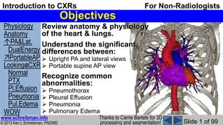
- 1. © 2013 Ken L Schreibman, PhD/MD www.schreibman.info Slide 1 of 99 Introduction to CXRs For Non-Radiologists Physiology Anatomy PA&Lat. DualEnergy PortableAP Looking@CXR Normal PTX Pl.Effusion Pneumonia Pul.Edema WOW Objectives Review anatomy & physiology of the heart & lungs. Understand the significant differences between: Upright PA and lateral views Portable supine AP view Recognize common abnormalities: Pneumothorax Pleural Effusion Pneumonia Pulmonary Edema Thanks to Carrie Bartels for 3D processing and segmentation!
- 2. © 2013 Ken L Schreibman, PhD/MD www.schreibman.info Slide 2 of 99 Introduction to CXRs For Non-Radiologists Physiology Anatomy PA&Lat. DualEnergy PortableAP Looking@CXR Normal PTX Pl.Effusion Pneumonia Pul.Edema WOW Disclaimer I am not Chest Radiologist This is my first CXR lecture I do read CXRs daily Mostly from the ER Reading CXR is something all radiologists should be able to do Looking at CXR something ALL health care providers should be willing to do
- 3. © 2013 Ken L Schreibman, PhD/MD www.schreibman.info Slide 3 of 99 Introduction to CXRs For Non-Radiologists Physiology Anatomy PA&Lat. DualEnergy PortableAP Looking@CXR Normal PTX Pl.Effusion Pneumonia Pul.Edema WOW Terminology: “Chest” X-Ray (CXR) X-Rays pass thru entire chest Superficial tissues Skin, breasts, sub-q fat Muscles Intercostals Bones Ribs, sternum, spine Heart&Mediastinum Trachea, Vessels, … Lungs mostly air
- 4. © 2013 Ken L Schreibman, PhD/MD www.schreibman.info Slide 4 of 99 Introduction to CXRs For Non-Radiologists Physiology Anatomy PA&Lat. DualEnergy PortableAP Looking@CXR Normal PTX Pl.Effusion Pneumonia Pul.Edema WOW Terminology: Chest “X-Ray” (CXR) This is a “Radiograph” Recall, “X-Rays” are: EM Spectrum energy Very short wavelength About size of an atom Man-made Emitted by a tube… …pass thru patient… …into a x-ray detector Film (old school) CR plate DR unit A,M 38yoF
- 5. © 2013 Ken L Schreibman, PhD/MD www.schreibman.info Slide 5 of 99 Introduction to CXRs For Non-Radiologists Physiology Anatomy PA&Lat. DualEnergy PortableAP Looking@CXR Normal PTX Pl.Effusion Pneumonia Pul.Edema WOW Radiographs Only Detect 4 Densities BicTM lighter = Smoker Air = Black Lungs (Air-filled alveoli) Soft Tissues = Gray Superficial tissues Heart, Mediastinum Bones = Lighter Gray Ribs, Spine Metal = White Outside/Inside patient Zipper= Clothed
- 6. © 2013 Ken L Schreibman, PhD/MD www.schreibman.info Slide 6 of 99 Introduction to CXRs For Non-Radiologists Physiology Anatomy PA&Lat. DualEnergy PortableAP Looking@CXR Normal PTX Pl.Effusion Pneumonia Pul.Edema WOW Convention: We look at CXR like we look at Pt… Q,J 23yoMH,C 64yoM Cardiac Apex Stomach Cardiac Apex Stomach Aortic Arch Right Aortic Arch Rare Normal Variant (0.01%)* Face-to-Face Tech’s marker, on detector, near patient’s Left shoulder *wikipedia.org
- 7. © 2013 Ken L Schreibman, PhD/MD www.schreibman.info Slide 7 of 99 Introduction to CXRs For Non-Radiologists Physiology Anatomy PA&Lat. DualEnergy PortableAP Looking@CXR Normal PTX Pl.Effusion Pneumonia Pul.Edema WOW PowerPoint Model: Lungs Bag of air Inhale: Fills Exhale:Empties (not completely) Pipes Bronchus Gr: “windpipe” etymonline.com
- 8. © 2013 Ken L Schreibman, PhD/MD www.schreibman.info Slide 8 of 99 Introduction to CXRs For Non-Radiologists Physiology Anatomy PA&Lat. DualEnergy PortableAP Looking@CXR Normal PTX Pl.Effusion Pneumonia Pul.Edema WOW Standard Convention: Look at CXR like we’re looking at a patient, from the front 2 Lungs 3 Pipes 2 Bronchi “Main Stem Bronchi” Stem off Main Pipe: 1 Trachea Meet at Carina RIGHT Bronchus LEFT Bronchus Trachea Carina PowerPoint Model: Lungs
- 9. © 2013 Ken L Schreibman, PhD/MD www.schreibman.info Slide 9 of 99 Introduction to CXRs For Non-Radiologists Physiology Anatomy PA&Lat. DualEnergy PortableAP Looking@CXR Normal PTX Pl.Effusion Pneumonia Pul.Edema WOW How Do The Lungs Work? The lungs don’t “work” (just bags of air) Lungs inflate passively in response to pressure gradients Carina Pressure > Lung Pressure Rescue Breathing On a ventilator Tracheal Tube Lung Pressure < Carina Pressure Negative pressure outside the lungs Created by the DIAPHRAGM - +
- 10. © 2013 Ken L Schreibman, PhD/MD www.schreibman.info Slide 10 of 99 Introduction to CXRs For Non-Radiologists Physiology Anatomy PA&Lat. DualEnergy PortableAP Looking@CXR Normal PTX Pl.Effusion Pneumonia Pul.Edema WOW Thought Experiment Balloon tied to a Straw Sealed inside glass jar Only way air gets into balloon is via end of straw outside jar Jar has sealed rubber bottom Pulling down on bottom… Increases size of sealed jar Decreases pressure in jar (PV=nrT) Jar Pressure < Atmospheric Balloon Inflates! Atmospheric Pressure - - --
- 11. © 2013 Ken L Schreibman, PhD/MD www.schreibman.info Slide 11 of 99 Introduction to CXRs For Non-Radiologists Physiology Anatomy PA&Lat. DualEnergy PortableAP Looking@CXR Normal PTX Pl.Effusion Pneumonia Pul.Edema WOW 2 Lungs (bags of air) Tracheobronchial tree Sealed within Thorax Chest Wall (layers) Skin/Sub-Q Ribs, Intercostal A/N/Muscles Inner Pleural Lining Diaphragm – Domed When it contracts it flattens Increases intra-thoracic volume Decreases intra-pleural pressure Lungs Work The Same - - -- Air enters via… Nose/ Mouth
- 12. © 2013 Ken L Schreibman, PhD/MD www.schreibman.info Slide 12 of 99 Introduction to CXRs For Non-Radiologists Physiology Anatomy PA&Lat. DualEnergy PortableAP Looking@CXR Normal PTX Pl.Effusion Pneumonia Pul.Edema WOW Not much in Thorax Lungs: Fill the Thorax From Apices Costo-Phrenic Angle (CPA) “Costo”: Ribs “Phrenic”: Diaphragm Tracheobronchial Tree Bronchioles branch off Heart Vessels (Mediastinum) Connect lungs to heart Connect heart to whole body Lungs Fill Pleural Space Lung Apex Lung Apex Media- stinum Heart CPA CPA
- 13. © 2013 Ken L Schreibman, PhD/MD www.schreibman.info Slide 13 of 99 Introduction to CXRs For Non-Radiologists Physiology Anatomy PA&Lat. DualEnergy PortableAP Looking@CXR Normal PTX Pl.Effusion Pneumonia Pul.Edema WOW Gas Exchange All human cells require O2, emit CO2 Red Blood Cells (RBC) transport gas Hemoglobin molecule (Hgb) Occurs at capillaries Diameter = 1 RBC (5µm) Walls: Very thin, semipermeable membranes Permits Diffusion of gasses RBC Capillary Thin Walls 1 RBC
- 14. © 2013 Ken L Schreibman, PhD/MD www.schreibman.info Slide 14 of 99 Introduction to CXRs For Non-Radiologists Physiology Anatomy PA&Lat. DualEnergy PortableAP Looking@CXR Normal PTX Pl.Effusion Pneumonia Pul.Edema WOW Diffusion Way of moving things for free(No energy expense) Related to a Fundamental Force: Entropy Things become disorderly It takes a lot less energy to knock down Jenga tower… Diffusion:Things move from high concentration (ordered) to lower concentrations (disordered) Capillary Thin Walls 1 RBC RBC O2 O2 O2 I have more O2 than you. Here’s some of mine I have more CO2 than you. Here’s some of mine Tissue CellCO2 CO2 CO2 CO2 CO2 CO2 CO2 CO2 CO2 CO2 O2 O2 O2
- 15. © 2013 Ken L Schreibman, PhD/MD www.schreibman.info Slide 15 of 99 Introduction to CXRs For Non-Radiologists Physiology Anatomy PA&Lat. DualEnergy PortableAP Looking@CXR Normal PTX Pl.Effusion Pneumonia Pul.Edema WOW alveolus [L]: “little cavity” Air sacs Bronchioles Looks like bunch of grapes. Interstitial spaces Outside alveoli Capillaries Where gas exchange occurs Gas Exchange: Alveoli Lungs C C C C C C C C C C C C C C C
- 16. © 2013 Ken L Schreibman, PhD/MD www.schreibman.info Slide 16 of 99 Introduction to CXRs For Non-Radiologists Physiology Anatomy PA&Lat. DualEnergy PortableAP Looking@CXR Normal PTX Pl.Effusion Pneumonia Pul.Edema WOW Gas Exchange: Alveoli Capillary 1 RBC Thin Walls 1 RBC RBC CO2 CO2 CO2 CO2 CO2 CO2 A I need O2 I need to get rid of CO2 I have plenty of O2 Room Air is 21% O2 O2 O2O2 O2 O2 O2 O2 O2 O2 O2 O2 O2 O2 O2 O2 O2 I have very little CO2 Room Air is 0.04% CO2 Diffusion!O2 CO2
- 17. © 2013 Ken L Schreibman, PhD/MD www.schreibman.info Slide 17 of 99 Introduction to CXRs For Non-Radiologists Physiology Anatomy PA&Lat. DualEnergy PortableAP Looking@CXR Normal PTX Pl.Effusion Pneumonia Pul.Edema WOW Circulatory System Lungs 1 RBC RBCCO2 CO2 CO2 CO2 CO2 CO2 CO2 1 RBC RBC O2 O2 O2 O2 O2 O2 Whole Body Transports O2-rich RBCs LungsWhole Body Transports CO2-rich RBCs Lungs Whole Body Heart
- 18. © 2013 Ken L Schreibman, PhD/MD www.schreibman.info Slide 18 of 99 Introduction to CXRs For Non-Radiologists Physiology Anatomy PA&Lat. DualEnergy PortableAP Looking@CXR Normal PTX Pl.Effusion Pneumonia Pul.Edema WOW Arteries carry blood from Heart Thick walls (>120mmHg) Veins carry blood to Heart Thin walls (<10mmHg) Left Ventricle Thick Wall >120mmHg The Heart: 4 Chambers Whole Body Right Ventricle Thin Wall <20mmHg Vena Cava RA LA Lungs
- 19. © 2013 Ken L Schreibman, PhD/MD www.schreibman.info Slide 19 of 99 Introduction to CXRs For Non-Radiologists Physiology Anatomy PA&Lat. DualEnergy PortableAP Looking@CXR Normal PTX Pl.Effusion Pneumonia Pul.Edema WOW Cardio-Pulmonary System Heart&Lungs intertwined also vessels, bronchi Anatomy of blood filled structures can be well demonstrated with CT-Angiography CT obtained immediately after IV contrast bolus Basic 3D processing… All blood/contrast filled structures (heart, vessels) have same density… Similar to bone density and with technologists skilled in segmentation processing we can add artificial colors!
- 20. © 2013 Ken L Schreibman, PhD/MD www.schreibman.info Slide 20 of 99 Introduction to CXRs For Non-Radiologists Physiology Anatomy PA&Lat. DualEnergy PortableAP Looking@CXR Normal PTX Pl.Effusion Pneumonia Pul.Edema WOW Left Ventricle (LV) Workhorse of the heart Thick muscular wall Pump blood entire body Generate pressure >120mmHg 5½ FEET of H2O! It doesn’t look like much here because this is a CTA, showing only the contrast in the blood INSIDE the chamber of the LV. Radiograph – LV: left cardiac border cardiac apex LV Cardiac Apex
- 21. © 2013 Ken L Schreibman, PhD/MD www.schreibman.info Slide 21 of 99 Introduction to CXRs For Non-Radiologists Physiology Anatomy PA&Lat. DualEnergy PortableAP Looking@CXR Normal PTX Pl.Effusion Pneumonia Pul.Edema WOW Red Blood: LV Aorta Body LV Anterior view LV Ascending Aorta Descending Aorta Arch Innominate Left Common Carotid Left Subclavian Right Common CarotidGreat Vessels: Right Subclavian Aorta Thick wall, 3 layers Intima (inner layer) Tear Dissection Media (muscle, elastic fibers) Tear Aneurysm Adventitia (outer, supportive layer) Tear ALL THREE Aortic Laceration Can’t see of this with CXR Need to use: CT, MR, US Oblique view
- 22. © 2013 Ken L Schreibman, PhD/MD www.schreibman.info Slide 22 of 99 Introduction to CXRs For Non-Radiologists Physiology Anatomy PA&Lat. DualEnergy PortableAP Looking@CXR Normal PTX Pl.Effusion Pneumonia Pul.Edema WOW Blue Blood: Body RA RV PulArt Right Ventricle (RV) Wraps around LV Thin muscular wall Only needs to pump blood to adjacent lungs (20mmHg) Pulmonary Artery (PA) Carries blood from RV to both lungs Only artery to carry deoxygenated blood RV LV PA Oblique view
- 23. © 2013 Ken L Schreibman, PhD/MD www.schreibman.info Slide 23 of 99 Introduction to CXRs For Non-Radiologists Physiology Anatomy PA&Lat. DualEnergy PortableAP Looking@CXR Normal PTX Pl.Effusion Pneumonia Pul.Edema WOW Blue Blood: Body RA RV PulArt Right Atrium (RA) Forms right heart border on CXR Right Ventricle (RV) Wraps around LV Does doesn’t contribute to heart shadow on frontal radiograph LV Anterior view RV RA PA
- 24. © 2013 Ken L Schreibman, PhD/MD www.schreibman.info Slide 24 of 99 Introduction to CXRs For Non-Radiologists Physiology Anatomy PA&Lat. DualEnergy PortableAP Looking@CXR Normal PTX Pl.Effusion Pneumonia Pul.Edema WOW Cardio-Pulmonary System Physiology requires heart & lungs work together Anatomy places heart & lungs adjacent so PA & PV can be short A lot of air & blood filled structures in the tight intra-thoracic space RV Posterior view RA PA LV LA PV PV PV PV Pulmonary Veins (PV) Carry blood lungs to LA Only veins w/ oxygenated blood There’s a lot to look at on CXR!
- 25. © 2013 Ken L Schreibman, PhD/MD www.schreibman.info Slide 25 of 99 Introduction to CXRs For Non-Radiologists Physiology Anatomy PA&Lat. DualEnergy PortableAP Looking@CXR Normal PTX Pl.Effusion Pneumonia Pul.Edema WOW Upright PA Chest Radiograph Standing Upright This is a big deal Permits full lung inflation Can’t fully inflate laying/sitting Makes use of gravity More blood flows bases Pleural effusions CP angles Pneumothoraces apices Pulls scapulae away Patient hugs detector Marty age 13 Detector CR plate DR array Grid
- 26. © 2013 Ken L Schreibman, PhD/MD www.schreibman.info Slide 26 of 99 Introduction to CXRs For Non-Radiologists Physiology Anatomy PA&Lat. DualEnergy PortableAP Looking@CXR Normal PTX Pl.Effusion Pneumonia Pul.Edema WOW Upright PA Chest Radiograph PA is Standard X-rays enter Posteriorly, X-rays exit Anteriorly Hence X-rays travel PosteriorAnterior,“PA” Detector is Anterior Allows patient to abduct scapulae away from chest Minimizes heart magnification X-ray tube
- 27. © 2013 Ken L Schreibman, PhD/MD www.schreibman.info Slide 27 of 99 Introduction to CXRs For Non-Radiologists Physiology Anatomy PA&Lat. DualEnergy PortableAP Looking@CXR Normal PTX Pl.Effusion Pneumonia Pul.Edema WOW Shadow-graphic Magnification Flashlight As my hand gets further from the wall, the shadow gets larger Similarly, as body parts get further from the detector, their radiographic shadow get larger
- 28. © 2013 Ken L Schreibman, PhD/MD www.schreibman.info Slide 28 of 99 Introduction to CXRs For Non-Radiologists Physiology Anatomy PA&Lat. DualEnergy PortableAP Looking@CXR Normal PTX Pl.Effusion Pneumonia Pul.Edema WOW Radiographic Magnification PA Projection Minimizes heart-detector distance Minimizes heart magnification AP Projection Increases heart-detector distance Increases heart magnification D E T E C T O R D E T E C T O R X-rays X-raysHeart is anterior in chest Heart is anterior in chest
- 29. © 2013 Ken L Schreibman, PhD/MD www.schreibman.info Slide 29 of 99 Introduction to CXRs For Non-Radiologists Physiology Anatomy PA&Lat. DualEnergy PortableAP Looking@CXR Normal PTX Pl.Effusion Pneumonia Pul.Edema WOW PA CXR is Backward Marty’s Left Shoulder Shot looking at patient’s back… Hung looking at patient’s front S,M 8yoM
- 30. © 2013 Ken L Schreibman, PhD/MD www.schreibman.info Slide 30 of 99 Introduction to CXRs For Non-Radiologists Physiology Anatomy PA&Lat. DualEnergy PortableAP Looking@CXR Normal PTX Pl.Effusion Pneumonia Pul.Edema WOW Importance of Scapular Abduction D,M 21yoF Scapula well abducted away from lung Scapulae overlap lungs Outpatient Clinic Edge Lung Apex Pneumo- thorax! 1 hour later, Emergency Room Can’t see the PTX! Scapula overlaps lung
- 31. © 2013 Ken L Schreibman, PhD/MD www.schreibman.info Slide 31 of 99 Introduction to CXRs For Non-Radiologists Physiology Anatomy PA&Lat. DualEnergy PortableAP Looking@CXR Normal PTX Pl.Effusion Pneumonia Pul.Edema WOW Upright Lateral Chest Radiograph Importance of multiple views Pt standing Lungs inflated Left side next to the detector Minimize heart magnification Tradition: Hung backwards Arms Elevated! S,M 8yoM
- 32. © 2013 Ken L Schreibman, PhD/MD www.schreibman.info Slide 32 of 99 Introduction to CXRs For Non-Radiologists Physiology Anatomy PA&Lat. DualEnergy PortableAP Looking@CXR Normal PTX Pl.Effusion Pneumonia Pul.Edema WOW Importance of Arm Elevation D,R 37yoM Repeat Lateral ViewInitial Lateral View Arms not elevated Can barely even see the heart Now we can see heart… & lung in front of it With good arm elevation…
- 33. © 2013 Ken L Schreibman, PhD/MD www.schreibman.info Slide 33 of 99 Introduction to CXRs For Non-Radiologists Physiology Anatomy PA&Lat. DualEnergy PortableAP Looking@CXR Normal PTX Pl.Effusion Pneumonia Pul.Edema WOW Importance of Lateral View S,M 8yoM Subtle density Overlaps heart Obvious density Behind heart = LLL Pneumonia!
- 34. © 2013 Ken L Schreibman, PhD/MD www.schreibman.info Slide 34 of 99 Introduction to CXRs For Non-Radiologists Physiology Anatomy PA&Lat. DualEnergy PortableAP Looking@CXR Normal PTX Pl.Effusion Pneumonia Pul.Edema WOW CXR is NOT a Simple Exam Requires patient able to: Stand upright Sitting in a chair is not the same Propped up on a cart, not the same Elevate their arms, abduct scapulae Take in a big breath, hold it 1 second Requires technologist to: Make sure patient is optimally positioned Look at images to access optimal quality Repeat images as needed
- 35. © 2013 Ken L Schreibman, PhD/MD www.schreibman.info Slide 35 of 99 Introduction to CXRs For Non-Radiologists Physiology Anatomy PA&Lat. DualEnergy PortableAP Looking@CXR Normal PTX Pl.Effusion Pneumonia Pul.Edema WOW Dual Energy CXR: Separate Lungs/Bones The lungs are hard to see on CXR as they are nearly radiolucent, compared to the overlying bones which are relatively radiopaque. Wouldn’t it be nice if we could remove calcified ribs on a CXR… generating a 2nd CXR of the lungs without the overlying ribs… and even generate a 3rd CXR showing just calcified structures! S,J 58yoM At UW, our standard PA CXR is Dual Energy Requires investment in dedicated DR unit
- 36. © 2013 Ken L Schreibman, PhD/MD www.schreibman.info Slide 36 of 99 Introduction to CXRs For Non-Radiologists Physiology Anatomy PA&Lat. DualEnergy PortableAP Looking@CXR Normal PTX Pl.Effusion Pneumonia Pul.Edema WOW Digital Radiography CR (Computed Radiography) Similar to old-fashioned film radiography Technologist places a cassette under the patent Contains photostimulable phosphor plate, rather than film Runs cassette through IIP(ImageInformationProcessor) Laser scanner, rather than darkroom chemical developer Used for bones, abdomen, portables DR (Direct Radiography) Similar to modern digital camera Captures directly to image detector, no cassette Can take 2 images, rapidly, at 2 energies (DE) First only text slide
- 37. © 2013 Ken L Schreibman, PhD/MD www.schreibman.info Slide 37 of 99 Introduction to CXRs For Non-Radiologists Physiology Anatomy PA&Lat. DualEnergy PortableAP Looking@CXR Normal PTX Pl.Effusion Pneumonia Pul.Edema WOW CR: Can use old film equipment Outpatient CR room Same film equipment Same X-ray tube Same cassette holder Same cassette holder Same art
- 38. © 2013 Ken L Schreibman, PhD/MD www.schreibman.info Slide 38 of 99 Introduction to CXRs For Non-Radiologists Physiology Anatomy PA&Lat. DualEnergy PortableAP Looking@CXR Normal PTX Pl.Effusion Pneumonia Pul.Edema WOW CR: Film, cassettes, dark room replaced CR cassettes containing the phosphor plates IIP plate reader
- 39. © 2013 Ken L Schreibman, PhD/MD www.schreibman.info Slide 39 of 99 Introduction to CXRs For Non-Radiologists Physiology Anatomy PA&Lat. DualEnergy PortableAP Looking@CXR Normal PTX Pl.Effusion Pneumonia Pul.Edema WOW DR: Requires all new equipment Outpatient DR room It looks a lot like the CR room New dual energy X-ray tube New DR detector built into table New DR detector built into upright chest unit
- 40. © 2013 Ken L Schreibman, PhD/MD www.schreibman.info Slide 40 of 99 Introduction to CXRs For Non-Radiologists Physiology Anatomy PA&Lat. DualEnergy PortableAP Looking@CXR Normal PTX Pl.Effusion Pneumonia Pul.Edema WOW Technically: Perfect! Lungs: Well Inflated Patient: Not Rotated Scapulae: Abducted Dual Energy CXR: Helps See Pneumonia D,K 63yoM Hard to see subtle density overlapping ribs… Standard PA view Dual Energy Lung view
- 41. © 2013 Ken L Schreibman, PhD/MD www.schreibman.info Slide 41 of 99 Introduction to CXRs For Non-Radiologists Physiology Anatomy PA&Lat. DualEnergy PortableAP Looking@CXR Normal PTX Pl.Effusion Pneumonia Pul.Edema WOW Dual Energy CXR: Helps See Nodules A,S 75yoM Nipple Marker Standard view Lung view Ca++ view ? Scapula overlaps lungs Better scapula abduction !!! Lung view
- 42. © 2013 Ken L Schreibman, PhD/MD www.schreibman.info Slide 42 of 99 Introduction to CXRs For Non-Radiologists Physiology Anatomy PA&Lat. DualEnergy PortableAP Looking@CXR Normal PTX Pl.Effusion Pneumonia Pul.Edema WOW Dual Energy CXR: Shows Ca++ Nodules L,T 61yoM Standard view Lung view Ca++ view Nipple Marker Tiny Nodule Hard to see Easy to see Calcified Granuloma
- 43. © 2013 Ken L Schreibman, PhD/MD www.schreibman.info Slide 43 of 99 Introduction to CXRs For Non-Radiologists Physiology Anatomy PA&Lat. DualEnergy PortableAP Looking@CXR Normal PTX Pl.Effusion Pneumonia Pul.Edema WOW Portable Supine AP CXR Used in cases when patient cannot stand for upright PA & Lateral views
- 44. © 2013 Ken L Schreibman, PhD/MD www.schreibman.info Slide 44 of 99 Introduction to CXRs For Non-Radiologists Physiology Anatomy PA&Lat. DualEnergy PortableAP Looking@CXR Normal PTX Pl.Effusion Pneumonia Pul.Edema WOW Supine AP: Can’t make use of Gravity PTX Supine AP Upright PA Pl.Ef. Pl.Ef. Normal Lung Normal Lung PTX rises above normal lung PTX layers anterior to normal lung Pleural Effusion flows below normal lung Pleural Effusion layers out posterior to normal lung Vertical beam PARALLEL to gravity Horizontal beam PERPEN- DICULAR to gravity Complex Slide Warning
- 45. © 2013 Ken L Schreibman, PhD/MD www.schreibman.info Slide 45 of 99 Introduction to CXRs For Non-Radiologists Physiology Anatomy PA&Lat. DualEnergy PortableAP Looking@CXR Normal PTX Pl.Effusion Pneumonia Pul.Edema WOW Problems with Supine AP vs PA X-ray beam not perpendicular to gravity Hard to see PTX air rising anteriorly Hard to see pleural effusions layering out posteriorly Hard to assess increased blood flow to upper lobes Lungs never well inflated No lateral view No dual energy Hard to abduct scapulae Heart is magnified Harder to assess heart size (cardiomegaly) Second only text slide
- 46. © 2013 Ken L Schreibman, PhD/MD www.schreibman.info Slide 46 of 99 Introduction to CXRs For Non-Radiologists Physiology Anatomy PA&Lat. DualEnergy PortableAP Looking@CXR Normal PTX Pl.Effusion Pneumonia Pul.Edema WOW Portable AP CXR Never Fully Upright A,R 76yoM
- 47. © 2013 Ken L Schreibman, PhD/MD www.schreibman.info Slide 47 of 99 Introduction to CXRs For Non-Radiologists Physiology Anatomy PA&Lat. DualEnergy PortableAP Looking@CXR Normal PTX Pl.Effusion Pneumonia Pul.Edema WOW Portable AP vs Upright PA C,C 54yoF Semi-upright AP Upright PA Heart looks big Lots of pulmonary interstitial markings Extending to periphery! Dx: Resembles Pulmonary Edema? Rec: Upright PA & Lat. Lungs better inflated Heart less magnified Normal pulmonary markings Dx: Normal
- 48. © 2013 Ken L Schreibman, PhD/MD www.schreibman.info Slide 48 of 99 Introduction to CXRs For Non-Radiologists Physiology Anatomy PA&Lat. DualEnergy PortableAP Looking@CXR Normal PTX Pl.Effusion Pneumonia Pul.Edema WOW Portable AP vs Upright PA O,T 54yoM Semi-upright AP Upright PA
- 49. © 2013 Ken L Schreibman, PhD/MD www.schreibman.info Slide 49 of 99 Introduction to CXRs For Non-Radiologists Physiology Anatomy PA&Lat. DualEnergy PortableAP Looking@CXR Normal PTX Pl.Effusion Pneumonia Pul.Edema WOW Portable AP vs Upright PA K,R 50yoM Supine AP Upright PA OK to travel to DR Chest room with EKG electrodes attached
- 50. © 2013 Ken L Schreibman, PhD/MD www.schreibman.info Slide 50 of 99 Introduction to CXRs For Non-Radiologists Physiology Anatomy PA&Lat. DualEnergy PortableAP Looking@CXR Normal PTX Pl.Effusion Pneumonia Pul.Edema WOW Portable AP vs Upright PA & Lat J,K 63yoM Semi-upright AP Upright PA Lungs very underinflated Dx: Negative? Rec: Upright PA & Lat. Lungs well inflated Density R heart border Dx: RLL Pneumonia Dual Energy Lung view Lateral
- 51. © 2013 Ken L Schreibman, PhD/MD www.schreibman.info Slide 51 of 99 Introduction to CXRs For Non-Radiologists Physiology Anatomy PA&Lat. DualEnergy PortableAP Looking@CXR Normal PTX Pl.Effusion Pneumonia Pul.Edema WOW C,W 83yoM What is the Portable AP CXR Good For? Checking tubes and lines Is Endo Tracheal (ET) Tube above the carina? Is the Central Line (CL) in the SVC? Is the Naso Gastric (NG) Tube stomach? ET Carina CL NG in Left Bronchus!
- 52. © 2013 Ken L Schreibman, PhD/MD www.schreibman.info Slide 52 of 99 Introduction to CXRs For Non-Radiologists Physiology Anatomy PA&Lat. DualEnergy PortableAP Looking@CXR Normal PTX Pl.Effusion Pneumonia Pul.Edema WOW What is the Portable AP CXR Good For? M,M 75yoM In the ER trauma room Is there a LARGE pneumothorax? Is the Chest Tube in the right place? After placing Chest Tube Edge Left Lung Pneumo- thorax! Chest Tube Left lung much better inflated
- 53. © 2013 Ken L Schreibman, PhD/MD www.schreibman.info Slide 53 of 99 Introduction to CXRs For Non-Radiologists Physiology Anatomy PA&Lat. DualEnergy PortableAP Looking@CXR Normal PTX Pl.Effusion Pneumonia Pul.Edema WOW What is the Portable AP CXR Good For? In the ER trauma room Is there a LARGE pleural effusion? Is there a LARGE pulmonary density? (Pneumonia, Contusion) Should get Upright PA & Lateral whenever possible G,S 40yoMK,C 64yoF Large Right Pleural Eff. Air space density throughout Left Lower Lobe
- 54. © 2013 Ken L Schreibman, PhD/MD www.schreibman.info Slide 54 of 99 Introduction to CXRs For Non-Radiologists Physiology Anatomy PA&Lat. DualEnergy PortableAP Looking@CXR Normal PTX Pl.Effusion Pneumonia Pul.Edema WOW Break… then Key things to look for… S,J 12yM
- 55. © 2013 Ken L Schreibman, PhD/MD www.schreibman.info Slide 55 of 99 Introduction to CXRs For Non-Radiologists Physiology Anatomy PA&Lat. DualEnergy PortableAP Looking@CXR Normal PTX Pl.Effusion Pneumonia Pul.Edema WOW Schreibman’s Says… 1st Rule of Radiology: Know what you’re looking at! Is this the correct study? (YOU check every time) Correct Patient Correct Date/Time Correct Study (CXR ≠ CT ≠ MR ≠ US) Correct Body part Is this a good study? (This takes experience) Is it upright PA & lateral, or portable supine AP? Is this a good quality study? Patient well positioned? Lungs well inflated? Third only text slide
- 56. © 2013 Ken L Schreibman, PhD/MD www.schreibman.info Slide 56 of 99 Introduction to CXRs For Non-Radiologists Physiology Anatomy PA&Lat. DualEnergy PortableAP Looking@CXR Normal PTX Pl.Effusion Pneumonia Pul.Edema WOW Is the Patient fully upright? M,S 20yoF PA view Lateral view Stomach bubble under Left hemidiaphragm Stomach bubble under Left hemidiaphragm Right hemidia. Air-Fluid Level Air-Fluid Level Right hemidiaphragm
- 57. © 2013 Ken L Schreibman, PhD/MD www.schreibman.info Slide 57 of 99 Introduction to CXRs For Non-Radiologists Physiology Anatomy PA&Lat. DualEnergy PortableAP Looking@CXR Normal PTX Pl.Effusion Pneumonia Pul.Edema WOW Air Below Diaphragm Common below LEFT Stomach Colon (Splenic flexure) Uncommon below RIGHT That’s where the liver lives Rarely (0.1%) loop of colon (hepatic flexure) Chilaiditi’s (“Ky-La-Ditty”) Free air below diaphragm is Abnormal Recent (past week) surgery, laparoscopy Bowel Perforation! (Pneumoperitoneum) learningradiology.com B,C 57yoF Stomach Colon Liver
- 58. © 2013 Ken L Schreibman, PhD/MD www.schreibman.info Slide 58 of 99 Introduction to CXRs For Non-Radiologists Physiology Anatomy PA&Lat. DualEnergy PortableAP Looking@CXR Normal PTX Pl.Effusion Pneumonia Pul.Edema WOW Air Below RIGHT Diaphragm L,A 77yoF …19 days later Colon Colon Free Air!
- 59. © 2013 Ken L Schreibman, PhD/MD www.schreibman.info Slide 59 of 99 Introduction to CXRs For Non-Radiologists Physiology Anatomy PA&Lat. DualEnergy PortableAP Looking@CXR Normal PTX Pl.Effusion Pneumonia Pul.Edema WOW W,D 28yoM Is the Patient fully upright?Are the Lungs fully included? Apex Apex R CPA L CPA R CPA L CPA From Apices To CP Angles on Lateral! L CPA R CPA
- 60. © 2013 Ken L Schreibman, PhD/MD www.schreibman.info Slide 60 of 99 Introduction to CXRs For Non-Radiologists Physiology Anatomy PA&Lat. DualEnergy PortableAP Looking@CXR Normal PTX Pl.Effusion Pneumonia Pul.Edema WOW Are Lungs Well Inflated? G,J 47yoM Patient is instructed: “Breathe in… hold it…” Tech shoots picture “… and breathe…” We want lungs maximally inflated Old Radiology Lore: The lungs are well inflated if you can see 10 ribs. (I can’t find a reference) InspirationExpirationInspiration 1 2 34 5 6 7 8 9 10 11
- 61. © 2013 Ken L Schreibman, PhD/MD www.schreibman.info Slide 61 of 99 Introduction to CXRs For Non-Radiologists Physiology Anatomy PA&Lat. DualEnergy PortableAP Looking@CXR Normal PTX Pl.Effusion Pneumonia Pul.Edema WOW Good Inspiration (even on Portable Supine AP) Density LLL Pneumonia? Mass? C,J 70yoM With better inflation, density nearly resolves. It was just Atelectasis!
- 62. © 2013 Ken L Schreibman, PhD/MD www.schreibman.info Slide 62 of 99 Introduction to CXRs For Non-Radiologists Physiology Anatomy PA&Lat. DualEnergy PortableAP Looking@CXR Normal PTX Pl.Effusion Pneumonia Pul.Edema WOW “Atelectasis” wikipedia.org G,M 62yoF [Gr] ἀτελής: “incomplete” + ἔκτασις: “extension” Incomplete expansion Sub-segmental Entire segment/lobe Entire lung Can be caused by: Under inflation Airway obstruction (pneumonia) Pneumothorax Atelectasis ≠ Pneumothorax Right Upper Lobe Atelectasis
- 63. © 2013 Ken L Schreibman, PhD/MD www.schreibman.info Slide 63 of 99 Introduction to CXRs For Non-Radiologists Physiology Anatomy PA&Lat. DualEnergy PortableAP Looking@CXR Normal PTX Pl.Effusion Pneumonia Pul.Edema WOW Lung Anatomy: Lobes & Fissures V,A 19yoF Minor Fissure Major Fissure LUL LLL Major Fissure RUL RLL Left LungRight Lung LUL LLL Minor Fissure RML RUL RLL RML *[L] “little tongue”
- 64. © 2013 Ken L Schreibman, PhD/MD www.schreibman.info Slide 64 of 99 Introduction to CXRs For Non-Radiologists Physiology Anatomy PA&Lat. DualEnergy PortableAP Looking@CXR Normal PTX Pl.Effusion Pneumonia Pul.Edema WOW L,J 81yoF Is the Patient fully upright?Are the Lungs fully included?Are Lungs Well Inflated?Overinflated! (COPD?) Diaphragm relatively flat… rather than domed
- 65. © 2013 Ken L Schreibman, PhD/MD www.schreibman.info Slide 65 of 99 Introduction to CXRs For Non-Radiologists Physiology Anatomy PA&Lat. DualEnergy PortableAP Looking@CXR Normal PTX Pl.Effusion Pneumonia Pul.Edema WOW Look for sharp margins Heart borders Diaphragm CPAs “Should be sharp enough to pick your teeth!”Jannette Collins Lateral too.. A,M 38yoF
- 66. © 2013 Ken L Schreibman, PhD/MD www.schreibman.info Slide 66 of 99 Introduction to CXRs For Non-Radiologists Physiology Anatomy PA&Lat. DualEnergy PortableAP Looking@CXR Normal PTX Pl.Effusion Pneumonia Pul.Edema WOW Look behind heart/diaphragm A,M 38yoF A lot of disease occurs back here Pneumonia Aspiration Pleural Effusions Try to see lung marking behind heart/diaphragm May need to re-window
- 67. © 2013 Ken L Schreibman, PhD/MD www.schreibman.info Slide 67 of 99 Introduction to CXRs For Non-Radiologists Physiology Anatomy PA&Lat. DualEnergy PortableAP Looking@CXR Normal PTX Pl.Effusion Pneumonia Pul.Edema WOW Pneumothorax! Air in the thorax… outside the lung Air in the pleural space (Here illustrated in orange) There should be NOTHING pleural space Literally a vacuum The more the pleural space fills with air outside lungs there’s less air inside lungs
- 68. © 2013 Ken L Schreibman, PhD/MD www.schreibman.info Slide 68 of 99 Introduction to CXRs For Non-Radiologists Physiology Anatomy PA&Lat. DualEnergy PortableAP Looking@CXR Normal PTX Pl.Effusion Pneumonia Pul.Edema WOW PTX: Penetrating Trauma Air = Black Subcutaneous Air (Emphysema) M,M 75yoM Edge Left Lung Pneumo- thorax!
- 69. © 2013 Ken L Schreibman, PhD/MD www.schreibman.info Slide 69 of 99 Introduction to CXRs For Non-Radiologists Physiology Anatomy PA&Lat. DualEnergy PortableAP Looking@CXR Normal PTX Pl.Effusion Pneumonia Pul.Edema WOW PTX: Rib Fracture A,J 66yoM Standard view Lung view Ca++ view 4 Dual Energy Rocks! PTX
- 70. © 2013 Ken L Schreibman, PhD/MD www.schreibman.info Slide 70 of 99 Introduction to CXRs For Non-Radiologists Physiology Anatomy PA&Lat. DualEnergy PortableAP Looking@CXR Normal PTX Pl.Effusion Pneumonia Pul.Edema WOW PTX: Spontaneous S,M 18yoM Standard view Lung view
- 71. © 2013 Ken L Schreibman, PhD/MD www.schreibman.info Slide 71 of 99 Introduction to CXRs For Non-Radiologists Physiology Anatomy PA&Lat. DualEnergy PortableAP Looking@CXR Normal PTX Pl.Effusion Pneumonia Pul.Edema WOW Spontaneous Pneumothorax Primary Spontaneous PTX Absence of underlying lung disease Tall (76”), male (3-6x), smoker (22x) Secondary Spontaneous PTX Underlying lung disease Chronic Obstructive Pulmonary Disease (COPD): 70% Bullae rupture Death from PTX is uncommon (1:106)… except in TENSION PNEUMOTHORAX! wikipedia.org Bleb Bullae
- 72. © 2013 Ken L Schreibman, PhD/MD www.schreibman.info Slide 72 of 99 Introduction to CXRs For Non-Radiologists Physiology Anatomy PA&Lat. DualEnergy PortableAP Looking@CXR Normal PTX Pl.Effusion Pneumonia Pul.Edema WOW There is so much air in the pleural space in one half of the chest… it squishes the heart/ mediastinum into the other half of the chest. This can decrease blood flow fatally! PTX: Tension Pneumothorax E,K 16yoF Heart ETT Vents pleural air Heart/mediastinum returns to midline
- 73. © 2013 Ken L Schreibman, PhD/MD www.schreibman.info Slide 73 of 99 Introduction to CXRs For Non-Radiologists Physiology Anatomy PA&Lat. DualEnergy PortableAP Looking@CXR Normal PTX Pl.Effusion Pneumonia Pul.Edema WOW Pleural Effusion Just as PTX is air in pl. space Rises to apex on upright Pleural Effusion: fluid in pl. space Flows to bases/CPA on upright Can be due to: Lung disease Infection, PE, Malignancy Heart disease Congestive Heart Failure (CHF) Abdominal disease Hepatic cirrhosis, Nephrotic syndrome …
- 74. © 2013 Ken L Schreibman, PhD/MD www.schreibman.info Slide 74 of 99 Introduction to CXRs For Non-Radiologists Physiology Anatomy PA&Lat. DualEnergy PortableAP Looking@CXR Normal PTX Pl.Effusion Pneumonia Pul.Edema WOW K,C 64yoF Pleural Effusion: Meniscus Similar to water flowing up the glass.. Pleural effusions flow up chest wall. wikipedia.org
- 75. © 2013 Ken L Schreibman, PhD/MD www.schreibman.info Slide 75 of 99 Introduction to CXRs For Non-Radiologists Physiology Anatomy PA&Lat. DualEnergy PortableAP Looking@CXR Normal PTX Pl.Effusion Pneumonia Pul.Edema WOW Pleural Effusion: Silhouette Sign Basic Radiology Concept: We see edges when adjacent structures are different density We can see left heart border Heart (tissue density) / Lung (air density) We can see left hemidiaphragm Diaphr. (tissue density) / Lung (air density) We can’t see RIGHT heart border Heart / Pleural Effusion (both tissue den.) “The pleural effusion is silhouetting out the right heart border and hemidiaphragm” K,C 64yoF
- 76. © 2013 Ken L Schreibman, PhD/MD www.schreibman.info Slide 76 of 99 Introduction to CXRs For Non-Radiologists Physiology Anatomy PA&Lat. DualEnergy PortableAP Looking@CXR Normal PTX Pl.Effusion Pneumonia Pul.Edema WOW Pleural Effusion Fissures M,J 70yoM Left Pl. Effusion Silhouettes out: Heart/Diaphragm CP angle Right Pl. Effusion Fluid tracking into Minor fissure Major fissures
- 77. © 2013 Ken L Schreibman, PhD/MD www.schreibman.info Slide 77 of 99 Introduction to CXRs For Non-Radiologists Physiology Anatomy PA&Lat. DualEnergy PortableAP Looking@CXR Normal PTX Pl.Effusion Pneumonia Pul.Edema WOW Upright Lateral Pleural Effusion: Free Flowing H,R 44yoM Upright PA Decubitus view “Laying down” … on one side Left-side-down decubitus view Free Flowing Pleural Effusion Layering out
- 78. © 2013 Ken L Schreibman, PhD/MD www.schreibman.info Slide 78 of 99 Introduction to CXRs For Non-Radiologists Physiology Anatomy PA&Lat. DualEnergy PortableAP Looking@CXR Normal PTX Pl.Effusion Pneumonia Pul.Edema WOW Pleural Effusion: Loculated F,C 88yoF Upright PA Upright Lateral Left-side-down decubitus view Pleural Effusion NOT Layering out
- 79. © 2013 Ken L Schreibman, PhD/MD www.schreibman.info Slide 79 of 99 Introduction to CXRs For Non-Radiologists Physiology Anatomy PA&Lat. DualEnergy PortableAP Looking@CXR Normal PTX Pl.Effusion Pneumonia Pul.Edema WOW Pneumonia Pleural Effusion: Fluid outside lung CXR: Tissue density (Silhouettes heart/diaphragm) Dependent on upright & decubitus views Sharp meniscus border Pneumonia: Fluid inside lung (Alveoli) “Air Space Disease” CXR: Tissue density (Silhouettes heart/diaphragm) Lobar (segmental) distribution Fuzzy border Fourth only text slide
- 80. © 2013 Ken L Schreibman, PhD/MD www.schreibman.info Slide 80 of 99 Introduction to CXRs For Non-Radiologists Physiology Anatomy PA&Lat. DualEnergy PortableAP Looking@CXR Normal PTX Pl.Effusion Pneumonia Pul.Edema WOW Air Space Disease: Right Lower Lobe T,A 39yoF Upright PA Upright Lateral
- 81. © 2013 Ken L Schreibman, PhD/MD www.schreibman.info Slide 81 of 99 Introduction to CXRs For Non-Radiologists Physiology Anatomy PA&Lat. DualEnergy PortableAP Looking@CXR Normal PTX Pl.Effusion Pneumonia Pul.Edema WOW Air Space Disease: Left Lower Lobe S,M 8yoM Upright PA Upright Lateral
- 82. © 2013 Ken L Schreibman, PhD/MD www.schreibman.info Slide 82 of 99 Introduction to CXRs For Non-Radiologists Physiology Anatomy PA&Lat. DualEnergy PortableAP Looking@CXR Normal PTX Pl.Effusion Pneumonia Pul.Edema WOW Air Space Disease: Left Lower Lobe S,R 74yoM Upright PA Upright Lateral Density behind heart (Can’t see lung markings thru heart) Density over spine “Spine Sign” = LL ASD Importance of Lateral view!
- 83. © 2013 Ken L Schreibman, PhD/MD www.schreibman.info Slide 83 of 99 Introduction to CXRs For Non-Radiologists Physiology Anatomy PA&Lat. DualEnergy PortableAP Looking@CXR Normal PTX Pl.Effusion Pneumonia Pul.Edema WOW Air Space Disease: Right Middle Lobe B,D 49yoM Upright PA Upright Lateral
- 84. © 2013 Ken L Schreibman, PhD/MD www.schreibman.info Slide 84 of 99 Introduction to CXRs For Non-Radiologists Physiology Anatomy PA&Lat. DualEnergy PortableAP Looking@CXR Normal PTX Pl.Effusion Pneumonia Pul.Edema WOW G,M 62yoF Air Space Disease: Right Upper LobePost-Obstructive Pneumonia 3 months later… 6 weeks after that… Mass obstructing RMSB Peristent Airspace Disease Severe Atelectasis R Lung Right-shift heart/mediastinum PNEUMONIA NEEDS TO BE FOLLOWED UNTIL RESOLUTION!
- 85. © 2013 Ken L Schreibman, PhD/MD www.schreibman.info Slide 85 of 99 Introduction to CXRs For Non-Radiologists Physiology Anatomy PA&Lat. DualEnergy PortableAP Looking@CXR Normal PTX Pl.Effusion Pneumonia Pul.Edema WOW Pleural Effusion: Fluid outside lung Pleural Space Pneumonia: Fluid in lung Air Space (Alveoli) Pulmonary Edema: Fluid in lung Interstitial Space Fluid leaks from capillaries Interstitial Space Alveoli Pleural Space Pulmonary Edema: Spaces C C C C C C C C C C C C C C C
- 86. © 2013 Ken L Schreibman, PhD/MD www.schreibman.info Slide 86 of 99 Introduction to CXRs For Non-Radiologists Physiology Anatomy PA&Lat. DualEnergy PortableAP Looking@CXR Normal PTX Pl.Effusion Pneumonia Pul.Edema WOW Pulmonary Edema: Fluid Overload O,L 73yoF ER: Portable semi-upright AP ER: Portable semi-upright AP
- 87. © 2013 Ken L Schreibman, PhD/MD www.schreibman.info Slide 87 of 99 Introduction to CXRs For Non-Radiologists Physiology Anatomy PA&Lat. DualEnergy PortableAP Looking@CXR Normal PTX Pl.Effusion Pneumonia Pul.Edema WOW Pulmonary Edema: Fluid Overload T,P 81yoF ER: Portable semi-upright AP ER: Portable semi-upright AP Baseline (post-dialysis) 6 days later… (missed dialysis)
- 88. © 2013 Ken L Schreibman, PhD/MD www.schreibman.info Slide 88 of 99 Introduction to CXRs For Non-Radiologists Physiology Anatomy PA&Lat. DualEnergy PortableAP Looking@CXR Normal PTX Pl.Effusion Pneumonia Pul.Edema WOW Pulmonary Edema: Cardiogenic With Congestive Heart Failure (CHF) LV doesn’t pump well (doesn’t empty well) Blood gets backed up in LV LV dilates (chamber enlarges, wall thins) Blood backs up in LA (enlarges) Blood backs up in PVLungs Engorgement of pul. vessels Upper lobes Fluid leaks from capillaries Interstitial Space RA RV LV LA
- 89. © 2013 Ken L Schreibman, PhD/MD www.schreibman.info Slide 89 of 99 Introduction to CXRs For Non-Radiologists Physiology Anatomy PA&Lat. DualEnergy PortableAP Looking@CXR Normal PTX Pl.Effusion Pneumonia Pul.Edema WOW Multi Chamber Cardiomegaly B,L 77yoF Upright PA Upright Lateral
- 90. © 2013 Ken L Schreibman, PhD/MD www.schreibman.info Slide 90 of 99 Introduction to CXRs For Non-Radiologists Physiology Anatomy PA&Lat. DualEnergy PortableAP Looking@CXR Normal PTX Pl.Effusion Pneumonia Pul.Edema WOW Evolution of CHF B,L 77yoF Upright PA 2009: Baseline 2010: Needs O22011: Needs Pacemaker Upright PAPortable Semi-upright AP 2012: Pulmonary Edema
- 91. © 2013 Ken L Schreibman, PhD/MD www.schreibman.info Slide 91 of 99 Introduction to CXRs For Non-Radiologists Physiology Anatomy PA&Lat. DualEnergy PortableAP Looking@CXR Normal PTX Pl.Effusion Pneumonia Pul.Edema WOW What to Order When (WOW) Chest: PA & Lateral Upright views This is the standard exam for the lungs Lungs maximally inflated Heart magnification minimized Importance of multiple views Useful to see: Pneumonia (air space disease) Pneumothorax (+/- “tension”) Congestive Heart Failure (CHF) Pleural Effusion Cardiomegaly Increased interstitial markings Now just text slides…
- 92. © 2013 Ken L Schreibman, PhD/MD www.schreibman.info Slide 92 of 99 Introduction to CXRs For Non-Radiologists Physiology Anatomy PA&Lat. DualEnergy PortableAP Looking@CXR Normal PTX Pl.Effusion Pneumonia Pul.Edema WOW What to Order When (WOW) Chest: Portable Supine AP view NOT equivalent to PA & Lateral Much harder to see Pneumothorax Pleural Effusions Cardiomegaly Used ONLY when patient can’t be upright Used in ER/ICU To evaluate tube positions (ET, NG, central lines) Any findings on Portable CXR should be further imaged with another study Now just text slides…
- 93. © 2013 Ken L Schreibman, PhD/MD www.schreibman.info Slide 93 of 99 Introduction to CXRs For Non-Radiologists Physiology Anatomy PA&Lat. DualEnergy PortableAP Looking@CXR Normal PTX Pl.Effusion Pneumonia Pul.Edema WOW CXR: Charges UWHC 2013 Charges PA & Lateral views: $227 Dual Energy PA & Lat: $227 Portable AP view: $203 Now just text slides…
- 94. © 2013 Ken L Schreibman, PhD/MD www.schreibman.info Slide 94 of 99 Introduction to CXRs For Non-Radiologists Physiology Anatomy PA&Lat. DualEnergy PortableAP Looking@CXR Normal PTX Pl.Effusion Pneumonia Pul.Edema WOW What to Order When (WOW) Chest CT (Computed Tomography) Best way to see inside lungs Following up suspicious things seen on CXR Working up pulmonary nodules, tumors Can see tiny PTX, Effusions missed on CXR But are these tiny things really a problem??? Workup chronic interstitial lung disease Ordered by Pulmonologist Chest CT = 3 years background radiation Don’t want to order too many serial Chest CTs too close together Now just text slides…
- 95. © 2013 Ken L Schreibman, PhD/MD www.schreibman.info Slide 95 of 99 Introduction to CXRs For Non-Radiologists Physiology Anatomy PA&Lat. DualEnergy PortableAP Looking@CXR Normal PTX Pl.Effusion Pneumonia Pul.Edema WOW Pulmonary Emboli Life Threatening Condition Blood clots in legs veins are common Deep Venous Thrombosis (DVT) If a clot breaks loose, it travels up the veins, entering larger veins (won’t get stuck), entering the vena cava right heart, travels through main pulmonary artery, finally getting trapped in smaller arteriole. No gas exchange occurs off that arteriole If embolus is large enough, it will prevent gas exchange in a large percentage of the lung. Now just text slides…
- 96. © 2013 Ken L Schreibman, PhD/MD www.schreibman.info Slide 96 of 99 Introduction to CXRs For Non-Radiologists Physiology Anatomy PA&Lat. DualEnergy PortableAP Looking@CXR Normal PTX Pl.Effusion Pneumonia Pul.Edema WOW Pulmonary Emboli: WOW Can’t Diagnosis with CXR! CXR may show non-specific findings CXR may be normal! Nuclear Medicine (VQ Scan) Angiograms (Aorta, Pul.Art.) We don’t do these anymore Been replaced by CT-A (MR-A) Now just text slides…
- 97. © 2013 Ken L Schreibman, PhD/MD www.schreibman.info Slide 97 of 99 Introduction to CXRs For Non-Radiologists Physiology Anatomy PA&Lat. DualEnergy PortableAP Looking@CXR Normal PTX Pl.Effusion Pneumonia Pul.Edema WOW What to Order When (WOW) Chest: CT-A (CT-Angiography) Rapid IV contrast bolus to see blood vessels Increasingly frequently being ordered in ER to rule out life threatening conditions: Pulmonary Embolism Traumatic Aortic Laceration Aortic Dissection Also gives very nice look a lungs Don’t need to order a Chest CT if you are already getting a Chest CT-A 3 years radiation + large IV contrast bolus Now just text slides…
- 98. © 2013 Ken L Schreibman, PhD/MD www.schreibman.info Slide 98 of 99 Introduction to CXRs For Non-Radiologists Physiology Anatomy PA&Lat. DualEnergy PortableAP Looking@CXR Normal PTX Pl.Effusion Pneumonia Pul.Edema WOW What to Order When (WOW) Chest MRI / MR-A Ordered much less frequently than CT More issues with MR, longer scan time At progressive imaging centers, like UW, very good at seeing inside blood vessels Can be used to see Pulmonary Emboli! Without Radiation! Used in young women to avoid rad. to developing breasts (Birth Control Pills are a risk for forming Pulmonary Emboli) Without Iodine-Based Contrast! Used in patients with allergies or renal issues Now just text slides…
- 99. © 2013 Ken L Schreibman, PhD/MD www.schreibman.info Slide 99 of 99 Introduction to CXRs For Non-Radiologists Physiology Anatomy PA&Lat. DualEnergy PortableAP Looking@CXR Normal PTX Pl.Effusion Pneumonia Pul.Edema WOW Any Questions?