

Recommended
More Related Content
What's hot
What's hot (20)
Similar to Chest decubitus
Similar to Chest decubitus (20)
More from Self
More from Self (14)
Recently uploaded
Recently uploaded (20)
Chest decubitus
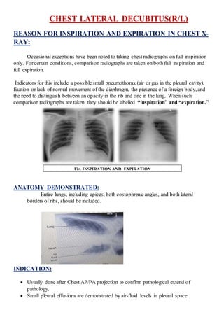
- 1. CHEST LATERAL DECUBITUS(R/L) REASON FOR INSPIRATION AND EXPIRATION IN CHEST X- RAY: Occasional exceptions have been noted to taking chest radiographs on full inspiration only. Forcertain conditions, comparison radiographs are taken on both full inspiration and full expiration. Indicators for this include a possible small pneumothorax (air or gas in the pleural cavity), fixation or lack of normal movement of the diaphragm, the presence of a foreign body, and the need to distinguish between an opacity in the rib and one in the lung. When such comparison radiographs are taken, they should be labelled “inspiration” and “expiration.” ANATOMY DEMONSTRATED: Entire lungs, including apices, both costophrenic angles, and both lateral borders of ribs, should be included. INDICATION: Usually done after Chest AP/PA projection to confirm pathological extend of pathology. Small pleural effusions are demonstrated by air-fluid levels in pleural space. Fig. INSPIRATION AND EXPIRATION
- 2. Small amounts of air in pleural cavity may demonstrate a possiblepneumothorax. Pneumoperitonium. TECHNIQUE: PATIENT POSITION: Place a cardiac board on the cart or radiolucent pad under patient. Patient will lying on right side for right lateraldecubitus and on left side for left lateral decubitus. Patient’s chin extended and botharms raised above head to clear lung field. Back of patient firmly against IR; cart secured to prevent patient from moving forward and possibly falling; pillow under patient’s head. Knees flexed slightly and coronal plane parallel to IR with no bodyrotation and is allowed to stay at leastfor 1 min for fluid/air to settle well. NOTEs:Place appropriate “decubitus marker” and R or L to indicate which side of chest is down. For possiblesmall amounts of air in the pleural cavity (pneumothorax), the affected side should be up side, and care must be taken not to cut off this side of the chest as in fig. below. For possiblefluid in the pleural cavity (pleural effusion), the suspected side should be down side and extend the lower limit of the cassette as down as possible upto the pathological area as in fig. below Pleural Effusion Pneumoperitonium
- 3. PART POSITION: Adjust height of IR to center thorax to IR . Adjust patient and cart to center midsagittal plane and T7 to CR (top of IR is approximately 1 inch [2.5 cm] above vertebra prominens). CR horizontal, directed to center of IR, to level of T7, 3 to 4 inches (8 to 10 cm) inferior to level of jugular notch. A horizontal beam must be used to show air-fluid level or pneumothorax. EXPOSURE CRITERIA: Minimum SID—72 inches (183 cm). IR size—35 × 43 cm (14 × 17 inches), crosswise(with respect to patient position). 60-75 kV range, SID-100cm, 10-12 mAs range. Use decubitus (decub) marker.
- 4. EXPOSURE CRITERIADIFFERENCE IN CHEST AP/PA AND DECUBITUS: If both air and fluid are present within a lung or within the pleural space, the heavier fluid, such as blood or pleural fluid resulting from infection or trauma, gravitates to the lowest position, whereas the air rises. In the recumbent/decubitus position, a pleural effusion spreads out over the posterior surface of the lung, producing a hazy appearance of the entire lung. In the upright position, free fluid is located near the base of the lung. The PA erect chest radiograph (Fig.) shows some fluid in the left lower thoracic cavity near the base of the lung. The supine radiograph taken on a different patient (Fig.) demonstrates a generalized hazy appearance of the entire right lung, resulting from the presence of fluid now spread throughout the right thorax. In chest AP/PA the fluid or air is settled below so it is not possible to rule out the exact level of the fluid or air extension. In chest lateral decubitus the fluid or air is settled below/above so it is possibleto rule out the exact level of the fluid or air extension. Fluid Level: Air Level: