Compare density gradient centrifugation, magnet-activated cell sorting
(MACS), and fluorescence-activated cell sorting (FACS) in the isolation of pure stem cell populations from a heterogeneous suspension
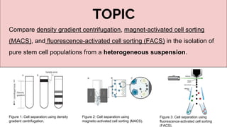
1. TOPIC
Compare density gradient centrifugation, magnet-activated cell sorting
(MACS), and fluorescence-activated cell sorting (FACS) in the isolation of
pure stem cell populations from a heterogeneous suspension.
Figure 1: Cell separation using density
gradient centrifugation.
Figure 2: Cell separation using
magnetic-activated cell sorting (MACS).
Figure 3: Cell separation using
fluorescence-activated cell sorting
(FACS).
2. Introduction
Why do we need to isolate stem cell from heterogenous suspension?
- To get pure stem cells.
How do we isolate them ?
Physical Parameters
Affinity
Size
Density
Chemical
Magnetic
Electrical
3. Density Gradient Centrifugation
Separation of components of a sample on the basis of their density, in a density gradient, in a centrifuge,
according to the centrifugal force they experience.
2 principle types
Rate zonal centrifugation Isopycnic Separation
● Separation of cells/particles based on the
differences in size, shape & density.
● involves carefully layering a sample
solution on top of preformed liquid density
gradient
● centrifuged until the desired degree of
separation is achieved
● Time dependent
● Separation of cells/particles solely by their density, not
by shapes, sizes & time
● Particle size only affect the rate at which particles
move until their density is the same as the
surrounding gradient medium.
● Used to separate particles of similar size but of
different density.
4. Rate zonal centrifugation Isopycnic Separation
Sample is layered as a narrow zone on the top of a
density gradient
Under centrifugal force, particles move at different
rates depending on their mass
Starting with a uniform mixture of sample and
density gradient.
Under centrifugal force, particles move until their
density is the same as the surrounding medium
Density Gradient Medium
Selection
Ideal density gradient media properties:
1. Sufficient solubility to produce the
range of densities required.
2. Does not form solutions of high
viscosity in the desired density
range.
3. Is not hyperosmotic or
hypoosmotic when the particles to
be separated are osmotically
sensitive.
4. Does not affect the biological
activity of the sample.
5. Non-toxic and not metabolized by
cells.
6. Does not interfere with assay
procedure or react with the
centrifuge tube.
7. Easily removed from the purified
product.
8. Autoclavable.
9. Low cost.
Eg: CsCl (isopycnic separation of DNA),
sucrose (rate-zonal separation of DNA),
Ficoll (separation of cells and subcellular
fractions
Gradient medium: to provide a
gradient of viscosity which improves
particle resolution while stabilizing the
column from convection currents
Gradient medium: density of gradient
media is higher than that of the
particles
6. Magnet Activated Cell Sorting
Utilization of microbeads that detect specific antigens and bind to them,
separation is then carried out by subjecting the sample to a magnetic field.
MicroBeads
● Superparamagnetic particles of approximately 50 nanometers in diameter
● Detect specific antigens and bind to them
● Conjugated to monoclonal antibodies
● Biodegradable
MACS Separators
● Powerful permanent magnets that induce a high-gradient magnetic field within
MACS Columns
MACS Columns
● MACS separation process occurs within the MACS Columns.
7. How Does It Work ?
Magnetic Labelling
Indirect labelling
Magnetic Separation Elution of labelled cell
fraction
Negative selection
● Magnetic field ON
● Untagged cells will elute
out.
● Cell of interest retain on
column.
Positive selection
● Magnetic field OFF
● Tagged cells will be eluted out
Direct Labelling
9. Fluorescence Activated Cell Sorting
Fluorescence-activated cell sorting is a specialized
type of flow cytometry.
Fluorescence-activated cell sorting able to sort
cells of heterogeneous suspension into different
containers according to
-light scattering
-fluorescent characteristic of each cell.
10. How Does It Works ?
1. Treat cells with fluorescent antibody
marker.
2. Cell mixture flow in stream and leave
nozzle as droplet
3. Laser beam strikes
4. Charge given to cells
5. Pass through electrically charged plate
6. Cells are separated
13. Emerging Method
Methods Aqueous Two Phase System SELEX Microfluidics
Principle - polyethylene glycol (PEG) (upper
phase) and dextran (lower phase) for
centrifugation
- resulting target cells form sediment
bands at the interface of the two
phases
- uses RNA, ssDNA, or
modified nucleic acids as
aptamers to selectively
capture target cells with
their high affinity
- Aims at miniaturization
- Mimic in vivo
microenvironment for cell
differentiation
- Chip based
Method - Uses temperature sensitive polymer
poly(N-isopropylacrylamide)
(PNIPAAm) which soluble at 20°C but
precipitate at 32-35°C
- Conjugated antibodies recognize
specific stem cells, capture and
precipitate out through switching of
temperature
(Beili.Z & Shashi.K, 2013)
- Incubate stem cells with
aptamers and remove
unbound aptamers
- The bound aptamers are
subsequently released from
surfaces of the stem cells
and are then further
amplified by RT-PCR for
SELEX cycle
- Antibodies have been
immobilized onto the luminal
surface of a parallel array of
hollow fibers
- Detachment of target cells
was performed in fluid flow
with a pre-defined shear
stress.
(Menachery. A et al, 2017)
14.
15. Density Gradient Centrifugation
Why this method ?
- Most simplified and cost effective.
- Label free from magnetic particles or antibodies.
- Ready to use (STEMCELL Technologies Inc., 2012).
Conclusion
16. Why this method ?
- Widely used in clinical settings for large scale processing. Due to its faster separation
compared to FACS.
- More cost effective compared to FACS (Pierzchalski et al. 2013).
Magnetic Activated Cell Sorting (MACS)
17. Fluorescence Activated Cell Sorting (FACS)
Why this method ?
- High sensitivity and precision compared to MACS (Zhu, B and Murthy, SK, 2013).
- Better separation of populations using antibodies (Flow Cytometer Facility, n.d.)
18. Conclusion
Which method to use ?
Most efficient Most simplified
Ready to use
High specificity
Generally, affinity-based approaches are most efficient & reliable, due to high
specificity. (Zhu, B and Murthy, SK, 2013)
Cost effective Precision
19. References
Asami, M, Higuchi, S, Shibata, N & Agata, K, 2006, ‘Isolation of planarian X-ray-sensitive stem cells by fluorescence-activated cell sorting’, Vol. 48, no. 6, pp.
371-380.
Catherine, M., Brian T, F. and Timothy C, F. 2010, Fluorescence-Activated Cell Sorting for CGMP Processing of Therapeutic Cells. 1st ed. [ebook] Sparks: BD
biosciences, p.7, viewed 20 May 2017,
<https://www.researchgate.net/profile/Timothy_Fong2/publication/228470167_FluorescenceActivated_Cell_Sorting_for_CGMP_Processing_of
_Therapeutic_Cells/links/55f6ef6e08ae07629dbb159e.pdf>
Flow Cytometer Facility, n.d., ‘FAQs for cell sorting’, viewed on 24 May 2017,
<https://med.virginia.edu/flow-cytometry-facility/resources/faqs/faqs-for-cell-sorting/>
Handgretinger, R, Lang, P, Schumm, M, Taylor, G, Neu, S, Koscielnak, E, Niethammer, D, L & Klingebiel, T, 1998,’Isolation and transplantation of autologous
peripheral CD341 progenitor cells highly purified by magnetic-activated cell sorting’, vol. 21, pp. 987-993.
Miltenyibiotec.com. (n.d.). MACS manual Cell Separation Columns - Miltenyi Biotec, online, viewed on 22 May 2017,
http://www.miltenyibiotec.com/en/products-and-services/macs-cell-separation/manual-cell-separation/columns.aspx
Miltenyi Biote, (n.d.) MACS Technolody Golden Standard in cell separation, viewed on 24 May 2017,
http://www.dartmouth.edu/~dartlab/uploads/MACS_Technology_Flyer.pdf
Oscar, Tom, Chen, WM, Lee, KD, Hsieh, SL & Chen, TH, 2003, ‘Isolation of multipotent mesenchymal stem cells from umbilical cord blood’, viewed on 20 May
2017, <http://www.bloodjournal.org/content/bloodjournal/103/5/1669.full.pdf?sso-checked=true>
20. Pierzchalski, A, Mittag, A, Bocsi, J, & Tarnok, A, 2013, ‘An Innovative Cascade System for Simultaneous Separation of Multiple Cell Types’, viewed on 24 May 2017,
<http://journals.plos.org/plosone/article?id=10.1371/journal.pone.0074745>
Shi, S and Gronthos, S, 2003, ‘Perivascular Niche of Postnatal Mesenchymal Stem Cells in Human Bone Marrow and Dental Pulp’, vol. 18, no. 4, pp. 696-704.
STEMCELL Technologies Inc., 2012, ‘How to Use SepMate™ to Isolate PBMCs from Whole Blood in Just 15 Minutes’, viewed on 20 May 2017,
<https://www.stemcell.com/how-to-use-sepmate-to-isolate-pbmcs-from-whole-blood-in-just-15-minutes.html>
Uchida, N, Buck, DW, He, D, Reitsma, MJ, Masek, M, Phan, TV, Tsukamoto, AS, Gage, FH, & Weissman, IL, 2000, ‘Direct isolation of human central nervous system
stem cells’, vol. 97, no. 26, viewed on 20 May 2017, <http://www.pnas.org/content/97/26/14720.abstract>
Zhu, B and Murthy, SK, 2013, ‘Stem Cell Separation Technologies’, vol. 2, no. 1, pp. 3-7.
References