Downloaded 4,700 times


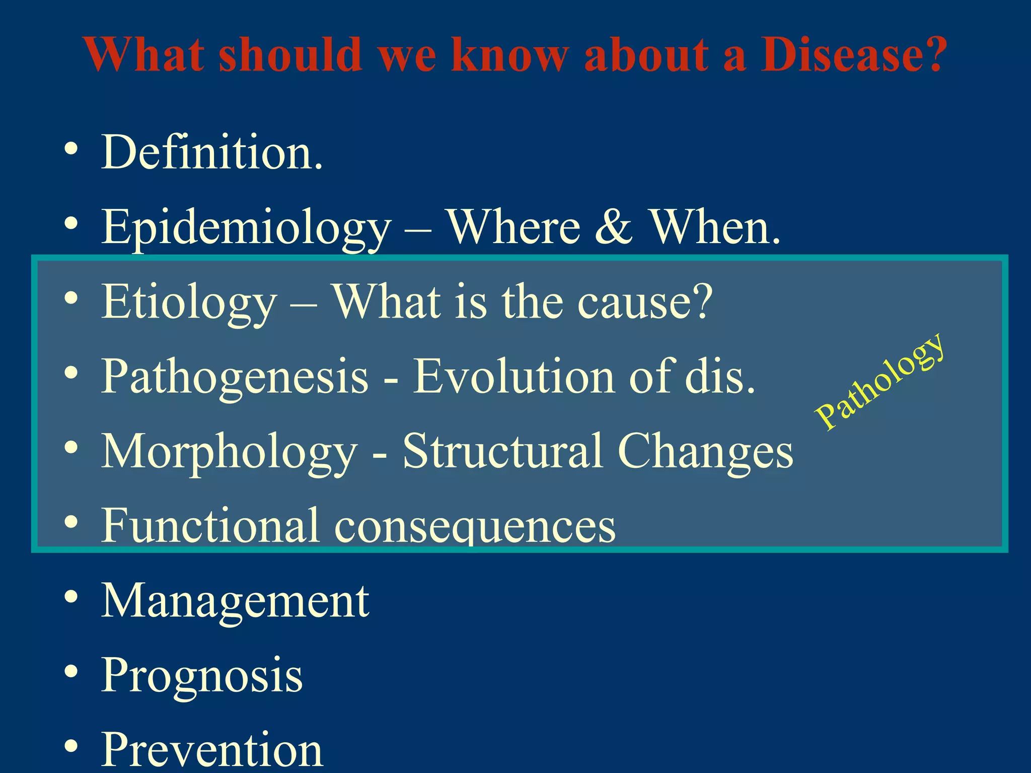
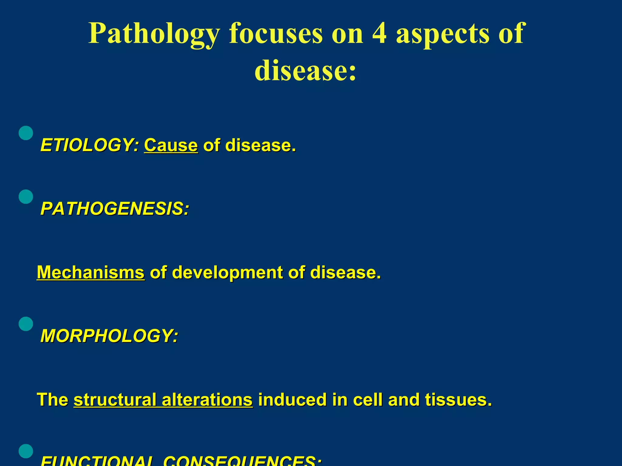


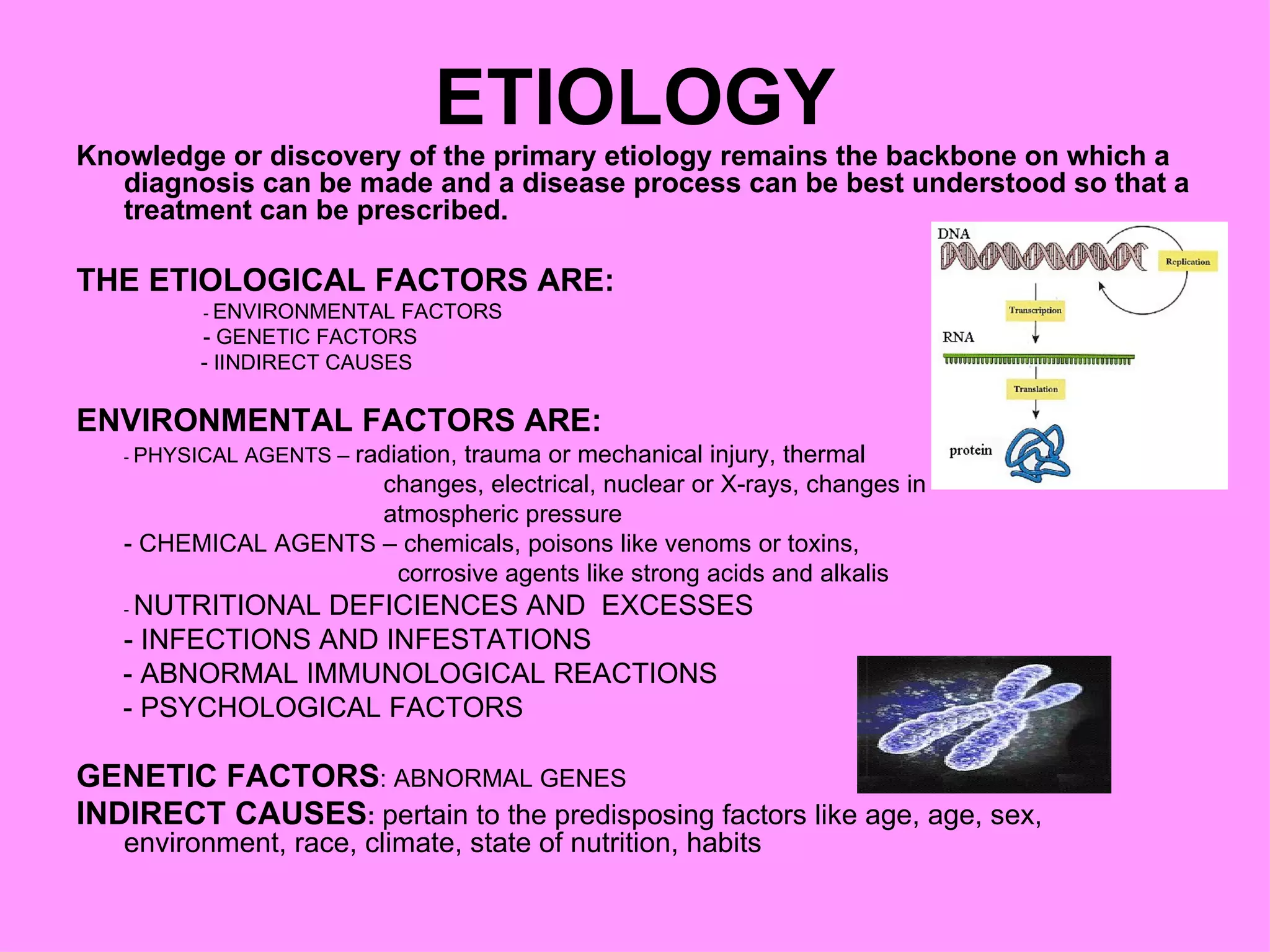


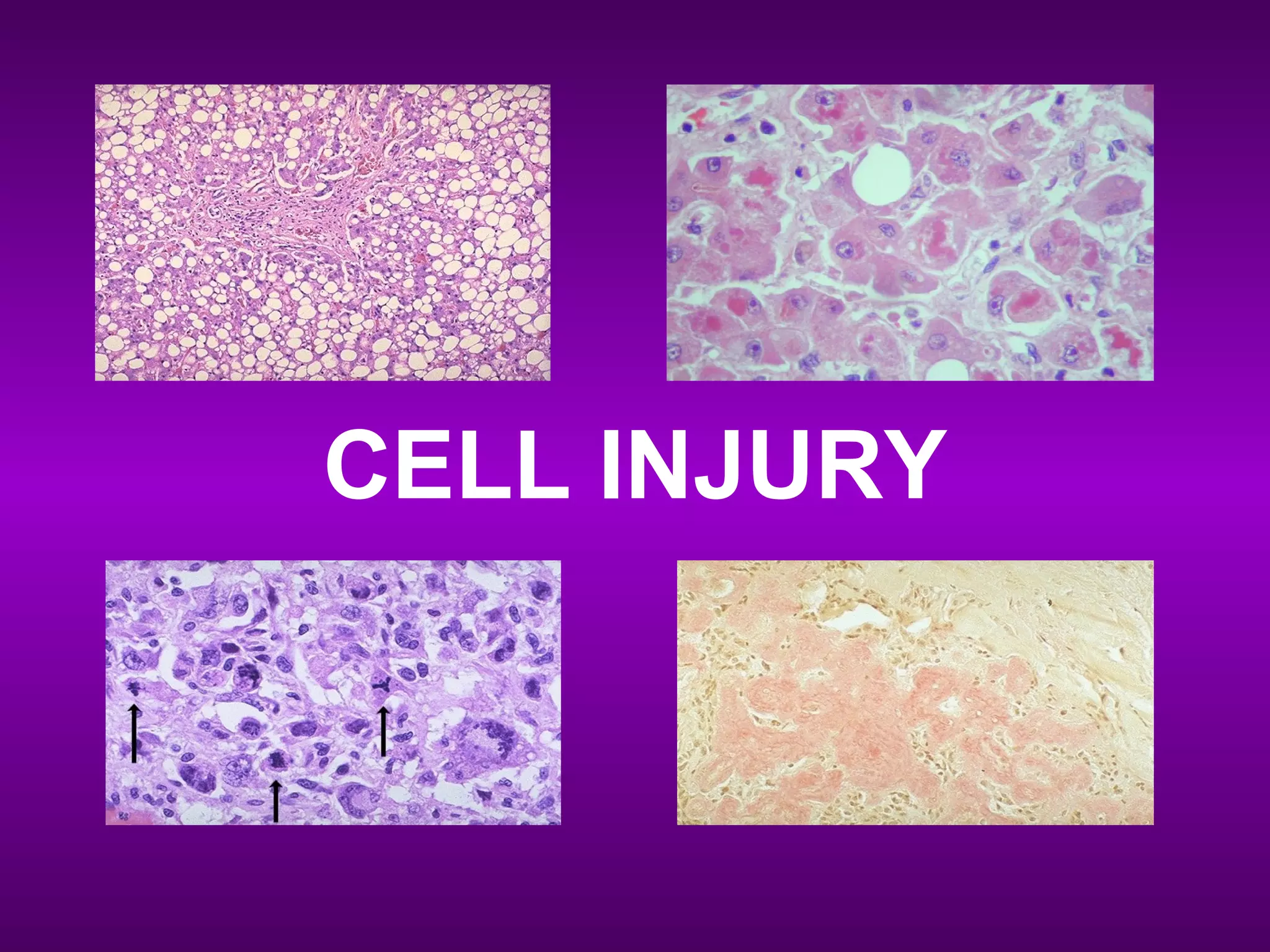

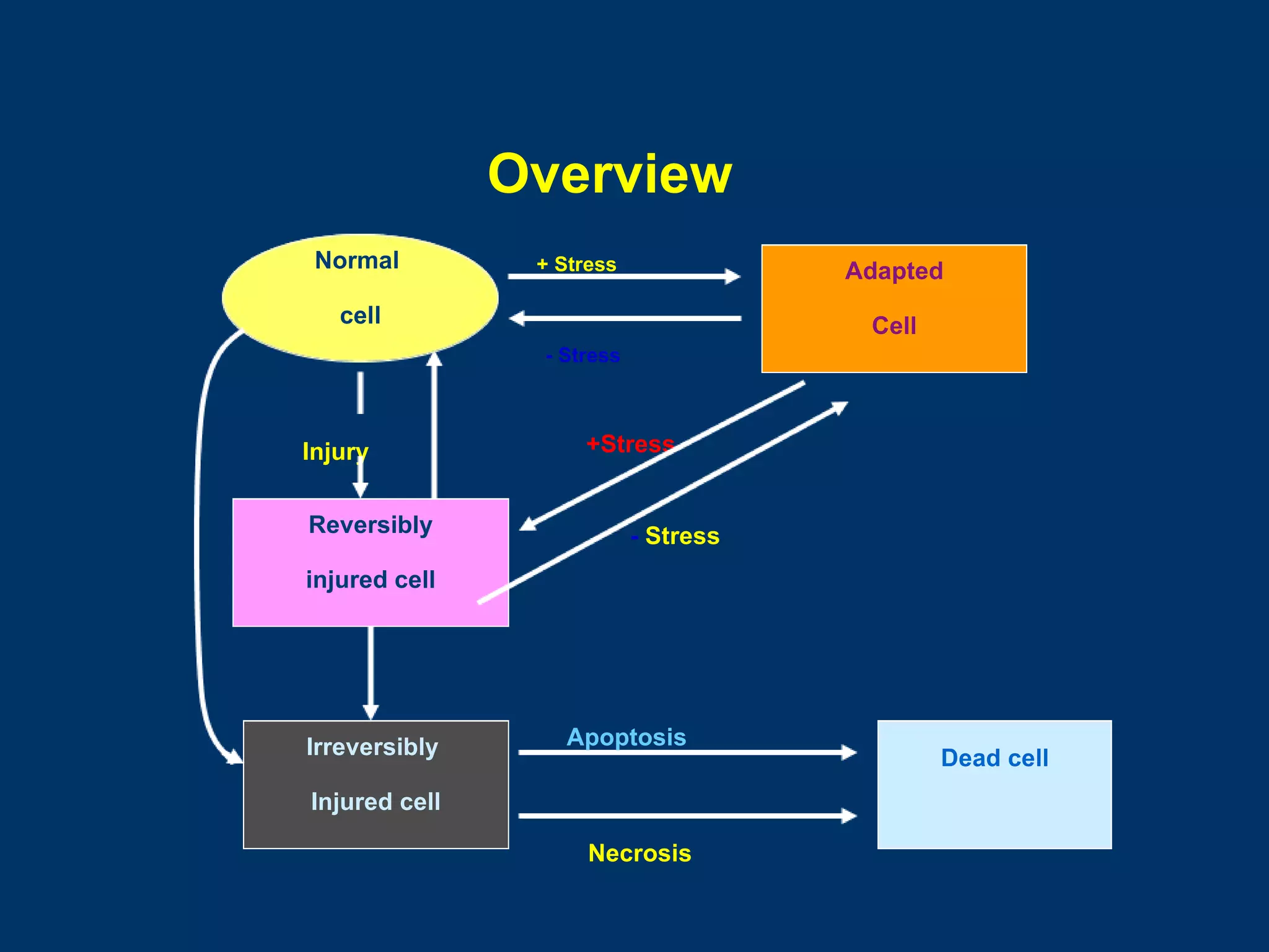

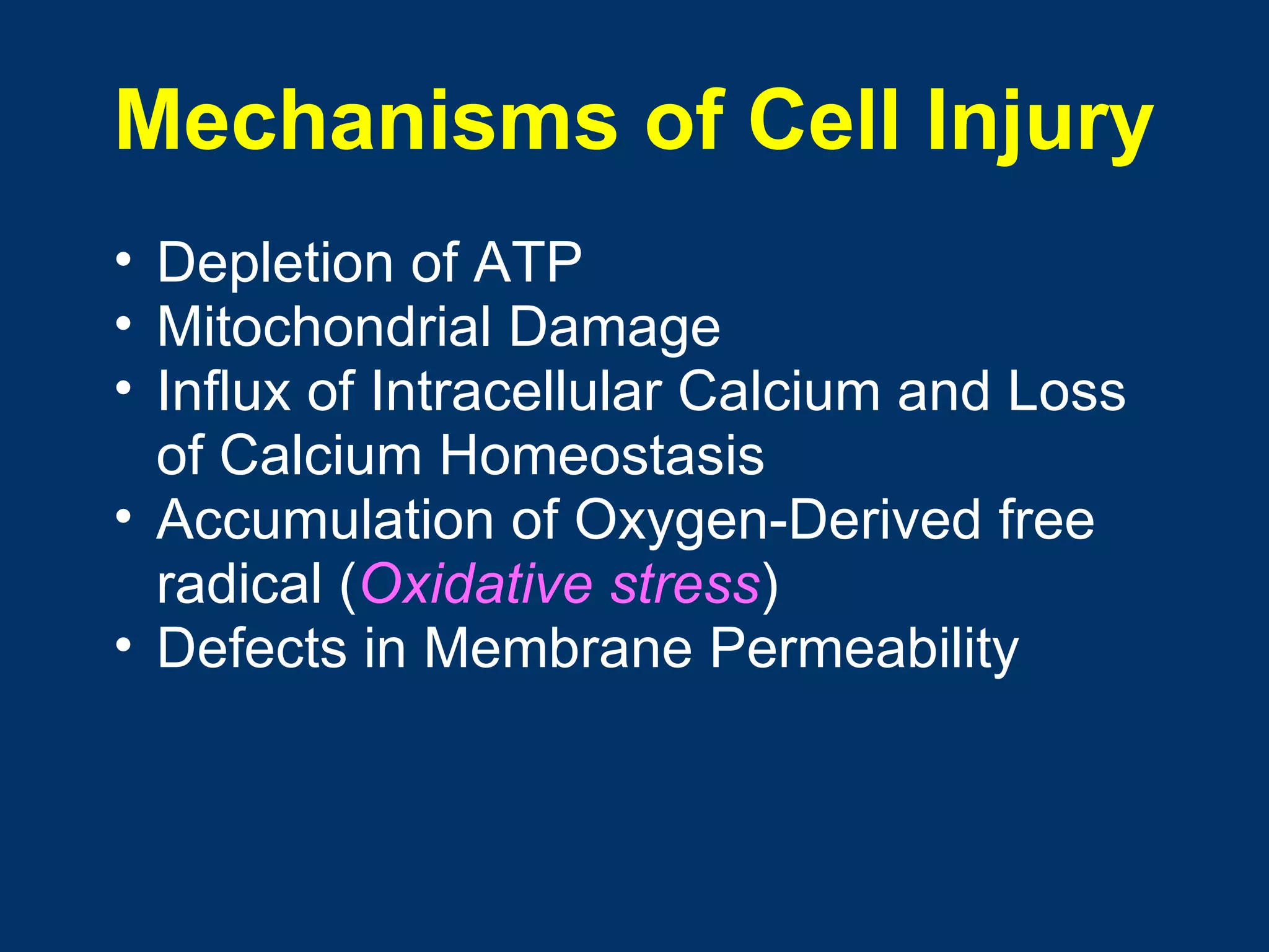

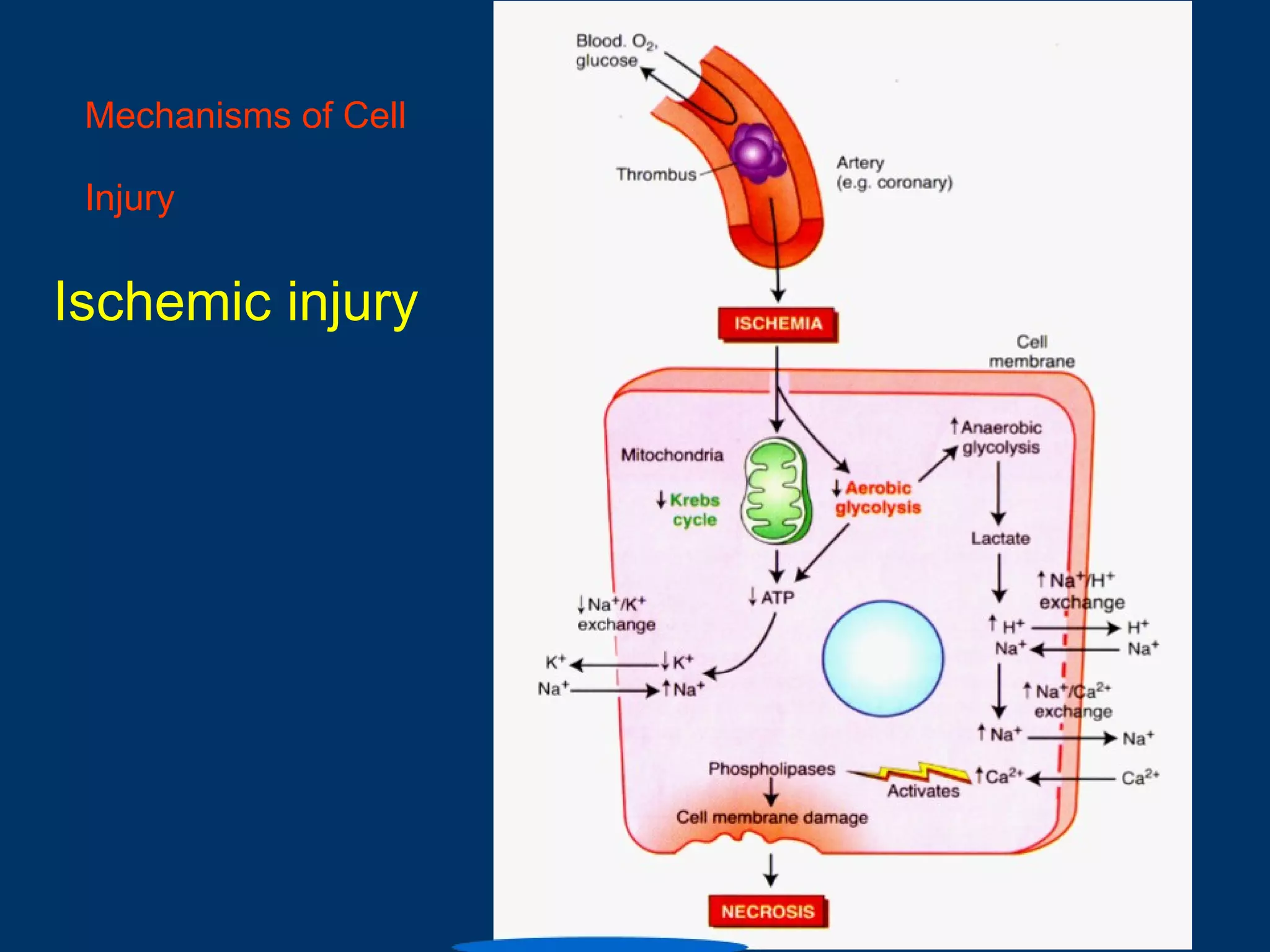
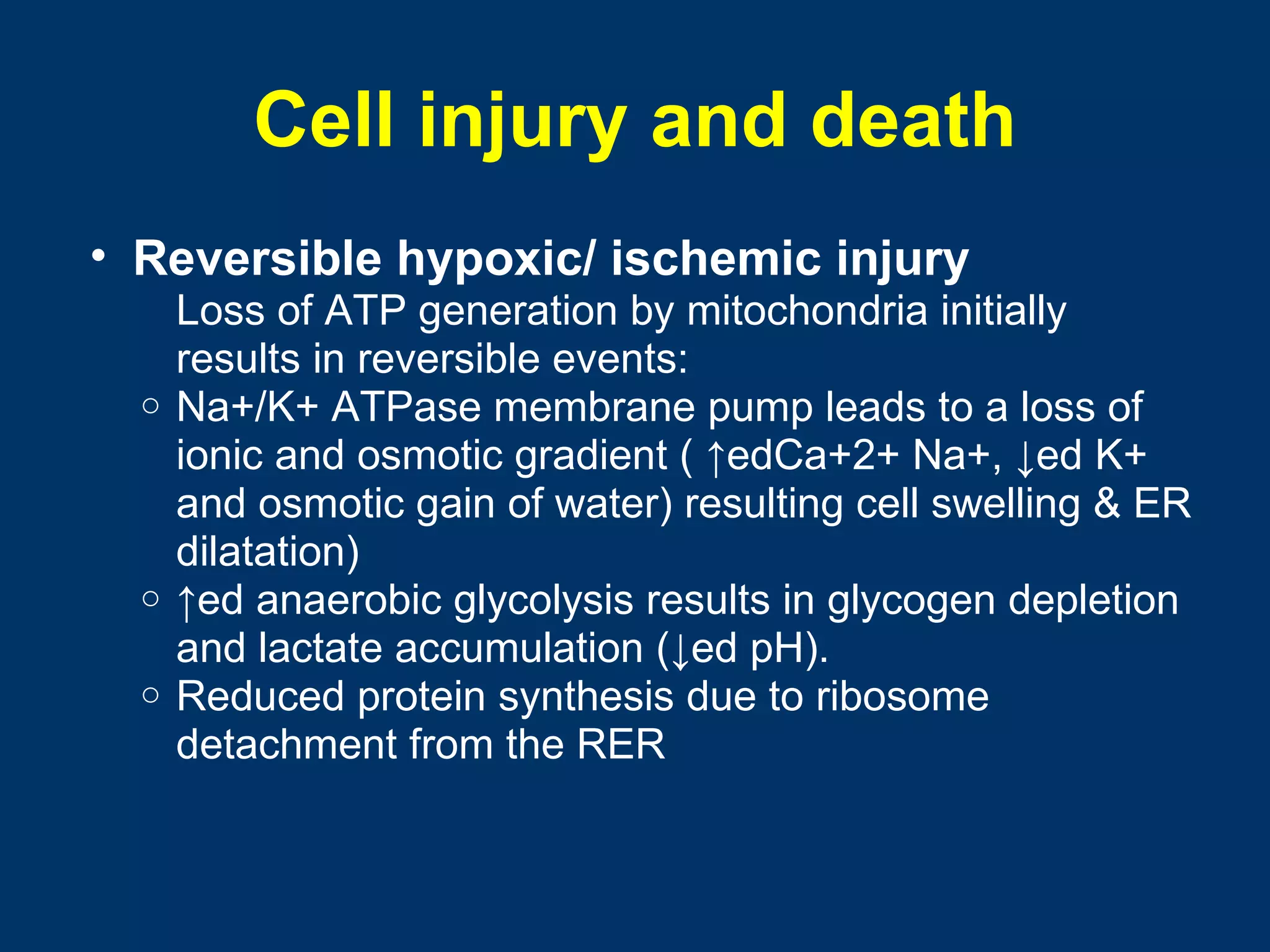


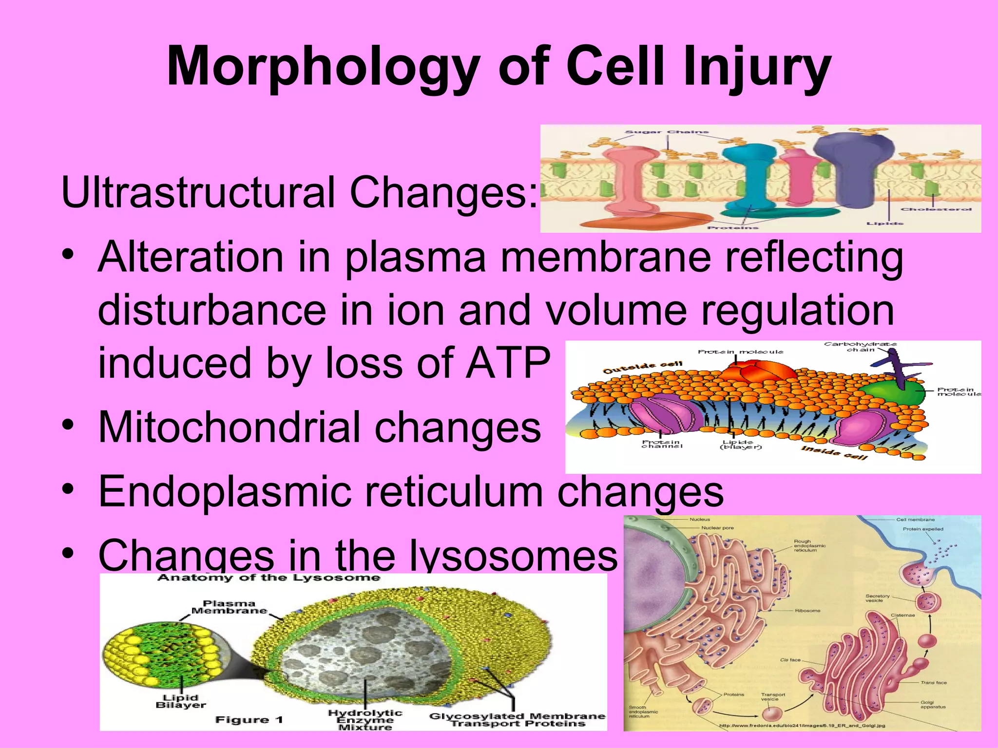


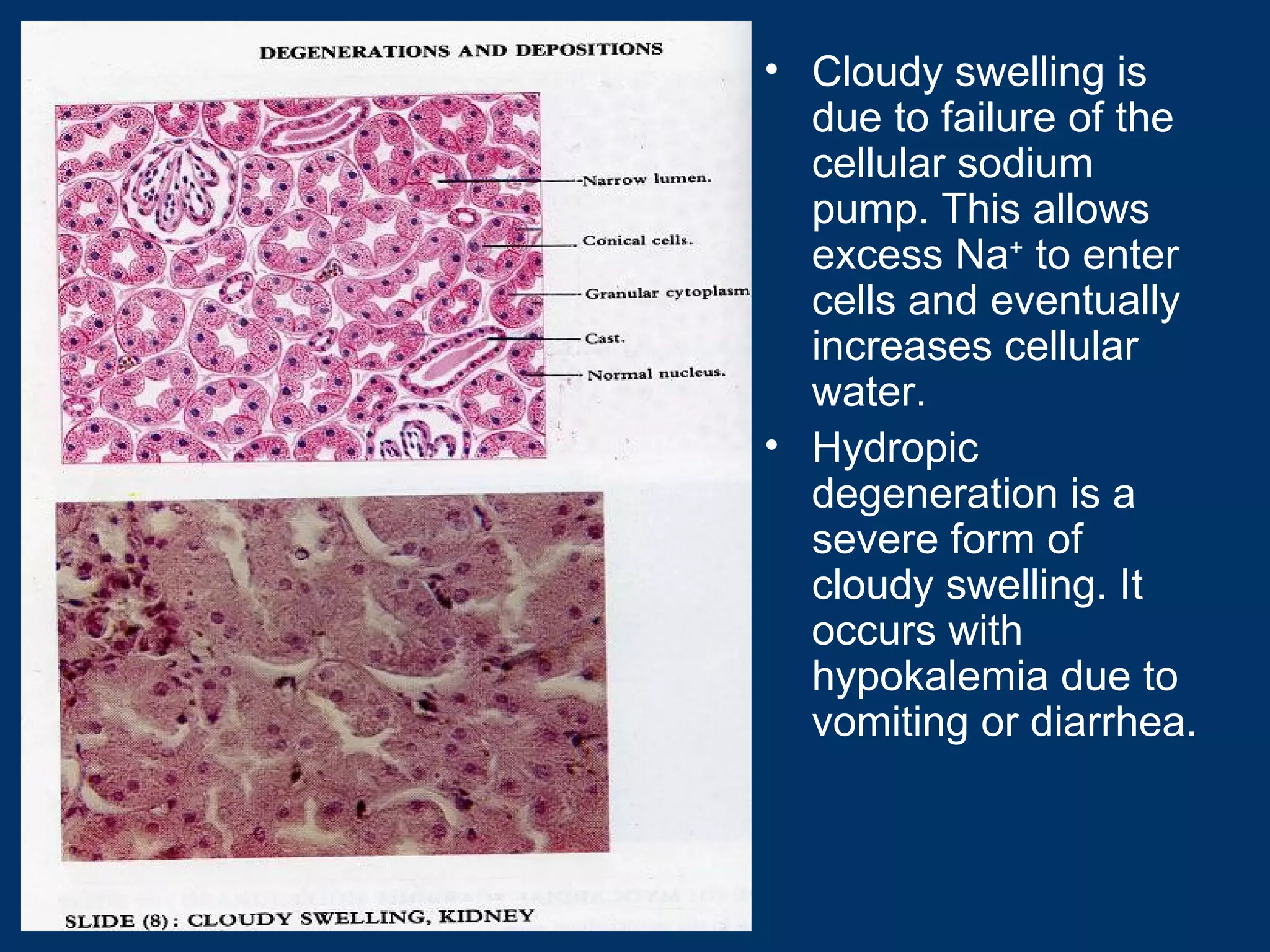


This document provides an overview of pathology and cell injury. It begins with definitions of pathology and discusses its focus on etiology, pathogenesis, morphology, and manifestations of disease. It then covers cell injury, describing the process from normal cell to reversible and irreversible injury. Specific types of cell injury are outlined like cloudy swelling, fatty change, and hyaline degeneration. The document concludes with examples of intracellular accumulations seen in various disease states.