
Recommended
More Related Content
What's hot
What's hot (20)
Similar to Membrane fusion
Similar to Membrane fusion (20)
More from obanbrahma
Recently uploaded
Recently uploaded (20)
Membrane fusion
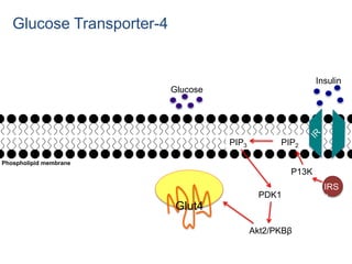
- 1. Glut4 P13K PIP3 PIP2 PDK1 Akt2/PKBβ IRS 1 Glucose Insulin Glucose Transporter-4 Phospholipid membrane
- 2. Membrane fusion • Can occur between cells, between intracellular compartments, between intracellular compartments and the plasma membrane, between lipid bound structures (such as viral particles) and cellular membranes • High curvature of the membrane promotes fusion
- 3. Fusion • Process by which 2 initially distinct lipid bilayers merge their hydrophobic cores resulting in one interconnected structure. • Contents can mix • If only one leaflet from each bilayer is involved in the fusion process the bilayer is hemifusion. -Inner layer remains distinct.
- 4. Hemifusion model • Believed that most if not all biological fusion proceeds through a hemifusion intermediate • Membrane fusion intermediates are regulated by proteins that bend and remodel membranes or by acting upstream to regulate the lipid or protein composition of the respective bilayers
- 5. Many membrane fusion events… • Exocytosis • Endocytosis • Fusion of egg to sperm • Transport of waste to lysozome • Entry of pathogens • Formation of myotubules • Synaptic vesicles
- 7. Challenge! Energy barriers have to be overcome • Bringing membranes in close proximity • Bringing together of repulsive membrane charges • Energy barrier of curvature formation for both hemifusion-stalk and fusion pore formation • Role of fusion proteins is to lower these barriers
- 8. 4 Steps need to happen for fusion to occur 1) Involved membranes must aggregate 2) bilayer needs to partially dehydrate otherwise they will repel each other 3) A destabilization must develop at one point b/w the bilayers, inducing a highly localized rearrangement of the two bilayers 4) This point defect grows and the components of the 2 bilayers mix and diffuse away from the site of contact
- 9. Divalent cations • Divalent cations play a critical role by binding to negatively charged lipids such as phosphatidylserine, phosphatidylglycerol and cardiolipin • One purpose of this is to shield negative charge on surface of bilayer and reduce electrostatic repulsion between bilayers
- 10. Fusion events need: • Molecules to tether and dock membranes and bring them in close proximity. • Molecules that locally disturb the lipid bilayers (ex. by inducing curvature) • Molecules that give direction to the process
- 12. Other factors • Lipid head group also affects dehydration • Independent of charge • PE binds water less tightly than PC • Size might also be a factor, according to the stalk hypothesis, a highly curved bridge must form. PE has smaller head group and can form inverted micelle phases
- 13. Fusion proteins • In vitro fusion is regulated by membrane associated proteins • First to be studied is the viral fusion proteins which allow an enveloped virus to insert its genetic material into the host cell
- 14. 2 classes of viral proteins • Acidic and pH independent • pH independent fusion proteins can function under neutral conditions and fuse with plasma membrane • HIV, measles, mumps • Acidic fusion proteins such as in influenza are only activated in low pH of acidic endosomes and must first be endocytosis.
- 15. Viral fusion 1) Transmembrane viral fusion proteins are kept in an inactive state on the viral surface 2) following exposure to an appropriate trigger the viral fusion proteins undergo dramatic conformation changes exposing fusion peptides or loops which insert into target 3) either concurrently or subsequently the fusion proteins undergo an additional conformational change that brings the transmembrane domains in the viral envelop into close proximity with the viral fusion peptides that are embedded in the target 4) membrane fusion occurs as a consequence of close proximity and bilayer disturbance
- 16. Viral surface fusion proteins: 3 classes • Class I mainly alpha helical • Class II mainly beta sheets • Class III are mixed secondary Often disrupt one layer and induce curvature
- 17. Mitochondrial fusion • Fusion and fission are necessary for normal mitochondrial function • Process unclear! • Homotypic fusion is unusual because it involves both outer and inner mitochondria • Dependent on dynamin superfamily GTPases, OPA1 (optin atrophy protein-1) and mitofusion • Members of this family are large, self-oligomerizing GTPases that mediate remodeling • Mitofusions can tether mitochondria to each other – mediated by c-terminal alpha helices from opposing mitochondra together form an antiparallel coiled structure
- 18. Cell-cell fusion • Essential during fertilization, development and immune responses • Little conservation from yeast to nematodes to insects to mammals likely involved separately • Lots unknown!
- 19. Assays to measure fusion: • Measure either mixing of membrane lipids or mixing of aqueous contents
- 20. Lipids mixing • NBD-Rhodamine Energy Transfer:membrane labeled with both NBD and Rhodamine combine with unlabeled membrane. When NBD and Rhodamine are within a certain distance, the Förster resonance energy transfer (FRET) happens. After fusion, resonance energy transfer (FRET) decreases when the average distance between probes increases, while NBD fluorescence increases. • Pyrene Excimer Formation: emission wavelength of monomer of pyrene is 400nm. The eximer is 470nm. Membrane labeled with pyrene combines with unlabeled membrane. Before fusion, majority of emission is excimer, after the distance between probes • Octadecyl Rhodamine B Self-Quenching:. Rhodamine dimers quench fluorescence. Fusion with unlabeled membranes resulting in dilution of the probe.
- 21. Contents mixing • Fluorescence quenching assays with ANTS/DPX: ANTS is a polyanionic fluorophore, while DPX is a cationic quencher. The assay is based on the collisional quenching of them. Separate vesicle populations are loaded with ANTS or DPX, respectively. • Fluorescence enhancement assays with Tb3+/DPA: This method is based on the fact that chelate of Tb3+/DPA is 10,000 times more fluorescent than Tb3+ alone.
- 22. Eukaryotic cells use different proteins • Best studied are SNAREs • Direct all vesicular intracellular trafficking • Debate on whether SNAREs are involved in early docking or participate late in the fusion process by facilitating hemifusion • Enormous diversity of structure and function within these classes and very few themes are conserved.
- 23. SNARE-dependent • SNARE motifs are the regions that contribute to the formation of a highly stable four-helix bundle called the SNARE complex • Each SNARE contributes one helix • Helices are all aligned in parallel • The folding of this bundle is thought to drive the fusion reaction
- 24. Fertilization • Few candidate protein for mediating fusion have been identified • CD9, a multiple transmembrane –domain protein on the egg surface • IZUMO, a single transmembrane protein with an exterior Ig-like domain on the sperm • CD9 localize in areas of extreme curvature called microvilla
- 25. Endosomal fusion • Endosomes fuse with late endosomes or lysosomes • SNAREs are essential • Syntaxin-6, syntaxin-13, VTI1A, VAMP4
- 26. Synaptic vesicle Synchronous release • Is the burst of small synaptic vesicle exocytosis after depolarization Asynchronous release • Following synchronous release, isolated exocytic events occur • Both are Ca2+ dependent • Molecules that are directly involved in fusion steps are synaptotagmins and SNAREs. • Accessory proteins are involved.
Editor's Notes
- Insulin initiates a signal cascade resulting in tyrosine kinase activity Pre-diabetes is a condition in which blood glucose levels are higher than normal but not high enough for a diagnosis of diabetes. This condition is sometimes called impaired fasting glucose (IFG) or impaired glucose tolerance (IGT),
- Membrane fusion in the secretory pathway and in the endosomal and lysosomal systems depends on SNAREs, which are assisted by tethering and regulatory factors that are generally required for efficient SNARE function. During Ca2+-dependent exocytosis the SNAREs are assisted by synaptotagmins, and in endosome fusion they are assisted by the Rab5 effector EEA1. SNAREs and tethering/regulatory factors are replaced by viral fusion proteins in enveloped viruses and by immunoglobulin (Ig)-domain-containing proteins in many cell–cell fusion events. The actin cytoskeleton has also been implicated in membrane fusion and in particular in cell–cell fusion, where it might stabilize the microvilli. Furthermore, multiple-C2-domain (MC2D) proteins such as tricalbin in yeast and myoferlin in mammals have been proposed to function during plasma-membrane repair during leaky cell–cell fusion. Yeast vacuole fusion requires SNAREs and the tethering factor HOPS (homotypic fusion and vacuole protein sorting). Mitochondrial fusion is mediated by the large GTPases mitofusin and OPA1 of the dynamin superfamily. Mgm1 and Fzo1 are the yeast orthologues of OPA1 and mitofusin, respectively. Plasma-membrane repair is initiated by the influx of Ca2+ into the cytoplasm and is mediated by the rapid and local fusion of small vesicles with each other and with the plasma membrane. The MC2D protein dysferlin has been shown to be required for this fusion. The involvement of SNAREs in plasma-membrane repair has not been explicitly shown. ER, endoplasmic reticulum.
- Come close together within several nanometers
- Proposed steps in synaptic vesicle fusion with the plasma membrane. Most membrane-fusion events are likely to follow a similar sequence. Steps might not be as temporally delineated as indicated, but the stepwise depiction helps to conceptualize the process. All the steps indicated are regulated by cellular proteins. In step 1, the vesicle is transported and tethered to the appropriate membrane by specific tethering factors, which mediate the long-range recognition between the membranes. In step 2, the loosely tethered state is converted to a tightly docked state, bringing the membranes into closer proximity. In some specialized cells docking is followed by a priming step, during which the fusion machinery is assembled such that it can rapidly respond to a trigger (for example, changes in the Ca2+ concentration). Docking should also entail the generation of protein-denuded membranes (Fig. 3). In step 3, the high-energy barrier must be lowered to initiate membrane fusion and some membrane stress, such as curvature stress, probably facilitates the reaction. In addition, the distance between the two membranes has to be further decreased in order to bring the membranes into direct contact. Indeed, bilayers have been proposed to fuse when they are separated by 1 nm152. In step 4, hemifusion occurs. Hemifusion is the defining step of this fusion model; in it apposing monolayers merge, whereas distal monolayers do not. In step 5, fusion-pore opening results from the further merger of the two distal monolayers and the release of vesicle content is initiated. In step 6, as a consequence of fusion-pore expansion, the vesicular membrane collapses into the plasma membrane and loses its identity.
- Uncharged headgroup PE increases fusion
- Enveloped viruses have a lipid billayer. Some viruses have only a protein coat.
- Much debate!
- 1)When content mixing happens, ANTS and DPX collide and fluorescence of ANTS monitored at 530 nm, with excitation at 360 nm is quenched. This method is performed at acidic pH and high concentration. 2) . In the Tb3+/DPA assay, separate vesicle populations are loaded with TbCl3 or DPA. The formation of Tb3+/DPA chelate can be used to indicate vesicle fusion. Terbium
- Diagram of the action of SNARE proteins docking a vesicle for exocytosis. Complementary versions of the protein on the vesicle and the target membrane bind and wrap around each other, drawing the two bilayers close together in the process
- Essential for neurotransmitter and hormrone release Fusion events are tightly coupled to to the influx of Ca+2 Synaptotagmins are Ca+2 sensors SNARES= SNAP-25 and Syntaxin, synaptobrevins DOC are calcium binding proteins ca levels are lower Synchronous is fast