More Related Content
Similar to Acs0705 Injuries To The Chest
Similar to Acs0705 Injuries To The Chest (20)
More from medbookonline (20)
Acs0705 Injuries To The Chest
- 1. © 2004 WebMD Inc. All rights reserved. ACS Surgery: Principles and Practice
7 TRAUMA AND THERMAL INJURY 5 INJURIES TO THE CHEST — 1
5 INJURIES TO THE CHEST
Edward H. Kincaid, M.D., and J.Wayne Meredith, M.D., F.A.C.S.
Most persons who experience torso trauma, whether blunt or pen- TUBE THORACOSTOMY: INDICATIONS AND TECHNIQUE
etrating, sustain some degree of associated injury to the chest. During initial resuscitation, chest tube placement can be both
Thoracic injuries are a primary or contributing cause of death in therapeutic and diagnostic.The two most common indications for
nearly half of all cases of torso trauma.1 Fortunately, many tho- tube placement in this setting are pneumothorax and hemothorax;
racic injuries can be treated effectively, and often definitively, by however, signs and symptoms of these conditions may not be
relatively simple maneuvers that can be learned and performed by readily apparent. In addition, when the patient is in shock, the sur-
most physicians involved in early trauma care. Approximately one geon often cannot afford to take the time to differentiate between
in six patients, however, has life-threatening injuries that necessi- various possible causative conditions. Because tube thoracostomy
tate urgent operative repair. These extremes in injury severity are is quick, relatively safe, and simple, it should be liberally used for
unique to the chest and require a correspondingly broad range of patients in extremis.
knowledge and skills on the part of the treating surgeon. Tension pneumothorax, the most common and easily treated
immediately life-threatening thoracic injury, results when blunt or
Initial Evaluation and Management penetrating trauma disrupts the respiratory system and allows air
to escape from the lung parenchyma or the tracheobronchial tree
PRIMARY SURVEY into the pleural space, thereby increasing intrathoracic pressure.
This increased pressure is transmitted to all the cardiac chambers
Initial evaluation and treatment of patients with thoracic
and retards venous return to the heart, resulting in hypotension.
injuries are guided by the same principles and priorities as initial
The classic signs of tension pneumothorax—decreased breath
evaluation and treatment of patients with other injuries. Eval-
sounds, tympany on the ipsilateral side, tracheal shift, and dis-
uation begins with an organized and rapid primary survey aimed
tended neck veins—commonly are absent or are incompletely
at recognizing and treating immediately life-threatening problems.
manifested in a busy emergency department. The diagnosis is
The first priority is to ensure an adequate airway. An airway
often suggested by the presence of shock accompanied by evi-
often can be established by clearing any blood or debris from the
oropharynx and pulling the mandible or the tongue forward. dence of adequate venous filling on physical examination and
Severely injured patients commonly require nasotracheal or oro- recognition of asymmetric motion of the two sides of the chest.
tracheal intubation, and some, especially those with severe max- Treatment of suspected tension pneumothorax should not be
illofacial trauma, require cricothyroidotomy or tracheostomy. delayed in patients with hemodynamic compromise.
The second priority is to ensure adequate ventilation. If the Chest tube placement in the trauma setting is a straightforward
patient is not breathing, he or she must be intubated promptly. If procedure. The chest is prepared and draped in a sterile fashion.
ventilation is inadequate because of open or tension pneumotho- A local anesthetic (e.g., 1% lidocaine) is not required in uncon-
rax, these problems should be addressed at this stage of care. scious patients but should be used in alert patients. On the midax-
The next priority is control of external hemorrhage and restora- illary line in approximately the fifth interspace, a scalpel is used to
tion of circulation. External hemorrhage is best controlled by make a 2 to 3 cm incision, oriented in the direction of the inter-
direct pressure. Inadequate perfusion generally results from either space, through all layers of the skin and subcutaneous tissue. A fin-
hypovolemia or pump (i.e., cardiac) problems. Hypovolemia from ger or a blunt clamp is inserted to penetrate the intercostal mus-
hemorrhage often must be treated operatively as part of the resus- cles and the parietal pleura.The wound is explored with the index
citative effort. Pump problems are signaled by distended neck finger (in adults) or the fifth finger (in children) to ensure that the
veins and are caused by one of four conditions: (1) tension pneu- pleural space has been entered and allow local exploration of the
mothorax, (2) pericardial tamponade, (3) coronary air embolism, chest cavity.
or (4) cardiac contusion or myocardial infarction (MI).These con- In adults, a 36 French chest tube is then inserted and directed
ditions are discussed in detail elsewhere (see below). At this early posteriorly and towards the apex for optimally effective drainage
stage of treatment, they should be addressed sufficiently to ensure of air and blood. The desired tube position is best achieved
adequate perfusion. through appropriate orientation of the skin incision relative to the
In most blunt trauma patients, urgent treatment of thoracic entrance into the chest cavity: the straight line between these two
injury is accomplished during the primary survey because the points defines the direction of the tube once it is in the chest.The
most common blunt chest injuries can be controlled with endo- tube is then attached to 20 cm of suction with a water seal and a
tracheal intubation or tube thoracostomy. In this setting, thoracot- collection chamber. Visual inspection of air passing through the
omy is indicated for cardiac tamponade, a massive hemothorax, or water seal yields an estimate of the size of the air leak—an impor-
uncontrolled massive air leaks. Neither pulmonary nor cardiac tant consideration with suspected airway injuries.
contusions should delay diagnosis or definitive treatment of Collection chambers for hemothoraces should be of the same
extrathoracic injuries resulting from blunt trauma. design. Those associated with autotransfusion devices (e.g., cell
- 2. © 2004 WebMD Inc. All rights reserved. ACS Surgery: Principles and Practice
7 TRAUMA AND THERMAL INJURY 5 INJURIES TO THE CHEST — 2
savers) have immense theoretical potential for rapid retrieval and thrive, are diagnostic. Analysis and culture of fluid obtained at tho-
processing of shed blood. In practice, however, their utility is lim- racocentesis or chest tube placement typically confirms the diag-
ited. For example, with a small to moderate-sized hemothorax (< nosis, but the fluid may be sterile if the patient is already receiving
1,000 ml), the red-cell yield of an autotransfusion device would be antibiotics.
small and not worth the associated time and expense.With a large Antibiotic therapy, either broad-spectrum or specifically direct-
hemothorax, the most important goal of therapy is control of ed against cultured organisms (usually gram-positive pathogens),
bleeding, and arranging for autotransfusion could delay or hinder is certainly an important component of therapy for empyema tho-
the achievement of this goal. In addition, products of autotrans- racis, but the primary goal is removal of the infection while the
fusion may contain harmful cytokines, damaged cells, and debris, fluid is still thin.When this goal is met, a more modest therapeu-
while lacking platelets and other important proteins and coagula- tic procedure can be performed, there is less risk that a restrictive
tion factors.2 pulmonary peel will develop, and the injured patient recovers
Antibiotic prophylaxis after tube thoracostomy is controver- faster overall. In the early stages, tube thoracostomy may suffice
sial.3 Most clinicians, however, would recommend use of a first- for treatment; however, if the infected pleural process cannot be
generation cephalosporin for 24 hours, ideally starting before the completely evacuated via chest tube because of thicker fluid, loc-
initial tube placement. ulations, or pleural adhesions, a formal thoracotomy with decor-
After the primary survey, less dramatic pneumothoraces may tication is generally required.
be recognized on diagnostic images, along with hemothoraces, on Decortication should not be undertaken in the face of severe
various imaging studies. Treatment of occult pneumothoraces sepsis. Instead, antibiotics and chest drainage (via tube thoracos-
(i.e., those seen only on computed tomography) deserves special tomy, CT-directed catheter placement, or open rib resection)
mention. In general, patients with occult pneumothoraces who should be employed until sepsis is controlled. In cases of early
require positive pressure ventilation, those who are hypotensive or empyema,VATS has been successfully used for lysis of adhesions
have respiratory distress of any etiology, and those who have asso- and removal of fluid.9,10 Because of the limited capacity for per-
ciated complex injuries or hemothorax should be treated with forming pleurectomy with this procedure, VATS should not be
tube thoracostomy. One group, however, has questioned the need used when thick peel or a trapped lung is present. In adult patients
for tube thoracostomy in patients requiring positive pressure ven- with posttraumatic empyema, there is no proven role for
tilation on the basis of findings from a small number of patients.4 intrapleural fibrinolytic therapy.
Patients with occult pneumothoraces who are treated without
EMERGENCY DEPARTMENT THORACOTOMY
tube thoracostomy should be observed for at least 24 hours.
ED thoracotomy is a drastic step in the treatment of the injured
Retained Hemothorax and Empyema patient. If possible, the patient should be stabilized and transport-
In treating a hemothorax with tube thoracostomy, the goal is ed to the operating room, where better facilities are available for
complete removal of blood. Complications such as atelectasis and definitive care.
empyema after chest trauma are clearly related to the presence of ED thoracotomy is best reserved for patients who arrive at the
residual blood, fluid, and air, as can occur secondary to improper ED and deteriorate rapidly and those who have undergone car-
positioning of the tube (i.e., within a fissure), obstruction of the diac arrest just before arrival.The results are dismal when it is per-
tube, or blood clot or loculated fluid within the chest. formed in patients who have undergone cardiac arrest some time
A persistent or clotted hemothorax is suggested by the presence before arrival and have required cardiopulmonary resuscitation
of a persistent opacification in the pleural space in a patient with for more than a few minutes. Blunt trauma victims who have sus-
a known previous hemothorax.This radiodensity can be confused tained cardiopulmonary arrest at the scene of injury should not be
with adjacent pulmonary contusion or atelectasis; chest CT con- subjected to thoracotomy, either at the scene or in the ED.11
firms the diagnosis. Because retained blood serves as a nidus for Similarly, patients who are found at emergency thoracotomy to
infection and empyema,5 aggressive attempts at removal are justi- have no cardiac activity have a dismal prognosis, as do those who
fied. Occasionally, removal can be accomplished by placing more do not respond to improvement of systolic blood pressure after
chest tubes, but often, an operative approach is needed. Video- aortic occlusion. Overall, the survival rate for patients undergoing
assisted thoracic surgery (VATS) [see 4:10 Video-Assisted Thoracic ED thoracotomy for blunt trauma is lower than 10%.The report-
Surgery] may be useful for managing small, clotted hemothoraces ed survival rate for patients undergoing ED thoracotomy for pen-
and free-flowing blood in patients who can tolerate single-lung etrating trauma ranges from 16% to 57%,11-13 and that for
ventilation6 ; however,VATS tends to limit the surgeon’s ability to patients with cardiac wounds ranges from 57% to 72%.11-13
control bleeding and perform definitive repair of injuries. In The technique of ED thoracotomy is straightforward. An anti-
patients who have ongoing bleeding or large, clotted hemotho- septic solution may be splashed on the chest, but skin preparation
races, posterolateral thoracotomy is generally required. is not required. An incision is made from the sternal border to the
Empyema thoracis is a relatively common complication after midaxillary line in the fourth intercostal space. A chest retractor is
chest trauma, occurring in 5% to 10% of patients.7,8 Possible caus- inserted and opened widely. The costochondral junctions of the
es include retained hemothorax, pneumonia with parapneumonic fifth, the fourth, and sometimes the third rib are divided quickly
effusion, persistent foreign body, ruptured pulmonary abscess, bron- with the scalpel to provide exposure. Attention is directed first to
chopleural fistula, esophageal leakage, and tracking through the the injury. If there is exsanguination from a great vessel, the hem-
intact or injured diaphragm from an abdominal source. Empyema orrhage is controlled with pressure. If air embolism is the cause of
may be difficult to diagnose and must be differentiated from pleur- the arrest, the hilum is clamped and air evacuated from the aorta.
al thickening, pulmonary contusion, and an uninfected effusion. Otherwise, the pericardium is opened anterior and parallel to the
Chest CT with intravenous contrast usually demonstrates a fluid phrenic nerve. The hemopericardium is evacuated, the cardiac
collection with loculations or an enhancing rim. Such findings, injury is controlled with digital pressure, and a temporary repair
coupled with a clinical scenario of low-grade sepsis or failure to is performed.
- 3. © 2004 WebMD, Inc. All rights reserved. ACS Surgery: Principles and Practice
7 TRAUMA AND THERMAL INJURY 5 INJURIES TO THE CHEST — 3
After the cause of the arrest has been addressed, the descend- Table 1 Surgical Approaches for Traumatic
ing thoracic aorta is occluded with a vascular clamp or digital pres- Injuries to Thoracic Structures
sure and intrathoracic cardiac compression is initiated. The
patient’s intravascular volume is restored, and electrolyte imbal- Incision
ances are corrected. If the patient can be saved, he or she is trans-
ported to the OR for definitive repair and closure. Site of Injury Right Left
Sternotomy Thoracotomy Thoracotomy
SECONDARY SURVEY AND DEFINITIVE DIAGNOSIS
Right atrium +++ ++ 0
The secondary survey should focus on more subtle evidence of
Right ventricle +++ + +
injury that may be detected on physical examination and chest x- Left atrium +++ + +
ray. Simple rib fractures are clinically relevant when associated Left ventricle ++ 0 +++
with pain and are better diagnosed by palpation than by most Superior vena cava +++ ++ 0
imaging studies. Pneumothorax and hemothorax often go unde- Azygos vein ++ +++ 0
tected during the primary survey but are common findings later in Inferior vena cava +++ ++ 0
the workup. Aortic root +++ + 0
Echocardiography can be an important adjunct for assessing Aortic arch +++ 0 ++
Right subclavian artery ++ ++ 0
proximity wounds to the heart or evaluating a new murmur.
Proximal right carotid artery +++ + 0
Because of continuing improvements in CT scanning technology,
Innominate artery +++ ++ 0
this modality is increasingly being used in the evaluation of the Left subclavian artery + 0 +++
widened mediastinum.14 Indeed, CT angiography is now routine- Proximal left carotid artery ++ 0 ++
ly employed for definitive diagnosis of aortic injuries, rendering Descending aorta 0 + +++
standard angiography unnecessary in most cases.15 Main pulmonary artery +++ 0 ++
Right pulmonary artery ++ +++ 0
Left pulmonary artery ++ 0 +++
Operative Considerations Right upper lobe ++ +++ 0
Right middle lobe ++ +++ 0
INDICATIONS FOR OPERATIVE MANAGEMENT Right lower lobe + +++ 0
Left upper lobe + 0 +++
Indications for operative treatment of thoracic injuries fall into Left lower lobe 0 0 +++
five broad categories: (1) hemorrhage, (2) major airway disrup- Right hilum ++ +++ 0
tion, (3) cardiac and vascular injuries, (4) esophageal disruption, Left hilum ++ 0 +++
and (5) diaphragmatic disruption. The extent and location of Pericardium +++ ++ ++
hemorrhage can sometimes be determined from open wounds but Right internal mammary artery ++ +++ 0
are more often established after chest tube insertion. If 1,500 ml Left internal mammary artery ++ 0 +++
of blood or more is obtained initially or ongoing bleeding at a rate Proximal esophagus 0 +++ 0
Distal esophagus 0 ++ +++
of 300 ml/hr or higher for 3 hours is noted, thoracotomy is indi-
Proximal trachea ++ + +
cated.16 Massive air leakage and the presence of gastric or esoph-
Carina 0 +++ +
ageal contents in the chest tube effluent also necessitate surgical Right main stem 0 +++ 0
intervention.The severity of an air leak can be estimated by exam- Left main stem 0 ++ ++
ining the amount of air traversing the water seal chamber. Inter- Right hemidiaphragm + +++ 0
mittent bubbling signifies a small leak, whereas a continuous stream Left hemidiaphragm + 0 +++
of bubbles signifies a large leak. A continuous leak seen in con- Cardiopulmonary bypass +++ ++ ++
junction with inability to expand the lung completely or with inad-
+++—preferred ++—acceptable +—site accessible with difficulty
equate tidal volumes is considered a massive air leak. In stable pa- 0—site inaccessible
tients with no evidence of bleeding, specific diagnostic measures
may be performed to evaluate the thoracic viscera. The likelihood
of associated intra-abdominal injuries must not be overlooked. eral, a median sternotomy provides the best exposure of the right-
side cardiac chambers, the ascending aorta, the aortic arch, and
CHOICE OF INCISION the arch vessels (excluding the left subclavian artery), and it pro-
The choice of thoracic incision obviously depends on many fac- vides adequate exposure of both lungs and both hemidiaphragms.
tors, including the indication for operation, the urgency of the sit- In the setting of exploratory surgery, a median sternotomy is the
uation, the presence of associated injuries, the mechanism of injury, best incision for mantle stab wounds and some precordial gunshot
and the results of preoperative studies. For injuries that are sus- wounds whose trajectory can be reliably determined. Its main lim-
pected or diagnosed preoperatively, the approach to the affected itation is that it does not provide exposure of the posterior medi-
thoracic structure is relatively straightforward [see Table 1]. For astinal structures.
exploratory surgery, the choice of incision should depend on the For exploration of lateral stab or gunshot wounds, a posterolat-
mechanism, the instrument, the location (of the entire injury, not eral thoracotomy on the side of the injury is the incision of choice.
just the entry site), and the symptoms. Stab wounds generally have Besides being the best incision for exploratory purposes, the fifth
a lower potential for deep penetration than missile injuries do. interspace thoracotomy is the most versatile approach to ipsilater-
A median sternotomy is one of the more versatile thoracic inci- al pulmonary and mediastinal pathologic states. Exposure can be
sions. It can be opened and closed more quickly than a thoracoto- markedly enhanced by removal of the fifth rib, which yields an
my, is associated with less postoperative pain, and may be less like- incision that is as long as the rib itself and extends as high as the
ly to result in contamination of the dependent hemithorax. In gen- fourth interspace and as low as the sixth interspace. In general, it
- 4. © 2004 WebMD Inc. All rights reserved. ACS Surgery: Principles and Practice
7 TRAUMA AND THERMAL INJURY 5 INJURIES TO THE CHEST — 4
is unwise to perform an exploratory thoracotomy below the fourth air, or hemoptysis), further evaluation with bronchoscopy in the
interspace or above the sixth. OR is indicated before a decision is made to replace the existing
A transverse anterior thoracotomy (clamshell incision) is occa- adequate airway. In the absence of massive air leakage, bronchial
sionally useful for undetermined or transmediastinal injuries in tearing, or hemorrhage into one mainstem bronchus, a double-
urgent situations.When exposure of both hemithoraces is required lumen tube should be advanced into the left mainstem bronchus.
in nonurgent situations, both staged bilateral posterolateral thora- Otherwise, the tube should be placed so as to protect the unin-
cotomies and median sternotomy provide better exposure; one or jured side.
the other is therefore preferred. When a patient with a double-lumen tube in place requires con-
The role of VATS in the trauma setting continues to evolve. In tinued intubation after operation, the tube generally must be
acute situations, VATS is useful for ruling out diaphragm injury replaced with a standard endotracheal tube or a tracheostomy
and may be preferable to laparoscopy insofar as it is less likely to because an adequate pulmonary toilet cannot be performed
cause tension pneumothorax.VATS may also have a role to play in through a double-lumen tube. In contrast, a bronchial-blocker
the management of persistent intercostal or internal mammary tube may be left in place after surgery because a suction catheter
artery bleeding, but this application demands some degree of can be passed down it. A disadvantage of the bronchial-blocker
experience with thoracoscopic surgery. Later after injury, VATS tube, however, is that intraoperative lung isolation may be less
can be employed for evacuation of retained hemothorax and for complete than that obtained with a double-lumen tube.
management of the early stages of empyema. When a patient requiring a thoracic operation presents with an
DAMAGE CONTROL TACTICS inadequate airway, specific airway management depends on the
nature of the injuries. Most of these patients can be intubated in
Whereas the concept of damage control is critically important the standard fashion. If intubation is unsuccessful, however,
in abdominal trauma, it is less important in thoracic trauma. cricothyroidotomy should be attempted without delay. In cases of
Although serious bleeding from most thoracic structures is unlike- tracheal transection, the distal segment must be controlled quick-
ly to be controlled with packing, severe coagulopathy occasionally ly through a neck incision and selectively intubated through the
prevents definitive repair and necessitates abbreviation of surgery wound. In cases of known or suspected thoracic airway injuries, an
and temporary closure of the chest by suturing or stapling the skin
endotracheal tube should be inserted over a bronchoscope past
incision only.17 The two most common locations of injury in these
the injury or into an uninjured mainstem bronchus.
scenarios are the lung and the chest wall.
Intraoperative management of the airway in patients with com-
Hemorrhage from lung lacerations in patients with metabolic
plex tracheobronchial injuries can be challenging and is discussed
exhaustion generally should not be treated with formal anatomic
in detail elsewhere [see Tracheobronchial Injuries, below]. Injuries
resection: stapled wedge resection, tractotomy, or simple suture
to the thoracic trachea may necessitate placement of temporary
repair is more appropriate. In patients with persistent chest wall
tubes within the operative field to provide ventilation [see Figure 1].
bleeding that is not associated with a major vessel, treatment with
After repair of a tracheobronchial injury, the patient should be
lung reexpansion for local tamponade and correction of coagu-
lopathy usually suffices. In rare circumstances, complex esophageal extubated if at all possible to prevent stress on the repair.
injuries may be associated with extensive loss of tissue, necessitat- The locations of arterial catheters should be considered as well.
ing rapid exclusion and proximal diversion. In most patients with In general, radial arterial lines should be placed in the extremity
any chance of survival, however, the surgeon should attempt pri- opposite the side of the intended thoracotomy and not (obvious-
mary closure of the injury, buttressing the repair with autologous ly) in vessels distal to anticipated cross-clamps. Placement of an
tissue, and employing wide drainage. Even with large defects, this epidural catheter for postoperative pain management [see 1:6
approach has a surprisingly high rate of ultimate success. Postoperative Pain] should also be considered in patients undergo-
ing nonurgent thoracotomy.
ANESTHETIC CONCERNS Control of body temperature is critical for both operative and
Airway management can be extremely complex in patients with nonoperative management. Most thoracic trauma patients are
thoracic injuries, especially when tracheobronchial injury is hypothermic and require a warm OR, warm I.V. fluids, and warm-
involved. Whenever operative management is required for a tho- ing blankets. Occasionally, controlled hypothermia is a useful
racic trauma patient, operative planning should begin with a dis- adjunct to procedures involving the thoracic aorta when spinal
cussion with the anesthesiology team about airway issues. cord injury and paraplegia are risks. For patients with severe coag-
In general, double-lumen and bronchial-blocker endotracheal ulopathy and a core temperature lower than 33.5° C, extracorpo-
tubes, which allow better exposure by partially or completely de- real warming can be lifesaving.This procedure involves placement
flating a selected lung, should be strongly considered for any thoracic of a 21 French femoral venous cannula and a 17 French internal
operation. For any given patient, the improved exposure achiev- jugular cannula and use of a centrifugal pump and a heat exchang-
able with such tubes must be weighed against the disadvantages— er. Heparin is not necessary.
namely, the additional time needed for placement and the require-
ment that single-lung ventilation be tolerable from a cardiopulmonary
Chest Wall Injuries
standpoint. Hemodynamic stability, therefore, is usually a prereq-
uisite for the use of these devices. The extent of the advantage
BLUNT
gained with lung isolation must also be considered. For example,
surgery on the mediastinum or the hilum is greatly facilitated by The greatest significance of certain chest wall injuries is their
lung deflation, whereas surgery on the chest wall is not. If neces- frequent association with other, more life-threatening injuries.
sary, these tubes can often be placed in the ED. These so-called sentinel injuries include first rib fractures, scapu-
If, at presentation, a patient with an otherwise adequate airway lar fractures, sternal fractures, bilateral rib fractures, and lower rib
has signs of airway injury (e.g., a massive air leak, subcutaneous fractures.
- 5. © 2004 WebMD Inc. All rights reserved. ACS Surgery: Principles and Practice
7 TRAUMA AND THERMAL INJURY 5 INJURIES TO THE CHEST — 5
Special mention should be made of rib fractures in the elderly.
The mortality associated with rib fractures is twice as high in
patients older than 65 years as in younger patients, and the rela-
tive increase in the incidence of pneumonia in older patients is
even higher, even after the increased comorbidity is taken into
account.18,19
Sternal Fractures
The vast majority of sternal fractures result from motor vehicle
accidents and are associated with the use of three-point restraints.
Isolated sternal fractures in this setting are relatively benign, hav-
ing a low incidence of associated cardiac, great vessel, and pul-
monary injuries. Sternal fractures in unrestrained occupants and
victims of crush injuries, however, are commonly associated with
underlying visceral injuries, which must be excluded.20
The diagnosis of sternal fracture is based on the presence of
severe pain, often associated with instability on sternal palpation.
In many cases, physical examination can clarify the nature of the
fracture. Sternal fractures are almost invariably transverse, with
the majority occurring at the sternomanubrial joint or in the mid-
body of the sternum. They may be characterized as simple (two
fragments) or comminuted (multiple fragments), as displaced or
aligned, or as stable or unstable.The fragments of an unstable frac-
Figure 1 Intraoperative place- ture move substantially with activity.
ment of an endotracheal tube is
useful for airway management
Management Initial management of sternal fracture is direct-
in patients with tracheo-
bronchial injuries. ed toward resuscitation and identification or exclusion of other life-
threatening injuries. In patients with isolated sternal fractures, a
normal electrocardiogram and a normal chest radiograph suggest
that associated serious injuries are unlikely. If the pain is controlled
with oral analgesics, these fractures can usually be managed on an
outpatient basis. Displaced fractures may be reduced by the simple
(albeit painful) maneuver of having the patient simultaneously raise
his or her head and legs from the bed. Such a position requires con-
traction of the rectus abdominis, which distracts the caudad seg-
ment inferiorly, and the sternocleidomastoid muscles, which retract
the cephalad segments superiorly. The physician can then depress
Simple Rib Fractures the anterior segment and allow the patient to relax. This measure
often suffices for alleviation of subsequent pain and sometimes
Rib fractures are the most common chest wall injuries resulting
constitutes adequate long-term treatment.
from blunt trauma. The main pathophysiologic consequences of
The vast majority of sternal fractures heal with nonoperative
rib fractures are pain, splinting, and prevention of adequate cough.
The diagnosis should be suspected if pain or splinting occurs on management. Those that are unstable or are displaced by more
deep inspiration, and it is confirmed by careful physical examina- than 1 cm of overlap are more likely to exhibit malunion or non-
tion, consisting of anterior-posterior and lateral-lateral manual union and subsequent chronic pain; they should be treated with
compression. If an alert patient feels no pain when these maneu- open reduction and internal fixation. Occasionally, a patient with
vers are done, clinically significant rib fractures can be excluded. a clinically stable, minimally displaced sternal fracture associated
Although rib fractures are often identified on routine chest radi- with lower extremity injuries who requires crutches for ambulation
ographs, they are more likely to be undetectable except on rib- experiences such disabling sternal pain during ambulation that
detail films, which are rarely indicated. A variant of rib fracture fracture repair is necessary.
that falls into the same physiologic category is costochondral or Sternal fractures may be repaired with either of two operative
costosternal separation. This condition is usually detected during techniques. In both, the sternum is approached via either a verti-
physical examination but is not seen on routine chest radiographs. cal midline incision or a sweeping transverse inframammary inci-
sion similar to that used for repair of pectus excavatum. The frac-
Management Isolated rib fractures can usually be adequate- ture is exposed, and the ends are mobilized and fixed with either
ly treated by giving oral analgesics and encouraging good pul- reconstruction plates or No. 6 sternal wires. Reconstruction plates
monary toilet.We mention chest wall strapping, taping, and brac- provide a more stable and less painful fixation, and they can be
ing only to condemn these practices. Binding devices generally used in the management of comminuted and crush fractures.Wire
restrict tidal volume and thus promote rather than prevent atelec- repair is unsatisfactory in patients with comminuted or crush frac-
tasis and pulmonary complications.Treatment of multiple rib frac- tures and in many patients requiring crutches for ambulation.
tures, costochondral separation, and costosternal separation is Both wire fixation and plate fixation are well tolerated and, in fact,
described more fully elsewhere [see Flail Chest, below]. greatly appreciated by properly selected patients.
- 6. © 2004 WebMD Inc. All rights reserved. ACS Surgery: Principles and Practice
7 TRAUMA AND THERMAL INJURY 5 INJURIES TO THE CHEST — 6
Flail Chest The mainstay of pain control in patients with flail chest is tho-
Flail chest is the most serious of the blunt chest wall injuries. It racic epidural anesthesia, in which a solution containing 0.002%
is common after any form of blunt thoracic trauma, and though it to 0.005% morphine sulfate and 0.075% to 0.2% bupivacaine is
may occur as an isolated finding, it is usually associated with other infused through a small catheter in the thoracic epidural space at
significant injuries.21 Flail chest represents a disruption of the sta- a constant rate of 0.15 to 0.75 mg morphine/hr. At this low dos-
bility and normal respiratory mechanics of the rib cage. It involves age, bupivacaine acts synergistically with morphine and does not
fractures of adjacent ribs, each of which is fractured in two or more exert a local anesthetic effect on the spinal cord; in addition, it
places, so that a panel of chest wall moves independently of, and generally does not give rise to the respiratory depression frequent-
in the opposite direction to, the remainder of the chest. When it ly observed with systemic narcotics. Epidural anesthesia provides
occurs in conjunction with separation of the costochondral or cos- immediate comfort, dramatically improves vital capacity and tidal
tosternal joints, the sternum can also be part of the flail segment, volume, and, most important, enables the patient to produce a
and the condition is termed a sternal flail chest. forceful cough.22
The following are the three components of the pathophysiolo- The most common serious adverse effect of epidural anesthesia
gy of flail chest: is transient hypotension at the time of insertion.This complication
can be prevented by providing adequate volume resuscitation
1. Alteration of chest wall mechanics.The paradoxical motion of before creating the chemical sympathectomy. Urinary retention
a large flail segment occasionally impairs the patient’s ability occurs in 30% to 50% of cases; in practical terms, this means that
to achieve an adequate tidal volume or an effective cough. most flail chest patients should not have their urinary catheters
2. Underlying pulmonary contusion. In the vast majority of seri- removed until the epidural analgesics are no longer required.
ous flail chest injuries, this is the most significant physiologic Patients who have head injuries and are thus at risk for increased
aberration. In the contused portion of the lung, there is intracranial pressure should not undergo epidural catheterization,
extravasation and accumulation of blood and fluid in the alve- because an unintentional dural puncture could alter cerebrospinal
olar air space, which results in sufficient shunting to produce pressure sufficiently to induce or contribute to cerebral herniation.
hypoxemia. Relative contraindications to epidural catheter placement include
3. Pain. The extreme pain of multiple rib fractures leads to pro- spine fractures and infection; however, fever may be a relative indi-
found splinting and diminution of tidal volume and prevents cation if it is thought to be secondary to splinting with atelectasis
adequate coughing and pulmonary toilet in most alert or pneumonia.
patients. The combination of depressed tidal volume and in- The decision-making process for management of flail chest
adequate coughing leads to hypoventilation, atelectasis, and should begin with assessment of the patient’s ability to cough [see
often pneumonia. Figure 2]. If the patient is able to clear tracheal secretions—that is,
actually cough them up into the oropharynx—then observation in
The diagnosis is typically suspected on the basis of the presence an acute care setting, in conjunction with small, infrequent doses of
of numerous adjacent rib fractures on a chest radiograph, but it narcotics, is appropriate. If the patient has no cough or has a very
can be conclusively confirmed only by the presence of a paradox- truncated cough that moves secretions but does not propel them
ical motion observed in the involved segment in a spontaneously into the oropharynx, an aggressive program to promote pulmonary
breathing patient. A flail segment may be overlooked in a patient toilet, including chest physiotherapy and postural drainage, should
undergoing positive pressure ventilation because there may be no be instituted. If a sufficiently vigorous cough cannot be achieved
paradoxical motion without inspiratory effort. Therefore, in an and there is no specific contraindication, an epidural catheter is
intubated patient, the diagnosis must be sought through careful inserted and the patient followed closely with frequent physical
examination and palpation of the rib cage for instability. examinations in the intensive care unit. Ambulation is encouraged,
and frequent coughing is required.
Management Proper management of flail chest hinges on There is no role for antibiotic prophylaxis in the management
the recognition that the injury is not a static condition but, rather, of flail chest or pulmonary contusion. Pneumonia is common in
an evolving process. Frequent reevaluation and timely, appropri- this setting, occurring in 25% to 50% of flail chest victims, but
ate intervention are essential. During the initial assessment, the prophylactic antibiotics do not reduce the incidence of this com-
patency of the airway and the adequacy of ventilation must be plication: they simply shift the spectrum of offending organisms to
established or confirmed. Immediate intubation is rarely required favor drug-resistant bacteria and fungi. Routine administration of
for patients with isolated flail chest injuries.When early intubation steroids also has no role in the treatment of flail chest.
is indicated, it is usually for associated injuries, most commonly to Given adequate pulmonary toilet, many flail chest victims can
the central nervous system. avoid intubation.23 However, any patient with flail chest who
In virtually all awake and alert patients, management without demonstrates further deterioration of pulmonary function and
intubation should be attempted.To this end, early and aggressive who becomes hypoxic or hypercarbic should undergo mechanical
pain management is essential. Pain cannot be eliminated entire- ventilation aimed at (1) ensuring a tidal volume adequate for
ly, but it usually can be diminished sufficiently to allow an ade- establishing normal chest wall excursion and (2) maintaining a
quate tidal volume and a forceful cough. Oral analgesics rarely respiratory rate adequate for achieving normocarbia. Hypoxia is
suffice for patients with even a small flail segment; stronger managed by increasing the fraction of inspired oxygen (FIO2) and
agents are required for all but the most stoic of patients. applying sufficient positive end-expiratory pressure to achieve ade-
Parenteral narcotics are effective, especially when administered quate oxygenation (usually defined as arterial oxygen saturation
in a patient-controlled analgesia (PCA) device. Intercostal nerve greater than 90%) with nontoxic levels of FIO2.
blocks occasionally provide dramatic pain relief, but only for A few patients with severe disruption of chest wall mechanics as
short periods. If the patient is encouraged to cough vigorously a result of flail chest continue to require positive pressure ventila-
during pain relief, intermittent nerve blocks may be helpful, tion even though adequate pain control has been achieved and the
despite the inherent risk of pneumothorax. pulmonary contusions are beginning to resolve. Some of them
- 7. © 2004 WebMD Inc. All rights reserved. ACS Surgery: Principles and Practice
7 TRAUMA AND THERMAL INJURY 5 INJURIES TO THE CHEST — 7
Patient has flail chest
Hemorrhage
Stab wounds and low-caliber gunshot wounds to the anterior
Assess airway status and chest are common in urban areas. Such injuries often can be man-
oxygenation.
aged with tube thoracostomy or observation alone, once serious
injury to intrathoracic organs has been excluded. Indications for
urgent thoracotomy are discussed elsewhere [see Operative
Airway is adequate, Airway is inadequate,
Considerations, Indications for Operative Management, above].
and patient is not or patient is hypoxic In patients with persistent hemorrhage from chest tubes who
hypoxic or hypercarbic or hypercarbic require thoracotomy, the most common source of the bleeding is
a lacerated internal mammary or intercostal artery. Attempts to
Assess ability to cough Intubate patient. control bleeding from these vessels nonoperatively usually fail.
and clear airway
secretions. Angiography to localize the bleeding vessel is unnecessary and
delays definitive care: coupled with embolization of the lacerated
vessel, it is more time-consuming than surgical intervention and
does not address associated injuries and hemothorax.
Penetrating wounds to the midportion of the pectoral muscle
Patient is unable to Patient is able to
clear secretions clear secretion occur with surprising frequency, possibly as a result of an
assailant’s erroneous conception of the location of the heart. Such
Initiate aggressive Administer small doses injuries often lacerate the pectoral branch of the thoracoacromial
pulmonary toilet: of oral narcotics for pain.
artery, which courses along the posterior surface of the pectoral
• Chest physiotherapy
• Postural drainage muscle. Control of this troublesome bleeding is extremely difficult
Give parenteral narcotics. to achieve if exploration is attempted directly through an extension
of the entrance wound, but it is a straightforward and simple mat-
ter if exploration is attempted through an oblique wound along the
lateral pectoral margin after entry into the subpectoral plane.
Effective cough Effective cough Open Chest Wounds
is not achieved is achieved
The diagnosis of an open chest wound is usually obvious, and
Insert epidural catheter their treatment depends on the size of the wound and the size of
into thorax. the chest wall defect. Most small open pneumothoraces can be
managed initially with occlusive dressings, but there is usually an
underlying pulmonary injury with air leakage, which necessitates
early tube thoracostomy to prevent tension pneumothorax. Once
Effective cough is Effective cough the patient’s condition is stable, the wound can be debrided and
not achieved is achieved
closed. Occasionally, primary skin closure must be delayed.
Larger chest wall defects pose a challenging therapeutic prob-
Evaluate frequently.
Risk of pneumonia is lem. Such wounds usually result from high-velocity missiles or
high, and mechanical shotguns fired at close range. Initial management is directed
ventilation is likely toward restoration of respiratory mechanics with early intubation
to be needed.
Observe patient. and mechanical ventilation.
The next priority is to address any underlying intrathoracic
Figure 2 Algorithm illustrates approach to management of flail injuries, which may range from mild pulmonary contusion to mas-
chest. sive hemorrhage in conjunction with severe lung or hollow viscus
injury. When associated intrathoracic injuries are present, the first
may benefit from internal fixation of the multiple rib fractures, step in the closure of the defect is to select an appropriate opera-
which restores chest wall stability and eliminates much of the frac- tive approach. Although the primary objective in this situation is
ture-related pain. In this procedure, the fractured ribs are exposed to provide excellent exposure for repair of what may be life-threat-
and a small orthopedic plate is affixed so as to stabilize the ribs and ening injuries, whenever possible, the thoracotomy should be per-
obtain compression osteosynthesis of each fracture site. To date, formed in such a way as to preserve the blood supply and muscle
internal fixation of rib fractures has not been widely studied in the mass of the chest wall adjacent to the defect.
United States, but reports involving a few carefully selected After definitive repair of intrathoracic injuries and debridement
patients suggest that it can provide excellent pain relief. It has also of devitalized chest wall tissue, the next step is to plan the closure.
been shown to improve tidal volume and pulmonary mechanics Such planning requires a degree of familiarity with current and
and reduce time spent on the ventilator.24,25 developing techniques and an understanding of pleural drainage,
respiratory mechanics, and techniques of tissue transfer.
PENETRATING Collaboration with plastic and thoracic surgeons is often helpful.
In most cases of penetrating thoracic trauma, the injury to the Most chest wall defects can be closed with viable autogenous tis-
chest wall is vastly overshadowed by the injury, or potential for sue, usually through rotation of local myocutaneous or myofascial
injury, to the intrathoracic structures. The notable exceptions to flaps of the pectoral muscle, the latissimus dorsi, or the rectus
this general rule are hemorrhage and open chest wounds. abdominis.
- 8. © 2004 WebMD Inc. All rights reserved. ACS Surgery: Principles and Practice
7 TRAUMA AND THERMAL INJURY 5 INJURIES TO THE CHEST — 8
Figure 3 Pulmonary tractotomy is
useful for controlling hemorrhage
associated with deep through-and-
through injuries to the lungs.
Pulmonary Injuries Selective repair of hilar structures is usually impractical in this set-
Because of their size, the lungs are commonly injured in cases ting, and lobectomy or pneumonectomy is the salvage procedure
of thoracic trauma. Mortality from pulmonary lacerations is of choice.
directly proportional to the amount of blood lost. A 1,500 ml CONTUSIONS
hemothorax, regardless of causative mechanism, should always
prompt exploration. Pulmonary contusions are bruises to the lung that usually are
caused by blunt trauma to the chest but sometimes result from
LACERATIONS penetrating injury by high-velocity weapons. The contused seg-
Bleeding pulmonary lacerations can be oversewn, resected, or ment of the lung has a profound ventilation-perfusion mismatch,
explored via pulmonary tractotomy. Bleeding from small or shal- which produces an intrapulmonary right-to-left shunt and hypox-
low lacerations can be controlled with an over-and-over repair ia. The bruise also may serve later as a source of sepsis. The diag-
using a continuous monofilament suture. Bleeding from deeper nosis is usually evident on initial chest x-ray or CT, but the bruise
lacerations is controlled with resection or tractotomy. Most pul- often is not fully developed until 12 to 24 hours after injury.
monary resections for trauma should be stapled, nonanatomic Most pulmonary contusions that are not complicated by
resections. Mortality is proportional to the amount of lung tissue excessive attempts at resuscitation or by superinfection resolve
resected: with suture repair alone, mortality is 9%; with tractoto- over 3 to 5 days. Cardiovascular and, if necessary, ventilatory
my, 13%; with wedge resection, 30%; with lobectomy, 43%; and support must be provided. In general, pulmonary contusion is
with pneumonectomy, 50%.26,27 treated in much the same fashion as flail chest. Patients with rib
Tractotomy is especially useful for deep through-and-through fractures and painful chest wall excursions must be given suffi-
injuries. In this technique, the injury tract is opened with a linear sta- cient analgesic support to allow them to produce a cough force-
pler [see Figure 3] or between two aortic clamps. If clamps are used, ful enough to maintain pulmonary toilet. Intubated patients
the cut lung edges are oversewn and the tract left open.Tractotomy should undergo suctioning frequently. Patients with pulmonary
exposes bleeding vessels and air leaks inside the tract and permits contusions who require substantial volume resuscitation should
selective ligation. Occasionally, it exposes an injury to a major vas- be considered for pulmonary arterial catheter monitoring.
cular or airway structure that must be treated with a formal resec- Steroids are not indicated, because they have no effect on the
tion. Because of the risk of exanguination or excessive devitalization development or resolution of the contusion and because they set
of lung tissue, tractotomy is not indicated when the injury traverses the stage for subsequent infection. Diuretics and prophylactic
the hilum or when the entire thickness of a lobe will be cut. antibiotics are also unnecessary.
Central lung injuries often cause massive hemorrhage. In addi-
tion, they may be sources of pulmonary venous air emboli when
Tracheobronchial Injuries
both a major pulmonary vein and a large airway are disrupted. A
common scenario involves an intubated patient on positive pres- Although tracheobronchial injuries are often lethal, a high index
sure ventilation who exhibits sudden deterioration of CNS and of suspicion for the existence of the injury, a timely diagnosis, and
cardiac status. Emergency thoracotomy must be performed and appropriate intervention can improve the chances of a successful
the pulmonary hilum clamped. The diagnosis is confirmed by outcome. The reported incidence of tracheobronchial injury in
visualizing air in the epicardial coronary arteries. Aspiration of air blunt chest trauma patients ranges from 0.2% to 8%.28-30 More
from the left-side cardiac chambers and elevation of central blood than 80% of tracheobronchial ruptures occur within 2.5 cm of the
pressure are also useful maneuvers. Most central lung injuries are carina. Mainstem bronchi are injured in 86% of patients and dis-
associated with extensive parenchymal injury in gravely ill patients. tal bronchi in only 9.3%, whereas complex injuries are seen in 8%.
- 9. © 2004 WebMD Inc. All rights reserved. ACS Surgery: Principles and Practice
7 TRAUMA AND THERMAL INJURY 5 INJURIES TO THE CHEST — 9
Knowledge of the anatomy of the trachea and the bronchial tree as abnormal migration of the tube tip and distention of the endo-
facilitates safe exposure and repair of these structures.The trachea tracheal tube balloon beyond the normal tracheal diameter.
is a tubular structure 10 to 13 cm long, with half of its length in the In only 30% of cases is a definitive diagnosis of tracheobronchial
neck and half in the thorax. The anterior two thirds of the trachea injury made within 24 hours. More often, the diagnosis is made
is protected by 18 to 22 U-shaped cartilages.The posterior wall of late, when pulmonary collapse and sepsis occur. As many as 10%
the trachea is membranous and in intimate contact with the esoph- of tracheobronchial tears give rise to no initial clinical or radiolog-
agus. The recurrent laryngeal nerve lies adjacent to the esophagus ic signs and are not recognized until months later, after stricture
and the trachea in the tracheoesophageal groove. At the level of the occurs. Immediate intubation of patients with multisystem trauma
fourth thoracic vertebra, the trachea ends and the mainstem can mask laryngeal or high cervical tracheal injuries and thereby
bronchi originate. The blood supply to the trachea is segmental delay the diagnosis. After tracheobronchial transection, the peri-
and approaches the trachea laterally. bronchial connective tissues sometimes remain intact and allow
When dissecting and mobilizing the trachea, one should avoid continued ventilation of the distal lung, in much the same way that
the lateral pedicles and mobilize only one tracheal ring circum- perfusion is maintained after traumatic aortic transection. If unrec-
ferentially so as to maintain an adequate vascular supply. One ognized, this injury heals with scarring and granulation tissue and
may resect a substantial length of the trachea (as much as 4 or 5 occasionally creates bronchial obstruction.
cm, or half) and still achieve a safe primary anastomosis. Because Concomitant injury is the rule rather than the exception, but the
the trachea is in contact with the esophagus along its entire pos- patterns of associated injury vary widely. Major vascular, cardiac,
terior length and is surrounded by vital structures (e.g., lungs, pulmonary, esophageal, bony thoracic, and neurologic injuries are
heart, and great vessels), associated injuries are common and common in tracheobronchial trauma and reflect the site, magni-
often fatal. tude, and mechanism of the trauma.The mechanism of injury may
Intrathoracic injury to the tracheobronchial tree is more com- alert the examiner to search for specific associated injuries. For
monly the result of blunt trauma but may also be caused by bullet example, transcervical and transmediastinal penetrating injuries
wounds. Blunt trauma may injure the trachea and the bronchi via pose a particular danger to the structures they traverse.The esoph-
several mechanisms, including direct blows, shear stress, and burst agus is the organ most frequently injured in conjunction with tra-
injury. Shear forces on the trachea cause damage at its relatively cheal trauma.31
fixed points—namely, the cricoid and the carina. Burst injury The diagnosis is typically suspected on the basis of the clinical
along the tracheobronchial tree often results in rapid anteroposte- history and the characteristic signs and symptoms [see Figure 4].
rior compression of the thorax. This compression causes a simul- The advent of spiral CT has led to renewed interest in the use of
taneous expansion in the lateral thoracic diameter, and the nega- this modality to evaluate tracheobronchial injury; however, there is,
tive intrapleural pressure stretches the lungs laterally along with the at present, no evidence that CT is adequate to exclude such injury
chest wall, thereby placing traction on the carina. When the plas- and render diagnostic bronchoscopy unnecessary. Indications for
ticity of the tracheobronchial tree is exceeded, the lungs are pulled bronchoscopy in this setting include a large pneumomediastinum,
apart and the bronchi avulsed. Closure of the glottis before impact a refractory pneumothorax, a large air leak, persistent atelectasis,
may convert the trachea into a rigid tube with increased intratra- and possibly marked subcutaneous emphysema.31 Bronchoscopy is
cheal pressure, and as a consequence, the impact may cause a lin- the most reliable means of establishing the diagnosis and determin-
ear tear or blowout of the membranous portion of the trachea or a ing the site, nature, and extent of the tracheobronchial disruption.
complex disruption of the trachea and the bronchi. Given the pro- Both rigid and flexible bronchoscopy have been employed in
tected nature of these structures, a high degree of energy transfer this setting; neither has been conclusively shown to be superior to
is usually required to injure them. the other. Rigid bronchoscopy must be performed with the patient
Various clinical presentations result after injury to the tracheo- under general anesthesia, and it requires a stable ligamentous and
bronchial tree, generally depending on the severity and the location bony cervical spine. On the other hand, it permits direct visualiza-
of the injury. In the neck, airway involvement may create severe tion and has the ability to provide ventilation. Flexible bron-
respiratory distress that causes death before emergency care can be choscopy may be performed without general anesthesia and allows
given. Alternatively, patients with cervical tracheal injuries may controlled insertion of a nasal or orotracheal tube while maintain-
present with stridor and severe respiratory distress or with hoarse- ing cervical stabilization; this maneuver may help prevent the need
ness, hemoptysis, or cervical subcutaneous emphysema. for emergency tracheostomy and the potential associated morbid-
The presentation of thoracic tracheobronchial injury depends ity. At the same time, the entire larynx and trachea can be visual-
on whether the injury is confined to the mediastinum or commu- ized with a flexible bronchoscope, as can the major lobar bronchi.
nicates with the pleural space. Thoracic tracheobronchial injuries Whichever type of scope is used, the signs and symptoms should
confined to the mediastinum usually present with a massive pneu- be correlated with the endoscopic findings.The most critical deter-
momediastinum. Pneumopericardium is occasionally described, minant seems to be the endoscopist’s level of experience and com-
but this description is usually mistaken, because air in the pericar- fort with the procedure. An experienced bronchoscopist can per-
dial sac is in fact rare. What is actually seen is air in the plane just form either technique with a high degree of sensitivity, specificity,
external to the pericardium, between it and the pleura. Injuries and accuracy.29 Often, the lesions are missed initially, or their
that do extend into the pleural space usually create an ipsilateral severity is underestimated, or they evolve into more obvious or
pneumothorax that may or may not be under tension. A pneu- severe lesions. For this reason, bronchoscopy should be liberally
mothorax that persists despite adequate placement of a thoracos- repeated as needed.
tomy tube and that leaks air continuously is suggestive of tracheo-
MANAGEMENT
bronchial injury and bronchopleural fistula. Dyspnea may actual-
ly worsen after insertion of the chest tube because of the loss of With tracheobronchial injuries, as with all injuries, airway man-
total volume via the tube. Several radiographic clues to possible agement is the first priority in treatment. If the patient is main-
airway injury may be observed with endotracheal intubation, such taining his or her own airway and is adequately ventilated, a cau-
- 10. © 2004 WebMD Inc. All rights reserved. ACS Surgery: Principles and Practice
7 TRAUMA AND THERMAL INJURY 5 INJURIES TO THE CHEST — 10
Patient has suspected tracheobronchial injury
lost. Paralytic medications should generally be avoided in this set-
ting, for the same reason. Furthermore, bronchoscopy requires a
Injury may present as level of expertise that is not always immediately available in an
• Pneumothorax • Pneumomediastinum emergency situation. Urgent tracheostomy or cricothyrotomy, per-
• Subcutaneous emphysema • "Fallen lung" formed in the OR, is advocated by many as the safest and securest
Perform tube thoracostomy. way of obtaining airway control. If the trachea is completely tran-
sected, the distal trachea can usually be found in the superior
mediastinum and grasped for insertion of a cuffed tube. Early tra-
cheostomy may prevent the damage often caused by these hurried,
Atelectasis or lung
blind attempts at emergency airway control, but inevitably, a num-
Lungs reexpand
collapse persists, or ber of these tracheostomies will prove to have been unnecessary.
persistent or massive The approach taken to airway control must vary with the resources
air leak is noted and expertise available at each institution. One must also keep in
mind that even after the airway is secured, it may still be possible
Perform bronchoscopy
Condition Patient experiences (repeated, if necessary) to exacerbate the injury by means of aggressive ventilation with
resolves late clinical to identify and locate high airway pressures. Tube thoracostomies should be appropri-
deterioration, with injury.
Remove tube. lobar collapse or
ately placed at this time and connected to suction, even though
persistent pneumonia dyspnea may worsen.
Once the airway is controlled, there is time for orderly identifica-
tion of concurrent injuries, esophagoscopy, laryngoscopy, arteriog-
raphy, transport to definitive care areas, and, if necessary, celiotomy.
Determine whether injury is amenable to nonoperative Nonoperative
management. Criteria for nonoperative management
include On occasion, asymptomatic tracheobronchial tears are found
• Small lesion (< one third of circumference) incidentally in the course of bronchoscopy or other imaging pro-
• Well-opposed wound edges cedures. Nonoperative management is reserved for highly selected
• No tissue loss patients with unsuspected small injuries of this type. Lesions suit-
• No associated injuries able for observation must involve less than one third of the cir-
• No need for positive pressure ventilation cumference of the tracheobronchial tree. For patients to be candi-
dates for nonoperative care, their lungs should be fully reexpand-
ed with tube thoracostomy, and any air leaks should stop soon after
insertion of the tube.There must be no associated injuries and no
need for positive pressure ventilation. Prophylactic antibiotics,
Injury can be managed Injury must be managed
nonoperatively operatively
humidified oxygen, voice rest, frequent suctioning, and close
observation for sepsis and airway obstruction are required.
Treat with Treat with Small penetrating wounds with well-opposed edges and no evi-
• Humidified air • Voice rest • Debridement dence of loss or devitalization of tracheal tissue may be effectively
• Frequent suctioning • End-to-end anastomosis, treated with temporary endotracheal intubation.32 The cuff of the
• Prophylactic antibiotics without tension, with care taken
endotracheal tube should be inflated below the level of injury and
• PPIs • Close observation to preserve blood supply
Perform follow-up bronchoscopy.
left undisturbed for 24 to 48 hours while the wound seals. If a con-
Perform follow-up bronchoscopy. servatively managed patient’s clinical condition deteriorates, bron-
choscopy should be liberally repeated. Even small tears may pro-
Figure 4 Algorithm illustrates approach to management of duce substantial amounts of granulation tissue upon healing,
suspected tracheobronchial injury. which may necessitate late endobronchial excision.
Operative
tious noninterventional approach is probably the best initial choice Like emergency airway management, intraoperative airway man-
until further diagnostic workup is performed or other life-threat- agement requires close coordination with the anesthesiologist. After
ening injuries are stabilized. Careless handling or mishandling of the airway is initially secured, manipulation during the repair cre-
the airway (e.g., inadvertently placing an endotracheal tube ates additional challenges. A sterile anesthesia circuit and tube must
through a transected or ruptured airway and into soft tissue) can be available to pass off the table once control of the airway at the
be disastrous and may compound the injury. ED tracheostomies level of transection has been regained and the peritracheal con-
are difficult and may be dangerous, to say the least. nective tissue has been disrupted or entered for repair. If orotra-
How best to secure an airway in a patient with neck trauma and cheal intubation is performed, either a single-lumen or a double-
possible tracheal injury is a matter of debate. With blind endotra- lumen endotracheal tube may be used. For proximal levels of rup-
cheal intubation, the path of the tube distal to the larynx is ture, a long single-lumen tube may be passed beyond the area of
unknown, and it is possible to lose the lumen or create a false pas- injury [see Figure 1]; for distal injuries, the tube may be advanced
sage.With intubation over a flexible bronchoscope, the tube can be into the contralateral mainstem bronchus for single-lung ventila-
visualized as it passes beyond the site of injury, and some of the tion. Intubation over a flexible bronchoscope improves the safety
dangers of blind intubation are thereby mitigated; however, some and diagnostic capability of the procedure.
degree of sedation is usually required, and if the patient is overse- If a tracheostomy is performed, it should be placed two to three
dated, the airway that was being spontaneously protected may be rings caudal to any high tracheal or laryngeal injuries and should
- 11. © 2004 WebMD Inc. All rights reserved. ACS Surgery: Principles and Practice
7 TRAUMA AND THERMAL INJURY 5 INJURIES TO THE CHEST — 11
be brought out through an incision separate from the surgical Tension may also be released with cervical flexion, which can be
repair wound.Tracheostomy proximal to an injury is probably not maintained postoperatively by securing the chin to the chest with
necessary to protect the suture lines after repair of the thoracic tra- a suture. Many investigators recommend performing the anasto-
chea or a major bronchus, and its prophylactic use is discouraged mosis with interrupted absorbable sutures; however, a continuous
for distal tracheobronchial injuries. Routine use of tracheostomy absorbable monofilament suture also offers a secure repair, is
may result in various complications, including pneumonia, medi- more readily visible during construction of the anastomosis, and
astinitis, wound infection, laryngeal and tracheal stenosis, and eliminates knots within the tracheal lumen.
postoperative dysphonia. The membranous portion may be repaired without tension and
In especially difficult cases, in which airway management is un- then brought together as repair of the cartilaginous portion is
satisfactory or the repair is complex, cardiopulmonary bypass or begun. Sutures may be placed around or through the cartilage
venovenous extracorporeal membrane oxygenation (ECMO) may but, in either case, must ensure approximation of mucosa to
be instituted. Both of these techniques require systemic anticoag- mucosa. Tying the suture knots on the outside of the lumen also
ulation, and their potential risks and benefits in multiply injured helps prevent suture granulomata and subsequent stricture. To
patients remain to be determined. prevent subsequent leakage and fistula formation, the suture line
Once the repair is complete, the endotracheal tube ideally should be reinforced with a patch of pericardium or a vascularized
should be removed immediately after the operation. Occasionally, pedicle from the pleura, an intercostal muscle, a strap muscle, or
there is a need for ongoing positive pressure ventilation, which may the omentum to protect the repair and assist bronchial healing.
require the surgeon to employ sophisticated techniques of critical For early repairs, the pleura is too flimsy to be suitable as rein-
care and ventilation, such as positioning of the endotracheal tube forcement. A vascularized pedicle of intercostal muscle offers
distal to the repair, dual-lung ventilation, high-frequency jet venti- both better protection and added healing potential. For this rea-
lation, and ECMO. Every effort must be made to improve lung son, the intercostal muscle should routinely be preserved during
compliance by providing good pulmonary toilet, appropriate fluid thoracotomy, along with the corresponding vein, artery, and
management, and aggressive treatment of pneumonia. nerve. This is accomplished by entering the chest through the
Intrathoracic tracheal, right bronchial, and proximal left main- bed of the rib; the rib itself may be either preserved or sacrificed.
stem bronchus injuries are best repaired through a right postero- An incision is made directly over the rib, and the periosteum is
lateral thoracotomy at the fourth or fifth intercostal space because stripped off. At the superior border of the rib, the incision is car-
this approach avoids the heart and the aortic arch. Complex or ried through the posterior layer of the periosteum to enter the
bilateral injuries are also approached through the right chest, for pleural space. The intercostal muscle is then divided from the
the same reason. Distal left bronchial injuries more than 3 cm ribs above and below and used as a flap to be wrapped around
from the carina are approached through a left posterolateral tho- and tacked to the trachea. In this manner, viable tissue is placed
racotomy in the fifth intercostal space. between the repair and the surrounding vital structures, and the
Optimal repair includes adequate debridement of devitalized blood supply in the area of the repair is increased, thus facilitat-
tissue (including cartilage) and primary end-to-end anastomosis ing healing [see Figure 5].
of the clean tracheal or bronchial ends. The anastomosis can be If diagnosis is delayed, repair should proceed as soon as the diag-
accomplished without tension by mobilizing the structures anteri- nosis is made or as soon as is practical after treatment of other life-
orly and posteriorly, thereby preserving the lateral blood supply. threatening injuries. Regardless of the length of the delay, recon-
Figure 5 Tracheal injury may be repaired by means of segmental resection, with the suture
line buttressed with an intercostal muscle flap.
- 12. © 2004 WebMD Inc. All rights reserved. ACS Surgery: Principles and Practice
7 TRAUMA AND THERMAL INJURY 5 INJURIES TO THE CHEST — 12
struction of the tracheobronchial tree should be attempted if there Table 2 Diagnostic Measures for Evaluating
is no distal suppuration. Total bronchial disruption often leads to
Suspected Esophageal Injuries
complete occlusion and sterile atelectasis and may be amenable to
repair years later. The stenotic segment is resected and repaired in
Sensitivity Specificity Accuracy
much the same manner as an acute injury or a benign stenosis Diagnostic Measure (%) (%) (%)
would be. Incomplete bronchial obstruction ultimately leads to
suppuration and irreversible pulmonary parenchymal destruction. Assessment of clinical signs and
80 64 72
Bronchography and sputum culture are useful for determining symptoms
whether pulmonary resection (pneumonectomy or lobectomy) is
Contrast esophagography 80–100 94–100 90–95
necessary for salvage. On occasion, bronchial sleeve resection,
lobectomy, or pneumonectomy is urgently required for more Rigid esophagoscopy 67–100 89–95 86–94
extensive or distal injuries to lobar or segmental bronchi.
Although bronchial rupture can be treated successfully in either Flexible esophagoscopy 67–100 67–100 82–97
the acute or the delayed phase, early diagnosis and treatment min-
imize the risk of infection and resection and shorten hospital stay.
mediastinum, sepsis ensues, and the resulting mortality is high.
Between 60% and 80% of esophageal injuries give rise to clini-
Esophageal Injuries cal signs or symptoms, the sensitivity and specificity of which
Injury to the esophagus, though relatively rare, poses particular depend on the location of the injury, the size of the perforation, the
problems for the treating physician because of the complexity of degree of contamination, the length of time elapsed after injury,
the presentation, the workup, and the treatment options. Despite and the presence of associated injury. Odynophagia, dysphagia,
diagnostic and therapeutic advances, the morbidity and mortality hematemesis, oropharyngeal blood, cervical crepitus, pain and ten-
associated with esophageal injury remain high. Diagnosis and derness in the neck or chest, resistance to passive motion, dyspnea,
management of esophageal injuries evoke strong, and widely vary- hoarseness, bleeding, cough, and stridor are commonly noted.
ing, opinions from surgeons. Preoperative evaluation is still a sub- Fever, subcutaneous emphysema, abdominal tenderness, and
ject of debate, as is the question of mandatory surgical exploration mediastinal crunching sounds (Hamman sign) may be observed.
versus selective nonoperative management. Currently, most Pain is the most common presenting symptom (71% of patients),
esophageal injuries are iatrogenic or endoluminal; however, we will followed by fever (51%), dyspnea (24%), and crepitus (22%).34
concentrate here on injuries specifically related to external trau- Overall, the signs and symptoms associated with esophageal
ma—namely, penetrating injury and blunt esophageal rupture. injury are fairly nonspecific, and a high index of suspicion must be
Whereas the cervical esophagus is relatively unprotected from maintained to make the diagnosis. As noted (see above), con-
external trauma, the thoracic esophagus, being surrounded by the comitant injuries to structures surrounding the esophagus are
bony thorax, is well protected.The esophagus lies in close proxim- common. Of these, tracheal and vascular injuries are most fre-
ity to the heart, the great vessels, and the entire membranous por- quently seen, occurring in as many as two thirds of victims. The
tion of the trachea. Consequently, simultaneous injury to several of associated injuries also determine the symptoms seen in patients
these intrathoracic organs is common, which greatly increases asso- with esophageal trauma, the course the diagnostic evaluation takes,
ciated morbidity and mortality.The esophagus has no serosal cov- and the treatment options considered.
ering; rather, it is entirely surrounded by loose areolar connective Evaluation of potential penetrating trauma to the esophagus is
tissue, which makes suture placement less secure and increases the based on the trajectory and path of the missile. For penetrating
overall difficulty of surgical repair.The planes of the paraesophageal injuries near the organ, it is necessary to prove that the esophagus
and prevertebral spaces communicate freely with the mediastinum. is uninjured. Generally, this is done in one of two ways: (1) surgi-
As a result, spillage from the esophagus readily tracks into the cal exploration demonstrates a missile path inconsistent with
mediastinum, leading to mediastinitis, causing sepsis, and account- esophageal injury, or (2) direct examination of the esophagus
ing for much of the increased morbidity and mortality seen with reveals no injury. If neither option is feasible, proof is obtained
delayed diagnosis and treatment of esophageal injuries. through diagnostic testing. Plain x-rays, CT scans, and contrast
The exact incidence of injury of the esophagus caused by exter- esophagograms have all been used for this purpose, with varying
nal trauma is unknown, but such injury accounts for fewer than 1% sensitivity and specificity [see Table 2]. Endoscopy may add to the
of injuries for which patients are admitted to hospitals. Most esoph- diagnosis, but the results depend on the operator’s technique and
ageal injuries result from penetrating trauma. If surgical exploration experience, and there is a risk that it may exacerbate an esophageal
is performed for all penetrating cervical wounds that violate the tear or further injure an unstable cervical spine.
platysma, the esophagus is found to be injured approximately 12% In an otherwise asymptomatic patient who is awake, alert, and
of the time. Penetrating intrathoracic esophageal injuries are less able to cooperate, a simple contrast swallow is usually sufficient to
common, however, occurring in about 0.7% of all penetrating chest exclude injury. Injuries resulting from low-velocity projectiles and
wounds.33 Blunt esophageal trauma is relatively uncommon; it may stab wounds do not cause large tissue defects; for these injuries,
develop when the organ suffers a direct blow or when increased intra- barium gives superior anatomic detail and is the agent of choice.
luminal pressure against a closed glottis causes a burst-type injury. Injuries from large-caliber or high-velocity projectiles usually
Intrathoracic esophageal injuries tend to occur just proximal to cause more damage, and thus, the contrast agent tends to spread
the esophagogastric junction on the left side, where the esophagus more widely throughout the mediastinum during a diagnostic
is less well protected. The injury is thought to be the result of swallow. If contrast studies are indicated in this setting, a water-
increased transmission of intra-abdominal pressure to the stom- soluble agent may be used first. Such agents cause pulmonary
ach, as is seen in postemetic esophageal rupture (Boerhaave syn- damage when aspirated, however, and should not be used if tra-
drome). Gastric contents are widely and violently expelled into the cheoesophageal fistula is suspected.
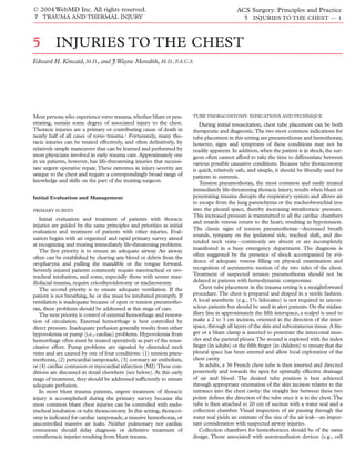
![© 2004 WebMD Inc. All rights reserved. ACS Surgery: Principles and Practice
7 TRAUMA AND THERMAL INJURY 5 INJURIES TO THE CHEST — 2
savers) have immense theoretical potential for rapid retrieval and thrive, are diagnostic. Analysis and culture of fluid obtained at tho-
processing of shed blood. In practice, however, their utility is lim- racocentesis or chest tube placement typically confirms the diag-
ited. For example, with a small to moderate-sized hemothorax (< nosis, but the fluid may be sterile if the patient is already receiving
1,000 ml), the red-cell yield of an autotransfusion device would be antibiotics.
small and not worth the associated time and expense.With a large Antibiotic therapy, either broad-spectrum or specifically direct-
hemothorax, the most important goal of therapy is control of ed against cultured organisms (usually gram-positive pathogens),
bleeding, and arranging for autotransfusion could delay or hinder is certainly an important component of therapy for empyema tho-
the achievement of this goal. In addition, products of autotrans- racis, but the primary goal is removal of the infection while the
fusion may contain harmful cytokines, damaged cells, and debris, fluid is still thin.When this goal is met, a more modest therapeu-
while lacking platelets and other important proteins and coagula- tic procedure can be performed, there is less risk that a restrictive
tion factors.2 pulmonary peel will develop, and the injured patient recovers
Antibiotic prophylaxis after tube thoracostomy is controver- faster overall. In the early stages, tube thoracostomy may suffice
sial.3 Most clinicians, however, would recommend use of a first- for treatment; however, if the infected pleural process cannot be
generation cephalosporin for 24 hours, ideally starting before the completely evacuated via chest tube because of thicker fluid, loc-
initial tube placement. ulations, or pleural adhesions, a formal thoracotomy with decor-
After the primary survey, less dramatic pneumothoraces may tication is generally required.
be recognized on diagnostic images, along with hemothoraces, on Decortication should not be undertaken in the face of severe
various imaging studies. Treatment of occult pneumothoraces sepsis. Instead, antibiotics and chest drainage (via tube thoracos-
(i.e., those seen only on computed tomography) deserves special tomy, CT-directed catheter placement, or open rib resection)
mention. In general, patients with occult pneumothoraces who should be employed until sepsis is controlled. In cases of early
require positive pressure ventilation, those who are hypotensive or empyema,VATS has been successfully used for lysis of adhesions
have respiratory distress of any etiology, and those who have asso- and removal of fluid.9,10 Because of the limited capacity for per-
ciated complex injuries or hemothorax should be treated with forming pleurectomy with this procedure, VATS should not be
tube thoracostomy. One group, however, has questioned the need used when thick peel or a trapped lung is present. In adult patients
for tube thoracostomy in patients requiring positive pressure ven- with posttraumatic empyema, there is no proven role for
tilation on the basis of findings from a small number of patients.4 intrapleural fibrinolytic therapy.
Patients with occult pneumothoraces who are treated without
EMERGENCY DEPARTMENT THORACOTOMY
tube thoracostomy should be observed for at least 24 hours.
ED thoracotomy is a drastic step in the treatment of the injured
Retained Hemothorax and Empyema patient. If possible, the patient should be stabilized and transport-
In treating a hemothorax with tube thoracostomy, the goal is ed to the operating room, where better facilities are available for
complete removal of blood. Complications such as atelectasis and definitive care.
empyema after chest trauma are clearly related to the presence of ED thoracotomy is best reserved for patients who arrive at the
residual blood, fluid, and air, as can occur secondary to improper ED and deteriorate rapidly and those who have undergone car-
positioning of the tube (i.e., within a fissure), obstruction of the diac arrest just before arrival.The results are dismal when it is per-
tube, or blood clot or loculated fluid within the chest. formed in patients who have undergone cardiac arrest some time
A persistent or clotted hemothorax is suggested by the presence before arrival and have required cardiopulmonary resuscitation
of a persistent opacification in the pleural space in a patient with for more than a few minutes. Blunt trauma victims who have sus-
a known previous hemothorax.This radiodensity can be confused tained cardiopulmonary arrest at the scene of injury should not be
with adjacent pulmonary contusion or atelectasis; chest CT con- subjected to thoracotomy, either at the scene or in the ED.11
firms the diagnosis. Because retained blood serves as a nidus for Similarly, patients who are found at emergency thoracotomy to
infection and empyema,5 aggressive attempts at removal are justi- have no cardiac activity have a dismal prognosis, as do those who
fied. Occasionally, removal can be accomplished by placing more do not respond to improvement of systolic blood pressure after
chest tubes, but often, an operative approach is needed. Video- aortic occlusion. Overall, the survival rate for patients undergoing
assisted thoracic surgery (VATS) [see 4:10 Video-Assisted Thoracic ED thoracotomy for blunt trauma is lower than 10%.The report-
Surgery] may be useful for managing small, clotted hemothoraces ed survival rate for patients undergoing ED thoracotomy for pen-
and free-flowing blood in patients who can tolerate single-lung etrating trauma ranges from 16% to 57%,11-13 and that for
ventilation6 ; however,VATS tends to limit the surgeon’s ability to patients with cardiac wounds ranges from 57% to 72%.11-13
control bleeding and perform definitive repair of injuries. In The technique of ED thoracotomy is straightforward. An anti-
patients who have ongoing bleeding or large, clotted hemotho- septic solution may be splashed on the chest, but skin preparation
races, posterolateral thoracotomy is generally required. is not required. An incision is made from the sternal border to the
Empyema thoracis is a relatively common complication after midaxillary line in the fourth intercostal space. A chest retractor is
chest trauma, occurring in 5% to 10% of patients.7,8 Possible caus- inserted and opened widely. The costochondral junctions of the
es include retained hemothorax, pneumonia with parapneumonic fifth, the fourth, and sometimes the third rib are divided quickly
effusion, persistent foreign body, ruptured pulmonary abscess, bron- with the scalpel to provide exposure. Attention is directed first to
chopleural fistula, esophageal leakage, and tracking through the the injury. If there is exsanguination from a great vessel, the hem-
intact or injured diaphragm from an abdominal source. Empyema orrhage is controlled with pressure. If air embolism is the cause of
may be difficult to diagnose and must be differentiated from pleur- the arrest, the hilum is clamped and air evacuated from the aorta.
al thickening, pulmonary contusion, and an uninfected effusion. Otherwise, the pericardium is opened anterior and parallel to the
Chest CT with intravenous contrast usually demonstrates a fluid phrenic nerve. The hemopericardium is evacuated, the cardiac
collection with loculations or an enhancing rim. Such findings, injury is controlled with digital pressure, and a temporary repair
coupled with a clinical scenario of low-grade sepsis or failure to is performed.](data:image/gif;base64,R0lGODlhAQABAIAAAAAAAP///yH5BAEAAAAALAAAAAABAAEAAAIBRAA7)