Polytrauma part 2 (ards)
•Download as PPT, PDF•
4 likes•888 views

Acute respiratory distress syndrome (ARDS) is a Sudden failure of the respiratory system. It Can occur in anyone over the age of one who is critically ill. It is a Life- threatening because normal gas exchange does not take place due to severe fluid buildup in both lungs. Prevention can be achieved by Limiting Blood Loss so decreasing transfusion requirements, Early Stabilization Of unstable Fractures and Early prophylactic mechanical Ventilation. Established cases with ARDS is treated in the Intensive Care Unit By Mechanical ventilation and Oxygen therapy through a ventilator, Fluids through an IV line to improve blood flow and provide nutrition and medicine to prevent and treat infections and to relieve pain.

Recommended
More Related Content
What's hot
What's hot (20)
Similar to Polytrauma part 2 (ards)
Similar to Polytrauma part 2 (ards) (20)
More from fathi neana
More from fathi neana (20)
Recently uploaded
Recently uploaded (20)
Polytrauma part 2 (ards)
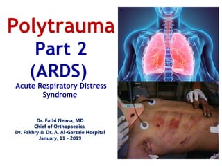
- 1. Dr. Fathi Neana, MD Chief of Orthopaedics Dr. Fakhry & Dr. A. Al-Garzaie Hospital January, 11 - 2019 Polytrauma Part 2 (ARDS) Acute Respiratory Distress Syndrome
- 3. Trauma Mortality Causes 3 Peaks First peak (24 hours) Second peak (2-7 days) Third peak – delayed ( <7 days)
- 4. Trauma Mortality (3 Peaks) FIRST PEAK (24 hours) Early phase - immediate death severe brain injury, disruption of great vessels, cardiac disruption Second phase – within 24 hours subdural, epidural hematomas, hemopneumothorax, severe abdominal injuries, multiple extremity injuries (bleeding) SECOND PEAK (2-7 Days) Acute Respiratory Distress Syndrome (ARDS) 40 - 50% Mortality THIRD PEAK – DELAYED ( <7 Days) Multisystem organ failure, Sepsis (Septic Death) 28% Mortality
- 5. • Define ARDS, FES, Thromboembolic Disease, Septic Shock (Septic Death), DIC, etc… • Understand Etiology & Pathophysiology of each Condition • Understand Prevention, Diagnosis, Treatment, Outcomes Trauma Mortality Objectives
- 6. 3 Rights Right Patient, Right Hospital, Right Time Delayed arrival increase Infection rate (3rd peak of death) Delayed or Inappropriate Surgery increase ARDS rate (2nd peak of Death) Trauma centre vs. usual G. hospital Treating Team (senior orthopedic trauma) Trauma Mortality Factors Affecting Mortality
- 7. Ventilation and Gas exchange under the control of the autonomic nervous system from parts of the brain stem, the medulla oblongata and the pons Immune functions Metabolic and endocrine functions of the lungs Vocalization Temperature control Coughing and sneezing Air & Oxygen are free to all The Respiratory system
- 8. 18 If you try to reckon up Allah's blessings, you cannot count them. Indeed, He is Forgiving and Compassionate, (16:18) Alnahl Air and Oxygen are free to all
- 9. The right lung is slightly larger than the left. Hairs in the nose help to clean the air we breathe as well as warming it. The highest recorded "sneeze speed" is 165 km per hour. The surface area of the lungs is roughly the same size as a tennis court. The capillaries in the lungs would extend 1,600 kilometers if placed end to end. We lose half a liter of water a day through breathing. This is the water vapour we see when we breathe onto glass. A person at rest usually breathes between 12 and 15 times a minute. The breathing rate is faster in children and women than in men. Amazing Facts about the Respiratory System
- 10. Lung Compliance • Measurement of how easy or hard it is to inflate the lungs. • COMPLIANCE IS RECIPROCAL OF ELASTANCE. • Compliance = ∆ Volume ∆ Pressure • Normal Values – Lung 0.2 L/cm H20 – Thorax 0.2 L/cm H20 – Total 0.1 L/cm H20
- 11. Increased Lung Compliance • Easier to inflate the lung • Lower inflation pressures • Decreased Elastance • Causes – Emphysema Lung Compliance Decreased Lung Compliance • Higher inflation pressures – Higher airway pressures on the ventilator – Increased complications (barotrauma, decreased CO) • Harder to inflate the lung • “Stiff lung” • Increased Elastance • Causes – Atelectasis – Fibrosis – Pneumothorax – ARDS
- 13. Acute Respiratory Distress Syndrome (ARDS) Acute Respiratory Distress or Failure 40 - 50% Mortality rate Big Cats Tigers and Lions know how to suffocate their prey?
- 14. • Acute respiratory distress syndrome (ARDS) is a life-threatening lung condition that prevents enough oxygen from getting into the blood. • Acute respiratory distress syndrome was first described in 1967 by Ashbaugh and colleagues. • ARDS is also referred with variety of terms like • Stiff Lung • Shock lung • Wet lung • Post traumatic lung • Adult respiratory distress syndrome • Adult hyaline membrane disease • Capillary leak syndrome & • Congestive atelectasis. Mr. sanjay. M. Peerapur, Principal, KLES Institute of Nursing Sciences, Hubli Acute Respiratory Distress Syndrome (ARDS) Introduction
- 15. Acute Respiratory Distress Syndrome (ARDS) Definition Acute Respiratory Distress Syndrome (ARDS) is a clinical syndrome: - Severe dyspnea of rapid onset - Hypoxemia -Alveolar capillary membrane damage -Diffuse pulmonary infiltrates Inflammatory cells and proteinaceous fluid accumulate in the alveolar spaces leading to a decrease in diffusing capacity and hypoxemia - Respiratory failure
- 16. Lung Injury • Acute Lung Injury (ALI) – Normal barriers to fluid movement within the lungs is disrupted. – If it is severe enough to cause acute hypoxemic respiratory failure it is termed: • Acute respiratory distress syndrome (ARDS) – Most severe gas exchange abnormalities – Multiple Organ Dysfunction Syndrome (MODS)
- 17. Criteria Differentiating ALI and ARDS • PaO2/FiO2 ratio • Example – 100 mm Hg/.21 = 476 – As the number decreases, oxygenation is getting worse • ALI is when the ratio is LESS THAN 300 • ARDS the ratio LESS THAN 200
- 18. Acute lung injury (ALI) and acute respiratory distress syndrome (ARDS) ALI is the term used for patients with significant hypoxemia (PaO2/FiO2 ratio of ≤ 300) ARDS is the term used for a subset of ALI patients with severe hypoxemia (PaO2/FiO2 ratio of ≤ 200) Normal → ALI → ARD Clinical Spectrum
- 19. Tissue injury Infection Inflammatory Mediators (Decompensated SIRS) Organ Injury ARDS is related to Multi organ dysfunction (MOD) Persistent release of (inflammatory mediators) results in Systemic inflammatory response syndrome (SIRS). Inefficient compensatory anti inflammatory response syndrome (CARS) leads to Multi organ dysfunction (MOD) including Lungs (ARDS) Acute Respiratory Distress Syndrome (ARDS) Pathophysiology
- 20. Multi organ Failure in ARDS (ARDS) is related to Multi organ dysfunction (MOD)
- 21. Systemic Inflammatory Mediators Damage to Endothelial Lining Increased Capillary Permeability Fluid Extravasation Alveolar Collapse Decreased Pulmonary Compliance Ventilation Perfusion Abnormalities Arteriolar Hypoxemia Acute Respiratory Distress Syndrome (ARDS) Pathophysiology
- 22. • Injury of the lung • Destruction of the alveolar-capillary membrane (the barrier) • Engorged capillaries • Interstitial and alveolar edema • Hemorrhage • Decreased surfactant with atelectasis • Hyaline Membrane formation • • Fibrosis Acute Respiratory Distress Syndrome (ARDS) Pathophysiology
- 23. acute respiratory distress syndrome (ARDS) A normal alveolus (left) and a damaged injured alveolus in the acute phase of acute respiratory distress syndrome (right).
- 25. The Inflammatory Response Inflammation is an Adaptive response to noxious conditions (Infection and Tissue injury) an attempt to restore homeostasis 1- Induced by immune recognition of infection or tissue damage (usually good). But if the immune recognition is hypersensitive to environmental or auto inflammatory components (autoimmune disease) 2- Inflammation depends upon the Immune Recognition of infection or tissue damage. a very Balanced process, Normally end with Recovery but Errors, in recognition - Hypo or Hyper sensitivity end by Death from the Disease or Inflammation 3- Opsonisation, Antibody opsonization is the process by which the pathogen is marked for ingestion and eliminated by the phagocytes. 4- Phagocytosis—Process in which phagocytes engulf and digest the (Opsonised) microorganisms and cellular debris. 5- Recruitment, The Complement (Opsonin) and the Phagocytes (Exist mainly in blood). A mechanism to recruit these elements to the invaded tissue led to inflammatory response (inflammation)
- 26. Vasodilatation .. increase blood flow .. Capillary permeability .. Passage large immune cells + proteins Increase in body heat .. swing the balance of chemical reactions in favour of the host The tissues become red. warm, swollen, painful , Organ dysfunction Inflammatory process once started it continues until infection is eradicated Phagocytes continue to consume and destroy bacteria The acquired immune system binds & disposes harmful toxins Pus is produced The Effects of the Inflammatory Response The Effects of the Inflammatory Response Cornelius Celsus in De Medicina, 1st century A.D. rubor et tumor cum calore et dolore” (Redness and Swelling with Heat and Pain) later “functio laesa” (Disturbance of Function) was added
- 27. When the infection has been eradicated, and there is no more foreign antigen The helper T cells stop emitting the stay-alive signal, thus allowing the cells involved in the inflammatory response to die off (apoptosis) If foreign antigen is not eradicated, or if the immune cells receive the stay-alive signal from another source, then chronic inflammation may develop How does the inflammatory response end ?
- 28. The inflammatory response Conclusion Acute inflammation: influx of white blood cells and fluid from blood to fight infection and aid tissue repair Chronic inflammation: inducer of inflammation is not removed Leads to tissue damage and loss of tissue function (joint destruction, lung fibrosis, etc.) Current view: aggressively fight inflammation in certain chronic diseases to decrease / delay progressive loss of function Current research suggests that inflammation may play an important role in common chronic diseases including atherosclerosis, type 2 diabetes, neurodegeneration, and cancer
- 30. Sudden failure of the respiratory system Can occur in anyone over the age of one who is critically ill Life- threatening because Normal gas exchange does not take place Due to severe fluid buildup in both lungs What is Acute respiratory distress syndrome (ARDS)? Characterized by: Rapid breathing Difficulty getting enough air into the lungs Low blood oxygen levels Cough and fever (if caused by pneumonia) Low blood pressure Confusion & Extreme tiredness
- 31. Acute respiratory distress syndrome (ARDS) Clinical Definition – Acute onset of symptoms – Ratio of PaO2 to FIO2 of 200 mm Hg or less (PaO2 ) =Arterial partial pressure of oxygen = 95 mm Hg FIO2 = fraction of inspired oxygen". – Bilateral infiltrates on CXRs – Pulmonary arterial wedge pressure of 18 mm Hg or less or no clinical signs of left atrial hypertension – American-European Consensus Conference (AECC) on ARDS, 94
- 32. Acute respiratory distress syndrome (ARDS) in the post traumatic period • Incidence 5% – 8% after polytrauma • Much lower in isolated fracture • Mortality up to 40 -50% • Uncommon in Children and the Elderly Characterized by: • Decreased PaO2 And • Diffuse and often massive extravasations of fluid from the pulmonary vasculature to the interstitial space of the lungs
- 33. Acute respiratory distress syndrome (ARDS) Common Causes & risk factors (Multifactorial) • Trauma • Shock • Massive Transfusion • Embolism • Sepsis (bacterial infection of the blood) • Pneumonia or other lung infection • Aspiration , vomit , salt water • Breathing in harmful smoke or fumes • Prolonged LOC • Cardiopulmonary Bypass • Pancreatitis • Major Burns • Abdominal Distension • Narcotics • Sedatives • Overdoses of tricyclic antidepressants
- 34. Medical history, Physical exam and Medical tests Abnormal Breathing or Heart sounds Edema means heart or kidneys dysfunction Arterial blood gas test Chest x-ray Chest CT scan Blood tests for infection Heart tests look for heart failure ARDS usually diagnosed in a Hospitalized patient with a Critical illness such as Shock, Sepsis or Trauma Acute respiratory distress syndrome (ARDS) Diagnosis Chest Radiograph Autopsy Specimen Chest CT Scan
- 35. • Radiopaque – white • The more severe the ARDS, the whiter the lungs appear • Often described as “White Out” or diffuse “fluffy” infiltrates – Non-homogenous – “Baby Lungs” • Heart size is normal • Air Bronchograms • Honeycombing or Ground Glass Acute respiratory distress syndrome (ARDS) Chest X-ray
- 36. • Common complications are; – Nosocomial Infections pneumonia – Barotrauma – Renal failure • Other complications are : – O2 toxicity, – Stress ulcers, GI hemorrhage/gastric distension – Tracheal ulceration, – Blood clots, deep vein thrombosis & pulmonary embolism. – Sepsis, micro emboli – Disseminated Intravascular Coagulation (DIC), – Anemia – Pulmonary fibrosis – permanent disability – Cardiovascular Problems Acute respiratory distress syndrome (ARDS) Complications
- 37. Image shows subtle manifestations of barotrauma, pulmonary interstitial emphysema, and subcutaneous emphysema. This patient was being treated with noninvasive ventilation. Importantly, recognize that barotrauma can be associated with noninvasive ventilation.
- 38. • Limiting Blood Loss Decreasing Transfusion Requirements • Early Stabilization Of Unstable Fractures • Early Prophylactic Mechanical Ventilation Temporary Ex-Fix For Stabilization Acute respiratory distress syndrome (ARDS) Prevention
- 39. There is no specific treatment for ARDS ARDS is treated in the Intensive Care Unit By Mechanical ventilation & Oxygen therapy Treatments may include: Oxygen through a ventilator Fluids through an IV line to improve blood flow and provide nutrition Medicine to prevent and treat infections and to relieve pain Acute respiratory distress syndrome (ARDS) Management
- 40. • Ventilator Support – Acceptable ABG’s Normal Value, Range. pH, 7.4, 7.35 to 7.45. Pa02, 90mmHg, 80 to 100 mmHg. – Avoid further alveolar damage • Toxic FIO2 • Barotrauma • General Organ Support • Research – Optimal ventilator settings – Pharmacological agents Acute respiratory distress syndrome (ARDS) Management
- 42. • Reversal of underlying associated disorder • Oxygen & Diuretics (keep on dry side) • Steroids (after 7 days if no infection) • Antibiotics • Mechanical Ventilation • Proning or lateral rotation therapy • Nutritional support Acute respiratory distress syndrome (ARDS) Management • Minimize O2 demand by reducing metabolic rate – Control fevers – Control anxiety and pain • Altered Style of Ventilation – Bi-Level Ventilation (APRV) – High frequency ventilation – Recruitment Maneuvers – Proning • Experimental Treatments: – Nitric Oxide – Extracorporeal membrane oxygenation (ECMO) – Exogenous Surfactant Administration
- 43. Proning Prone positioning was first proposed in the 1970s as a method to improve gas exchange • Should see improvement in 1 hour – Response should be significant – PaO2 improves from 60 – 100 mm Hg • Works better in indirect ARDS – Pancreatitis • May improve oxygenation but does not improve mortality • Facial Edema can be severe
- 45. Indications for Mechanical Ventilation • Acute Hypercapnic Respiratory Failure – Respiratory Acidosis • Acute Hypoxemic Respiratory Failure – PaO2 less than normal – FiO2 greater than 60% oxygen • Positive End Expiratory Pressure (PEEP) • Set PEEP at lower infection point to recruit alveoli and keep them open • Shunting will improve with improved PaO2 • PEEP may decrease blood pressure and cardiac output so monitor both closely – Do not allow oxygen delivery to decrease
- 46. Ventilation Strategies • Use higher PEEP levels (10 – 15 cm H20) • Set low tidal volumes at 5-7 mL/kg • KEEP ALVEOLAR (PLATEAU) PRESSURE LESS THAN 30 – 35 cm H20 • This may result in permissive hypercapnia.
- 47. 1. Ineffective breathing pattern related to decreased lung compliance, decreased energy as characterized by dyspnea, abnormal ABGs, cyanoisis & use of accessory muscles. 2. Impaired gas exchange related to diffusion defect as characterized by hypoxia (restlessness, irritability & fear of suffocation), hypercapnia, tachycardia & cyanosis. 3. Risk for decreased Cardiac output related to positive pressure ventilation 4. Ineffective protection related to positive pressure ventilation, decreased pulmonary compliance & increased secretions as characterized by crepitus, altered chest excursion, abnormal ABGs & restlessness. 5. Impaired physical mobility related to monitoring devices, mechanical ventilation & medications as characterized by imposed restrictions of movement, decreased muscle strength & limited range of motion. 6. Risk for impaired skin integrity related to prolonged bed rest, prolonged intubation & immobility. 7. Knowledge deficit related to health condition, new equipment & hospitalization as characterized by increased frequency of questions posed by patient and significant others. Nursing care and diagnosis
- 49. Acute respiratory distress syndrome (ARDS) Outcome Mortality : • Significant Cause of Mortality • Polytrauma second peak 0f death (2-7 Days) 40 - 50% Mortality rate • Major Cause of Death in Patients with the Lowest ISS scores – Mortality Rate Slowly Decreasing with Changing & Improving Therapy – Mortality rates have declined over the past two decades to 30 – 40%
- 50. Recovery: • Extent of recovery depends on: – Severity of the initial lung injury – Influence of secondary forms of injury • Oxygen toxicity • Nosocomial infections • DIC • Barotrauma with mechanical ventilation • Normal lung function after 1 year • Residual effects may result in decreased flow rates and DLCO (Diffusing capacity of the lungs for carbon monoxide (DLCO) Acute respiratory distress syndrome (ARDS) Outcome
- 51. Acute respiratory distress syndrome (ARDS) Outcome Survivors: • Good function in many • Diffuse pulmonary fibrosis and pulmonary hypertension in severe cases • Prognostic markers: – Extreme pO2 decrease – fibrosis
Editor's Notes
- First phase is immediate death or shortly thereafter due to severe brain injury, or disruption of the heart, aorta, or great vessels. Second phase: Death occurs minutes to a few hours. Deaths are due to lifethreating injuries such as subdural, epidural hematomas, hemopneumothoraces, severe abdominal injuries, long bone fractures, multiple injuries associated with blood loss. This is the group that with rapid evacuation , resuscitation and proper early management can be given the best chance for survival and improved outcomes. Third phase: Patients die days to weeks from multiple system organ failure and sepsis. Specialized centers with their expertise and experience can reduce mortality in the second and third phases
- First phase is immediate death or shortly thereafter due to severe brain injury, or disruption of the heart, aorta, or great vessels. Second phase: Death occurs minutes to a few hours. Deaths are due to lifethreating injuries such as subdural, epidural hematomas, hemopneumothoraces, severe abdominal injuries, long bone fractures, multiple injuries associated with blood loss. This is the group that with rapid evacuation , resuscitation and proper early management can be given the best chance for survival and improved outcomes. Third phase: Patients die days to weeks from multiple system organ failure and sepsis. Specialized centers with their expertise and experience can reduce mortality in the second and third phases
- First phase is immediate death or shortly thereafter due to severe brain injury, or disruption of the heart, aorta, or great vessels. Second phase: Death occurs minutes to a few hours. Deaths are due to lifethreating injuries such as subdural, epidural hematomas, hemopneumothoraces, severe abdominal injuries, long bone fractures, multiple injuries associated with blood loss. This is the group that with rapid evacuation , resuscitation and proper early management can be given the best chance for survival and improved outcomes. Third phase: Patients die days to weeks from multiple system organ failure and sepsis. Specialized centers with their expertise and experience can reduce mortality in the second and third phases