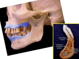
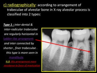
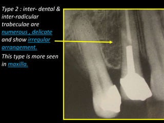
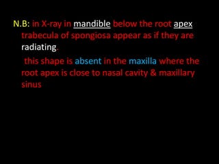
5. Definitions:
1)Alveolar process: its bone of jwas containing the teeth.
Alveolar process has alveolar margin & alveolar crest.
A) alveolar margin: its free borders of alveolus.
B) alveolar crest : its coronal portion of alveolar bone ,
usually occurs opposite the cervical portion of the root
about 2mm apical to cervical line.
Alveolar process also called
functional bone:
because its susceptible
to functional changes
(its lost after tooth extraction)
7. 2)Basal bone: its unchanged part of jaws.
N.B: no distinct demarcation between alveolar process &
basal bone. both are covered by the same periosteum.
surfaces of alveolar process
1- facial surface: correspond to facial s. of root inveted in it
2- lingual s.: correspond to lingual s. of root invested in it.
3-alveolar septa include(interdental septa & intraradicular septa)
a) inter dental septa : between the adjacent teeth.
b) intraradicular septa: between the roots of the same tooth.
8.
9. Development: at the end of 8 W.i.U life both jaws
form groove that open toward the surface of oral
cavity.
this grooves formed by : growth of outer & inner
plates of the body of jaws.
Tooth germs & alveolar nerves , vesseles are
contained in this grooves.
Gradually bony septa developed between the
adjacent tooth germs.
10. Structure of alveolar process:
adult alveolar process is composed of 2 parts:
1)supporting bone (cortical & spongy).
2) alveolar bone proper.
Recently component of alveolar process described as follow:
1) facial & lingual cortical plates: which are compact bone.
2)central spongiosa: its spongy bone fills the space between
cortical plates & alveolar bone
3) alveolar bone : from inner side facing P.D.L .
N.b: alveolar bone & cortical plate merges at alveolar
process crest (1.5 to 2mm below the level of C.E.J)
12. 1) The cortical plates: (anatomically & histologically)
a)anatomically:
1- in anterior teeth of both jaws: labilal cortical
plate is thinner than the lingual cortical plate.
(cortical plate & alveolar bone directly fused & no central spongiosa)
2-in mandibular posterior teeth: buccal cortical plate is
thicker & denser than the lingual cortical plate
because the external oblique line mask the buccal
cortical plate laterally.
3-in maxillary posterior teeth: the buccal cortical plate
is thinner than the lingual cortical plate
13. N.B: the buccal cortical plate of max. Post. Teeth either
thin , perforated or even missed.
Buccal cortical plates are perforated by volkmann’s canals & may
show defect in posterior teeth region which expose part of the
root but alveolar margin remain intact & this doesn’t impair the
firm attachment , function of the teeth because the P.D.L on this
side is attached directly to covering mucosa.
Fenestration: its defect exposing part of the root with alveolar margin
intact.
Dehiscence: its defect include the alveolar margin.
b) histologically: cortical plate consist of lamellar bone supported by
haversian system (osteon).
14.
15. 2) The central spongiosa (trabecular bone):
(anatomically & histologically & radiografically)
a)anatomically:
* fills the space between cortical plate &alveolar bone
* its degree of development is related to
force of mastication.(its formation increased as the force increased)
* it form the main bulk of alveolar septa.
* in some location its minimal and may be
missed (as in anterior teeth & mandibular bi cuspid).
b) histologically: its formed of spongy bone.
16. c) radiographically: according to arrangement of
trabeculae of alveolar bone in X-ray alveolar process is
classified into 2 types:
Type 1 : inter-dental &
inter-radicular trabeculae
are regularly horizontal in
ladder-like arrangment
and inter connected by
shorter , finer trabeculae .
this type is more seen in
mandibule.
N.B: this arrangement more
resistance to force of mastication
17. Type 2 : inter- dental &
inter-radicular
trabeculae are
numerous , delicate
and show irregular
arrangement.
This type is more seen
in maxilla.
18. N.B: in X-ray in mandible below the root apex
trabecula of spongiosa appear as if they are
radiating.
this shape is absent in the maxilla where the
root apex is close to nasal cavity & maxillary
sinus
19. 3) Alveolar bone (cribriform plate & lamina dura): its
thin lamella of bone surrounds the root of the tooth &
gives attachment to principal fibers of P.D.L
(anatomically & histologically & radiographically).
a)anatomically: its thin lamella perforated by
volkmanns canals that carry nerves,vessels
from marrow space to P.D.L
and that’s why its called crepriform plate.
21. b) radiographically: in X-ray its called lamina dura.
lamina dura: appears as radiopaque (white)
line adjacent to root and separated from it by
radiolucent (dark) line representing the space of
P.D.L
Its appear rdiopaqu because cribriform plate has no
trabeculation that X-ray penetrate it . And not to
any increased in mineral contents.
24. c) histologically: its consist of 2 types of bone
(lamellar and bundle bone).
its called bundle bone because of bundle fipers
of perioeteum & P.D.L continue in the bone as
sharpey’s fibers .
Woven bone found in sockets at areas where there
is formation of new bon.
25. Age changes of alveolar bone
• Decrease water contents thus increase the brittleness.
• Thinning of trabecula of spongiosa thus Widening of marrow
space.
• Marrow Is transformed into fatty marroe except (condylad
head , mandibular angle & maxillary tuberosity)
Thus Decrease in bone marrow,thus vascularity
• Osteoprosis: decrease in bone due to loss of some
elements like Ca .
• Bone loss specially if there are extraction of tooth..
• Mental foramen become closer to upper border of the
mandibule.
• Approximation to the floor of maxillary sinus.
• Distal sloping of crest of alveolar septa in PM , M region.
• Sockets appear noched , scalloped