Periodontium in Health and Disease
•
0 likes•299 views

Describing and Discussing more about: What is Healthy Periodontium? What is Gingivitis? What is Periodontitis? What are the Stages of Periodontal Diseases?

Recommended
More Related Content
What's hot
What's hot (20)
Similar to Periodontium in Health and Disease
Similar to Periodontium in Health and Disease (20)
Recently uploaded
Recently uploaded (20)
Periodontium in Health and Disease
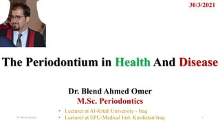
- 1. The Periodontium in Health And Disease 30/3/2021 Dr. Blend Ahmed Omer M.Sc. Periodontics Dr. Blend Ahmed 1 • Lecturer at Al-Kitab University - Iraq • Lecturer at EPU-Medical Inst. Kurdistan/Iraq
- 2. 2 Three Basic State of the Periodontium ➢ The periodontium exists in three basic states: • Health • Gingivitis, and • Periodontitis Dr. Blend Ahmed
- 3. 3 Periodontium in Health 1. Clinical Picture of Healthy Gingiva A. Color: - Pink, may be pigmented B. Gingival Margin: 1. Scalloped outline. 2. Located coronal to the cementoenamel junction (CEJ). C. Interdental Papillae: - Firm and occupy the embrasure spaces . D. Absence of Bleeding: - No bleeding upon probing. E. Sulcus: - Probing depths range from 1 to 3 mm. Dr. Blend Ahmed
- 4. 4 Periodontium in Health 2. Microscopical Picture of Healthy Gingiva A. Junctional Epithelium: ✓ Coronal to the CEJ. B. Gingival Fibers: ✓ intact Supragingival fibers. C. Alveolar Bone: ✓ the crest of AB is intact. D. Periodontal Ligament Fibers: ✓ Intact periodontal ligament fiber. Dr. Blend Ahmed
- 6. 6 Gingivitis —ReversibleTissue Damage 1. Characteristics of Gingivitis • Gingivitis is a type o periodontal disease characterized by changes in the color, contour, and consistency of the gingival tissues. A. Onset of Gingivitis. - Gingivitis is observed clinically from 4 to 14 days after plaque biofilm accumulates in the gingival sulcus. 1. Acute gingivitis is a gingivitis that lasts a short period of time. 2. Chronic gingivitis is a gingivitis that lasts months or years. B. Tissue Enlargement (TE). - Gingival enlargement may be caused by swelling (acute gingivitis) or fibrosis (chronic gingivitis). 1. TE causes the GM to cover more of the crown of the tooth and results in deeper probing depths. 2. TE of the gingival tissue is produce a gingival pocket. 3. A gingival pocket has a sulcus depth over 3 mm.
- 7. 7 Gingivitis —ReversibleTissue Damage 1. Characteristics of Gingivitis (Cont.) C. Reversible Tissue Damage. The tissue damage in gingivitis is reversible—that is, with good patient self -care the body can repair the damage. D. Duration o Gingivitis. In many cases, gingivitis may persist or years without ever progressing to the next stage, periodontitis. In some cases, a combination of risk factors may result in gingivitis progressing to periodontitis.
- 8. Dr. Blend Ahmed 8 2. Clinical Picture of Gingivitis A. Color: the gingival tissue usually is red or reddish blue in color. 1. The blood flow increases in the gingival connective tissue and the gingival blood vessels become engorged with blood, causing the gingiva to appear red. 2. If the gingivitis persists, the gingival blood vessels may become congested. This slow-moving blood flow causes the gingiva to have a bluish color. B. Gingival Margin & Interdental Papillae 1. The gingival margin is swollen and loses its knife-edge adaptation to the tooth. 2. Gingival tissue may cover more of the crown of the. 3. The interdental papillae often are swollen. C. Bleeding. There is bleeding upon gentle probing. E. Sulcus. Probing depths may be greater than 3 mm due to swelling of the tissues. Gingivitis —ReversibleTissue Damage
- 9. Dr. Blend Ahmed 9 3. The Microscopic Picture of Gingivitis A. Junctional Epithelium: JE coronal to the CEJ B. Gingival Fibers. Damage/Destruction of supragingival fiber bundles. C. Alveolar Bone. The bacterial infection has not progressed into the alveolar bone. There is no destruction of alveolar bone. D. Periodontal Ligament Fibers. The bacterial infection has not progressed into the periodontal ligament fibers. Gingivitis —ReversibleTissue Damage
- 11. 11 Dr. Blend Ahmed Periodontitis —PermanentTissue Damage 1. Characteristics of Periodontitis A. Extent of Tissue Destruction 1. Periodontitis is a type o periodontal disease that is characterized by the: (1) apical migration of the junctional epithelium, (2) loss of connective tissue attachment, and (3) loss of alveolar bone. 2. The tissue damage of periodontitis is permanent. B. Process of Tissue Destruction 1. The tissue destruction of periodontitis is not a continuous process. 2. Tissue destruction progresses at different rates throughout the mouth. Destruction does not occur in all parts o the mouth at the same time.
- 12. 12 Dr. Blend Ahmed Periodontitis —PermanentTissue Damage 2. Clinical Picture of Periodontitis A. Color: 1. Edematous tissue (spongy tissue)—bluish red or purplish red with a smooth, shiny appearance. 2. Fibrotic tissue ( firm, nodular tissue)—light pink with a leathery consistency. B. Gingival Margin 1. The gingival margin may be swollen or fibrotic and does not have a close knife edged adaptation to the tooth. 2. The position o the gingival margin is apical to the CEJ (recession) resulting in a portion of the root being visible in the mouth.
- 13. 13 Dr. Blend Ahmed Periodontitis —PermanentTissue Damage 2. Clinical Picture of Periodontitis (Cont.) C. Interdental Papillae The interdental papillae may not fill the interdental embrasure spaces. D. Bleeding There often is bleeding upon probing, and suppuration (a discharge of pus) may be visible. E. Pocket Probing depths are 4 mm or greater in depth
- 14. 14 Dr. Blend Ahmed 3. The Microscopic Picture of Periodontitis A. Junctional Epithelium The junctional epithelium is located on the cementum B. Gingival Fibers widespread destruction of the supragingival fiber bundles C. Alveolar Bone. There is permanent destruction of the alveolar bone that supports the teeth. Tooth mobility may be present. E. Periodontal Ligament Fibers. There is permanent destruction of some or all o the periodontal ligament fiber bundles. Periodontitis —PermanentTissue Damage
- 16. 16 Dr. Blend Ahmed Stages of Periodontal Disease
- 17. Dr. Blend Ahmed 17
- 18. Dr. Blend Ahmed 18 in Healthy Gums: 1. the gingival or gum tissue is a pink or coral color. 2. The tissue is firm and resilient and there will be minimal crevice pocket depth.
- 19. Dr. Blend Ahmed 19 in Gingivitis: 1. The gingival or gum tissue will be inflamed at the neck of the tooth. 2. There will be some pocket depth and gingival bleeding on probing (BOP). 3. There will not be any destruction of supporting tooth structure.
- 20. Dr. Blend Ahmed 20 1. Inflammation of periodontal ligaments and minor loss of attachment or pocket development. 2. No tooth mobility at this stage. 3. No connective tissue loss. in Early Periodontitis:
- 21. Dr. Blend Ahmed 21 1. Moderate loss of attachment and/or moderate to deep pocket formation. 2. (30%-50%) loss of bone support and slight tooth mobility. in Moderate Periodontitis:
- 22. Dr. Blend Ahmed 22 1. Advanced breakdown of supporting periodontal tissues. 2. Severe pocket depth or significant gingival recession. 3. Severe loss of attachment. 4. Greater than 50% loss of bone support and considerable tooth mobility. in Advanced Periodontitis:
- 23. Dr. Blend Ahmed 23
- 24. Dr. Blend Ahmed 24 Q1) Regarding the clinical pictures of normal periodontium which one is NOT true: Quiz 3 A) Pink in Color B) Scalloped outline of Marginal Gingiva C) Marginal Gingiva Located Apical to CEJ Q2) Acute gingivitis is a gingivitis that lasts a short period of time. A) True B) False Q3) in …………………. Probing depths are 4 mm or greater in depth A) Gingivitis B) Healthy Gingiva C) Periodontitis Q4) No tooth mobility at this stage: A) Moderate Periodontitis B) Early Periodontitis C) Advanced Periodontitis
- 25. Dr. Blend Ahmed 25 Q1) Regarding the clinical pictures of normal periodontium with one is NOT true: Quiz 3 A) Pink in Color B) Scalloped outline of Marginal Gingiva C) Marginal Gingiva Located Apical to CEJ Q2) Acute gingivitis is a gingivitis that lasts a short period of time. A) True B) False Q3) in …………………. Probing depths are 4 mm or greater in depth A) Gingivitis B) Healthy Gingiva C) Periodontitis Q4) No tooth mobility at this stage: A) Moderate Periodontitis B) Early Periodontitis C) Advanced Periodontitis Correct Answers 4/4