SlideShare a Scribd company logo
1 of 81
Mr. MaheshChand
Tutor
content
Reviewof anatomy and physiology of genitourinary system
Etiology, pathophysiology, clinical manifestations,diagnosis,
medical and surgical treatment modalities, alternative
therapies, dietetics and nursing management
Urological obstructions-
Urethral strictures
Renal calculi
Nephrosis
content
Disorders of kidney-
Nephrotic syndrome
Acute renal failure
Chronic renal failure
End stage renal disease
Dialysis, renal transplant
Cancer of kidney
Congenital disorder
content
Disorders of Ureters, urinary bladder andurethra-
UTI
Cystitis
Urinary incontinence
Urinary retention
Urinary bladdercalculi
Urethirtis
Urethral tumors
Ureteral calculi
Trauma of Ureters, bladder,urethra
Congenital disorders of ureters, bladder andurethra
anatomy and physiology of
genitourinary system
anatomy and physiology of
genitourinary system
physiology of genitourinary system
anatomy and physiology of
genitourinary system
Nursing assessment: history, and physicalexamination

urological obstructions
urethral strictures
A urethral stricture is a scar in or around
the urethra, which can block the flow of
urine, and is a result of inflammation,
injury or infection.
Anatomy of the Male Reproductive System
urological obstructions
Risk factors-
Urethral strictures are more common in men because
their urethras are longer than those in women.
Thus men's urethras are more susceptible to disease
or injury.
 A person is rarely born with urethral strictures and
women rarely develop urethral strictures.
urological obstructions
Etiology -
Stricture disease may occur anywhere from the
bladder to the tip of the penis.
 The common causes of stricture are trauma to the
urethra and infections such as sexually transmitted
diseases and damage from instrumentation.
Trauma such as straddle injuries, direct trauma to
the penis and catheterization can result in strictures
of the anterior part of the urethra.
urological obstructions
Etiology -
In adults, urethral strictures from instrumentation
trauma may occur after prostate surgery and urinary
catheterization.
 In children, urethral strictures most often follow
reconstructive surgery for congenital abnormalities
of the penis and urethra, cystoscopy and
occasionally may be congenital.
urological obstructions
Clinical features -
painful urination.
slow urine stream.
decreased urine output.
spraying of the urine stream.
blood in the urine.
abdominal pain.
urethral discharge.
urinary tract infections in men..
urological obstructions
Diagnostic evaluation-
• History Collection
• Blood test
• Physicalexamination.
• Urethral imaging (X-rays orultrasound).
• Retrograde urethrogram
Normal Urethrogram Obstructive Urethrogram
urological obstructions
Treatment-
• Enlarging the stricture by gradual stretching
(dilation).
• Urethrotomy: Cutting the stricture with a
endoscopic equipment
• Urethroplasty surgical repair of the stricture with
reconnection and reconstruction.
• Urethral Stents where a biocompatible hollow
tube is placed on the inside of the stricture to
allow for free passage of urine.
urological obstructions
renal calculi
Urolithiasis refers to stones (calculi) in the
urinary tract.
Stones are formed in the urinary tract when
concentrations of substances such as calcium
oxalate, calcium phosphate, and uric acid
increase.
urological obstructions
Types of stone-
Calcium stone
Uric acid stone
Cystiene stone
Struvite stone
urological obstructions
Types of stone-
Calcium stone:
• Most stones (75%) are composed mainly of calcium oxalate
crystals.
• Increased calcium concentrations in blood and urine promote
precipitation of calcium and formation of stones.
• Cellulose sodium phosphate (Calcibind) may be effective in
preventing calcium stones.
• Therapy with thiazide diuretics may be beneficial.
urological obstructions
Uric acidstones:
• 5% to 10% of all stones
• Gout
• Diet high in purines and abnormal purine metabolism.
• Low-purine diet such as shellfish, asparagus,
mushrooms, and organ meats are avoided.
• Allopurinol may be prescribed to reduce serum uric
acid levels and urinary uric acid excretion.
• Proteins may be limited in diet
urological obstructions
Struvite stones:
• 15% of urinary calculi
• Struvite stones are made of magnesium, ammonium and
phosphate
• Form in persistently alkaline, ammonia-rich urine
• Caused by the presence of urease splitting bacteria
such as Proteus, Pseudomonas, Klebsiella, Staphy-
lococcus, or Mycoplasma species.
urological obstructions
Cystine stones:
1% to 2% of all stones
Occur in patients with a rare inherited defect
in renal absorption of cystine (an amino acid).
Penicillamine is administered to reduce the
amount of cystine in the urine.
Low-protein diet
Urological obstrUctions
Causes and predisposing factors:
Chronic dehydration, poor fluid intake, andimmobility
Living in mountainous and desert areas
Infection, urinary stasis, and periods ofimmobility
Medications- antacids, acetazolamide (Diamox),
vitamin D, laxatives, and high doses of aspirin
Urological obstrUctions
Location of stones-
Kidney
Ureter
Bladder
Urethra
Urological obstrUctions
Site of obstruction-
Urological obstrUctions
Clinical features-
Pain that radiates to the lower abdomen and groin
Heamturia
Dysuria
Oedema
Pyuria
Nausea, vomiting, diarrhea, abdominal discomfort
Chills and fever (may)
Urological obstrUctions
renal calcUli
Diagnostic evaluation-
• History
• Physical examination
• Blood studies
• Urinalysis
• IVP
• Cystoscopy
• Radiographic studies
Urological obstrUctions
Management
General Principles
If small stone (< 4 mm) and able to treat as outpatient,
80% will pass stone spontaneously with hydration, pain
control, and reassurance.
Hospitalized for intractable pain, persistent vomiting,
high-grade fever, obstruction with infection, and solitary
kidney with obstruction.
Medical management
Surgical management
Nursing management
Urological obstrUctions
Medical management:
• Opioid analgesics or NSAIDs are administered.
• NSAIDs provide specific pain relief because they inhibit the
synthesis of prostaglandin E.
• Hot baths or moist heat to the flank areas may also be useful.
• Fluids are encouraged. This increases the hydrostatic
pressure behind the stone, assisting it in its downward
passage.
• A high, around-the-clock fluid intake reduces the
concentration of urinary crystalloids, dilutes the urine, and
ensures a high urine output.
Urological obstrUctions
renal calcUli
 N o n surgicalmanagement-
Ureteroscopy
ESWL (Extra Corporeal Shock wave lithotripsy)
Endoscopic procedures
Electrohydrolic lithotripsy
Chemolysis
Urological obstrUctions
Ureteroscopy
 Ureteroscopy involves visualizing the stone and then destroying it.
 Access to the stone is accomplished by inserting a ureteroscope into the
ureter and then inserting a laser, electrohydraulic lithotriptor, or
ultrasound device through the ureteroscope to fragment and remove the
stones.
 A stent may be inserted and left in place for 48 hours or more after the
procedure to keep the ureter patent
Urological obstrUctions
ESWL-
ESWL is a noninvasive procedure used to break up stones in
the calyx of the kidney
In ESWL, a high-energy amplitude of pressure, or shock wave,
is generated by the abrupt release of energy and transmitted
through water and soft tissues.
When the shock wave encounters a substance of different
intensity (a renal stone), a compression wave causes the
surface of the stone to fragment.
Repeated shock waves focused on the stone eventually reduce
it to many small pieces. These small pieces are excreted in the
urine, usually without difficulty.
Urological obstrUctions
ESWL-
Urological obstrUctions
Endoscopic procedures-
 A percutaneous nephrostomy or a percutaneous
nephrolithotomy may be performed, and a nephroscope is
introduced through the dilated percutaneous tract into the
renal parenchyma.
Depending on its size, the stone may be extracted with
forceps or by a stone retrieval basket. Alternatively, an
ultrasound probe may be introduced through the
nephrostomy tube.
Urological obstrUctions
Electrohydraulic lithotripsy-
an electrical discharge is used to create a
hydraulic shock wave to break up the stone.
 A probe is passed through the cystoscope, and
the tip of the lithotriptor is placed near
the stone
 This procedure is performed under
topical anesthesia.
Urological obstrUctions
Chemolysis-
Chemolysis, stone dissolution using infusions of
chemical solutions (eg, alkylating agents,
acidifying agents)
A percutaneous nephrostomy is performed, and
the warm irrigating solution is allowed to flow
continuously onto the stone.
Urological obstrUctions
Surgical management
 Nephrolithotomy - Incision into the kidney with
removal of the stone
 Nephrectomy –
 Pyelolithotomy -
Ureterolithotomy -
Cystostomy –
removal of kidney
removal of stone from renal pelvis
removal of stone from ureter
removal of stone from bladder
Cystolitholapaxy - an instrument is inserted through the
urethra into the bladder, and the stone is crushed in the
jaws of this instrument
DisorDers of kiDney
glomerUlonephritis
(acUte nephritic synDrome )
Definition –
Acute glomerulonephritis refers to a group of
kidney diseases in which there is an
inflammatory reaction in theglomeruli.
DisorDers of kiDney
Risk factors and Causes–
• Group A beta- hemolytic streptococcal infection in the
throat
• Impetigo (infection of the skin)
• Acute viral infections-URTI, mumps, varicella zoster virus,
Epstein-Barr virus, hepatitis B, and human immunodeficiency
virus [HIV] infection).
• Antigens outside the body (eg, medications, foreignserum)
• High blood pressure
• Diabetic kidney disease (diabetic nephropathy
DisorDers of kiDney
DisorDers of kiDney
Clinical features-
Hematuria - The urine may appear cola-colored be-
cause ofred blood cells (RBCs)
Edema andhypertension
Oliguria
Anemia from loss of RBCs into theurine
DisorDers of kiDney
Clinical features-
 I n the more severe form of the disease, patients
also complain of headache, malaise, and flank pain.
 Elderly patients may experience circulatory
overload with dyspnea, engorged neck veins,
cardiomegaly, and pulmonary edema.
Atypical symptoms include confusion, and
seizures, which are often confused with the
symptomsof a primary neurologicdisorder
DisorDers of kiDney
Diagnostic evaluation-
History
 O n examination- kidney is large, tender, edematous and
congested
Urinanalysis- protienuria, hematuria , oliguria
Blood studies-
Serum creatinine, BUN increased
Hypoalbuminemia, hyperlipidemia
 Antistreptolysin O titers are usually elevated in post
streptococcal glomerulonephritis
Electron microscopy analysis help identify the nature of the
lesion.
Kidney biopsy may be needed for definitive diagnosis.
DisorDers of kiDney
Complications-
Hypertensive Encephalopathy
Heart Failure
Pulmonary Edema
ESRD
DisorDers of kiDney
Management-
Goal-
To conserve renal function
To treat complication adequately
Types of management-
Non pharmacological management
Dietary management
Pharmacological management
Nursing management
DisorDers of kiDney
Non pharmacological management-
Complete bed rest – as excessive activity may increase
the proteinuria and hematuria. It should be encouraged
until the urine clears and BUN, creatinine and BP
return to normal.
Strict intake out put charting.
Fluid restrictions
Plasmapheresis to decrease the serum anti body level.
Dialysis if, uremic symptoms are severe.
DisorDers of kiDney
Dietary management-
Protein restricted diet as the level of BUN and
creatinine is high in blood
 L o w fat diet due tohyperlipidemia
Sodium restriction if hypertension, edema or
congestive heart failure are present.
 Increased carbohydrate diet to provide energy
and to prevent the catabolism of protein.
DisorDers of kiDney
Pharmacological management-
Residual streptococcal infection is suspected,
penicillin is the agent of choice.
Diuretics and antihypertensive agents may be
given to control hypertension.
Corticosteroids and cytotoxic agents are used to
reduce the inflammation.
 H 2 blockers (to prevent stress ulcers)
 Phosphate binding agents (to reduce phosphate
and elevate calcium).
DisorDers of kiDney
Nursing management-
Monitor vital signs, intake and output, and maintain
dietary restrictions during acute phase.
Encourage rest during the acute phase as directed until
the urine clears and BUN, creatinine, and blood pressure
normalize.
Administer medications as ordered, and evaluate
patient's response to antihypertensives, diuretics, H2
blockers, phosphate-binding agents, and antibiotics (if
indicated).
DisorDers of kiDney
Nursing management-
Regular monitoring of blood pressure, urinary
protein, and BUN concentrations
Encourage patient to treat any infection
promptly.
Tell patient to report any signs of decreasing
renal function and to obtain treatment
immediately.
DisorDers of kiDney
nephrotic synDrome
Definition-
Nephrotic syndrome is a clinical disorder characterized by
marked increase of protein in the urine (proteinuria),
decrease in albumin in the blood (hypoalbuminemia),
edema, and excess lipids in the blood (hyperlipidemia).
These occur because of increased permeability of the
glomerular capillary membrane.
Etiology:
Minimal change disease
Diabetic kidney disease
Systemic lupus erythematous
Heart failure ( right side HF)
Medications
Certain infections ( hepatitis B and C, HIV,malaria)
Clinical features-
Diagnostic evaluation-
Urinalysis- marked proteinuria, microscopic hematuria,
24-hour urine for protein (increased)
Protein electrophoresis and immunoelectrophoresis of
the urine to categorize the proteinuria
Needle biopsy of kidney for histologic examination of
renal tissue to confirm diagnosis.
Serum chemistry- decreased total protein and albumin,
normal or increased creatinine, increased triglycerides,
and altered lipid profile
DisorDers of kiDney
nephrotic synDrome
Management-
Treatment of causative glomerulardisease
Diuretics and angiotensinconverting enzyme
inhibitors to control proteinuria
Corticosteroids or immunosuppressant agents todecrease
proteinuria
General management ofedema
Sodium and fluid restriction; liberalpotassium
Dietary proteinsupplements
DisorDers of kiDney
nephrotic synDrome
Nursing Management-
Monitordaily weight, intake and output, and urine specific
gravity.
Monitor CVP (if indicated), vital signs, orthostaticblood
pressure, and heart rate to detecthypovolemia.
Monitor serum BUN and creatinine toassess renal function.
Administer diuretics or immunosuppressants asprescribed,
and evaluate patient's response.
Infuse I.V. albumin asordered.
Encourage bed rest for a few days to help mobilizeedema;
however, some ambulation is necessary to reduce risk of
thromboembolic complications.
DisorDers of kiDney
acute renal failure
Definition-
Acute renal failure is a sudden and almost complete
loss of kidney function caused by failure of renal
circulation or by glomerular or tubular dysfunction.
DisorDers of kiDney
acute renal failure
Etiology-
 P r e – renal (hypoperfusion of kidney)
Intra – renal (actual damage to the kidneytissue)
Post – renal (obstruction to urineflow)
DisorDers of kiDney
acute renal failure
Etiology-
 P r e –renal
Hemorrhage
Renal loses
GI losses
Impaired cardiac efficiency
sepsis
Anaphylaxis
Antihypertensive medications
DisorDers of kiDney
acute renal failure
Etiology-
Intra –renal
Prolonged renal ischemia
Nephrotoxic agents
Aminoglycosides agents
Radiopaque contrast agents
Heavy metals
Solvents and chemicals
DisorDers of kiDney
acute renal failure
Etiology-
Post –renal
Urinary tract obstructions
Renal calculi
Tumors
BPH
Blood clots
Strictutres
DisorDers of kiDney
acute renal failure
PHASES OFARF
Initiating phase
Oliguric phase
Diuretic phase
Recovery phase
DisorDers of kiDney
acute renal failure
PHASES OFARF
Initiating phase
Begins with the initial insult and ends when oliguria develops
Oliguric phase
Urine output less than 400 ml/day
Diuretic phase
Urine out put become normal but nitrogenous waste products
still remain elevated in blood
Recovery phase
It signifies the improvement of renal function
It takes 3-12 months to return normal
DisorDers of kiDney
acute renal failure
Clinical features-
Vomiting and/or diarrhea, which may lead to dehydration.
Nausea.
Weight loss.
pale urine.
Less frequent urination, or in smaller amounts thanusual,
with dark coloured urine
Haematuria.
Pressure, or difficulty urinating.
Itching.
DisorDers of kiDney
acute renal failure
Clinical features-
Bone damage.
Muscle cramps (caused by low levels of calcium whichcan
cause hypocalcaemia)
Muscle paralysis.
Swelling of the legs, ankles, feet, face and/or hands.
Shortness of breath due to extra fluid on the lungs
Pain in the back orside
Feeling tired and/or weak.
DisorDers of kiDney
acute renal failure
Clinical features-
Memory problems.
Difficulty concentrating.
Dizziness.
 L o w bloodpressure.
Anorexia
Seizures (if blood urea nitrogen level is very high)
DisorDers of kiDney
acute renal failure
Diagnostic evaluation-
History regarding the etiological factors and risk factors.
Physical symptoms
Urine output – scanty, bloody, and low specific gravity
Increased BUN and creatinine level in blood
Hyperkalemia
Metabolic acidosis
Hyperphoshatemia
Hypocalcemia
Anemia
DisorDers of kiDney
acute renal failure
Management-
• Antihypertensive agents
• Diuretics
• Sodium polystyrene sulfonate (Kayexalate)
• Fluid and electrolyte replacement
• Dialysis
• To correct dehydration.
• To Keep other body systems working properly
Chronic renal Failure
CRF or ESRD is a progressive, irreversible deterioration in
renal function in which the body’s ability to maintain
metabolic and fluid and electrolyte balance fails resulting in
uremia or azotemia.
Etiology
• Diabetes mellitus
• Hypertension
• SLE
• Sickle cell disease
• Glomerulonephritis
• Pyelonephritis
• ARF
• Infections
PATHPHYSIOLOGY
DUE TO ETIOLOGICAL FACTORS
DECREASED GFR
HYPERTROPHY OF REMAINING NEPHRONS
INABILITY TO CONCENTRATE URINE
FURTHER LOSS OF NEPHRON FUNCTION
LOSS OF NON-EXCRETORY AND EXCRETORY
FUNCTION
STAGES OF CRF
1) Reduced Renal reserve
- BUN is high or normal
- Client has no C/M
- 40 to 75 % loss of nephron function
2) Renal Insufficiency
-75 to 90 % loss of nephron function
-Impaired urine concentration
-Nocturia, mild anemia, increased
creatinine and BUN
3) Renal failure
- Severe azotemia
- Impaired urine dilution
- Severe anemia
-Electrolyte Imbalances
Hypernatremia
Hyperkalemia
Hyperphosphatemia
4) End Stage Renal Disease
-10 percentage nephrons functioning
-Multisystem dysfunction
Clinical Manifestations of CRF
Electrolyte and acid-base balance
-Anemia
-Bleeding Tendencies
-Infection
-Weight loss
-Nausea and vomitting
DIAGNOSTIC STUDIES
 History and physical examination
 Routine lab measurements
- BUN
- Serum Creatinine
- Serum Electrolytes
- Hematocrit and Hb levels
- Urine Analysis
- Urine Culture
Radiological:
- Renal Ultrasound
- Renal Scan
- C T Scan
- Renal Biopsy
Management
• Dialysis
• Renal Transplantation

More Related Content

What's hot

Ureteral stricture
Ureteral strictureUreteral stricture
Ureteral stricturesonia dagar
 
Disorders of urinary system
Disorders of urinary systemDisorders of urinary system
Disorders of urinary systemMd. Mynul Hasan
 
Chronic renal failure (CRF)
Chronic renal failure (CRF)Chronic renal failure (CRF)
Chronic renal failure (CRF)ROMAN BAJRANG
 
Acute renal failure and chronic renal failure
Acute renal failure and chronic renal failureAcute renal failure and chronic renal failure
Acute renal failure and chronic renal failureNEHA BHARTI
 
Bowel Incontinence / Fecal Incontinence
Bowel  Incontinence / Fecal IncontinenceBowel  Incontinence / Fecal Incontinence
Bowel Incontinence / Fecal IncontinenceAby Thankachan
 
Nephrotic syndrome
Nephrotic syndromeNephrotic syndrome
Nephrotic syndromeHari Nagar
 
Chronic Renal Failure (End Stage Renal Failure)
Chronic Renal Failure (End Stage Renal Failure)Chronic Renal Failure (End Stage Renal Failure)
Chronic Renal Failure (End Stage Renal Failure)Sachin Dwivedi
 
Benign prostate hyperplasia (BPH)
Benign prostate hyperplasia (BPH) Benign prostate hyperplasia (BPH)
Benign prostate hyperplasia (BPH) Sachin Dwivedi
 
Urinary incontinence
Urinary incontinenceUrinary incontinence
Urinary incontinenceindhu madhav
 
Hypospadias & epispadias
Hypospadias &  epispadiasHypospadias &  epispadias
Hypospadias & epispadiasROMAN BAJRANG
 
Urinary Tract Infection with Nursing Management
Urinary Tract Infection with Nursing ManagementUrinary Tract Infection with Nursing Management
Urinary Tract Infection with Nursing ManagementSwatilekha Das
 

What's hot (20)

Renal calculus
Renal calculusRenal calculus
Renal calculus
 
Epispadias
EpispadiasEpispadias
Epispadias
 
Ureteral stricture
Ureteral strictureUreteral stricture
Ureteral stricture
 
Orchitis
OrchitisOrchitis
Orchitis
 
Renal calculi ppt
Renal calculi pptRenal calculi ppt
Renal calculi ppt
 
Disorders of urinary system
Disorders of urinary systemDisorders of urinary system
Disorders of urinary system
 
Chronic renal failure (CRF)
Chronic renal failure (CRF)Chronic renal failure (CRF)
Chronic renal failure (CRF)
 
Acute renal failure and chronic renal failure
Acute renal failure and chronic renal failureAcute renal failure and chronic renal failure
Acute renal failure and chronic renal failure
 
Bowel Incontinence / Fecal Incontinence
Bowel  Incontinence / Fecal IncontinenceBowel  Incontinence / Fecal Incontinence
Bowel Incontinence / Fecal Incontinence
 
Nephrotic syndrome
Nephrotic syndromeNephrotic syndrome
Nephrotic syndrome
 
NEPHRITIS
NEPHRITISNEPHRITIS
NEPHRITIS
 
Chronic Renal Failure (End Stage Renal Failure)
Chronic Renal Failure (End Stage Renal Failure)Chronic Renal Failure (End Stage Renal Failure)
Chronic Renal Failure (End Stage Renal Failure)
 
Benign prostate hyperplasia (BPH)
Benign prostate hyperplasia (BPH) Benign prostate hyperplasia (BPH)
Benign prostate hyperplasia (BPH)
 
Urinary incontinence
Urinary incontinenceUrinary incontinence
Urinary incontinence
 
Epispadiasis
EpispadiasisEpispadiasis
Epispadiasis
 
Hypospadias & epispadias
Hypospadias &  epispadiasHypospadias &  epispadias
Hypospadias & epispadias
 
Urinary Tract Infection with Nursing Management
Urinary Tract Infection with Nursing ManagementUrinary Tract Infection with Nursing Management
Urinary Tract Infection with Nursing Management
 
Renal calculi
Renal calculiRenal calculi
Renal calculi
 
Acute renal failure
Acute renal failureAcute renal failure
Acute renal failure
 
Urology
UrologyUrology
Urology
 

Similar to Disorders of genitourinary

Similar to Disorders of genitourinary (20)

Urolithiasis ppt.pptx
Urolithiasis ppt.pptxUrolithiasis ppt.pptx
Urolithiasis ppt.pptx
 
Supra pubic cystostomy
Supra pubic cystostomySupra pubic cystostomy
Supra pubic cystostomy
 
urolithasis 2.pptx
urolithasis 2.pptxurolithasis 2.pptx
urolithasis 2.pptx
 
Urolithiasis by prof dr ahmed ragab
Urolithiasis by prof dr ahmed ragabUrolithiasis by prof dr ahmed ragab
Urolithiasis by prof dr ahmed ragab
 
Stones & instrumentation
Stones & instrumentationStones & instrumentation
Stones & instrumentation
 
Renal colic
Renal colicRenal colic
Renal colic
 
urolithasis.pptx for medical purposes...
urolithasis.pptx for medical purposes...urolithasis.pptx for medical purposes...
urolithasis.pptx for medical purposes...
 
Nephrolithiasis
NephrolithiasisNephrolithiasis
Nephrolithiasis
 
renal and bladder calculi
renal and bladder calculirenal and bladder calculi
renal and bladder calculi
 
Renal stone.pptx
Renal stone.pptxRenal stone.pptx
Renal stone.pptx
 
Renal stones
Renal stonesRenal stones
Renal stones
 
Dr.Manisha mani mbbs,fidm emergency department
Dr.Manisha mani mbbs,fidm emergency departmentDr.Manisha mani mbbs,fidm emergency department
Dr.Manisha mani mbbs,fidm emergency department
 
Upper urinary tract disorders
Upper urinary tract disordersUpper urinary tract disorders
Upper urinary tract disorders
 
Seminar on genitourinary system
Seminar on genitourinary systemSeminar on genitourinary system
Seminar on genitourinary system
 
Urinary Calculi.pptx
Urinary Calculi.pptxUrinary Calculi.pptx
Urinary Calculi.pptx
 
Urolithiasis
Urolithiasis Urolithiasis
Urolithiasis
 
Notes on urinary disorders 2
Notes on urinary disorders   2Notes on urinary disorders   2
Notes on urinary disorders 2
 
kidney.........................................
kidney.........................................kidney.........................................
kidney.........................................
 
Urology1
Urology1Urology1
Urology1
 
Urinary disorders watson
Urinary disorders  watsonUrinary disorders  watson
Urinary disorders watson
 

More from Mahesh Chand

How to develop self reported scale
How to develop self reported scaleHow to develop self reported scale
How to develop self reported scaleMahesh Chand
 
How to develop self reported scale ppt
How to develop self reported scale pptHow to develop self reported scale ppt
How to develop self reported scale pptMahesh Chand
 
Intercostal drainage tube insertion
Intercostal drainage tube insertionIntercostal drainage tube insertion
Intercostal drainage tube insertionMahesh Chand
 
Care of patient in a hospital settings
Care of patient in a hospital settingsCare of patient in a hospital settings
Care of patient in a hospital settingsMahesh Chand
 
Legal and ethical issues in icu
Legal and ethical issues in icuLegal and ethical issues in icu
Legal and ethical issues in icuMahesh Chand
 
Infection control in intensive care unit
Infection control in intensive care unitInfection control in intensive care unit
Infection control in intensive care unitMahesh Chand
 
Burn first aid ppt
Burn first aid pptBurn first aid ppt
Burn first aid pptMahesh Chand
 
Legal aspects in OT
Legal aspects in OTLegal aspects in OT
Legal aspects in OTMahesh Chand
 
Sterilization and disinfection
Sterilization and disinfectionSterilization and disinfection
Sterilization and disinfectionMahesh Chand
 
Pain management techniques
Pain management techniquesPain management techniques
Pain management techniquesMahesh Chand
 
Cardiac assessment
Cardiac assessmentCardiac assessment
Cardiac assessmentMahesh Chand
 
Organization and functions of nursing srvices and education
Organization and functions of nursing srvices and educationOrganization and functions of nursing srvices and education
Organization and functions of nursing srvices and educationMahesh Chand
 
Acute respiratory failure ppt
Acute respiratory failure pptAcute respiratory failure ppt
Acute respiratory failure pptMahesh Chand
 
Pulmonary tuberculosis
Pulmonary tuberculosisPulmonary tuberculosis
Pulmonary tuberculosisMahesh Chand
 
Chronic Obstructive pulmonary diasese
Chronic Obstructive pulmonary diaseseChronic Obstructive pulmonary diasese
Chronic Obstructive pulmonary diaseseMahesh Chand
 

More from Mahesh Chand (20)

IV cannulation
IV cannulationIV cannulation
IV cannulation
 
How to develop self reported scale
How to develop self reported scaleHow to develop self reported scale
How to develop self reported scale
 
How to develop self reported scale ppt
How to develop self reported scale pptHow to develop self reported scale ppt
How to develop self reported scale ppt
 
Intercostal drainage tube insertion
Intercostal drainage tube insertionIntercostal drainage tube insertion
Intercostal drainage tube insertion
 
Care of patient in a hospital settings
Care of patient in a hospital settingsCare of patient in a hospital settings
Care of patient in a hospital settings
 
Legal and ethical issues in icu
Legal and ethical issues in icuLegal and ethical issues in icu
Legal and ethical issues in icu
 
Infection control in intensive care unit
Infection control in intensive care unitInfection control in intensive care unit
Infection control in intensive care unit
 
Burn first aid ppt
Burn first aid pptBurn first aid ppt
Burn first aid ppt
 
Anesthesia ppt
Anesthesia pptAnesthesia ppt
Anesthesia ppt
 
Fumigation
FumigationFumigation
Fumigation
 
Legal aspects in OT
Legal aspects in OTLegal aspects in OT
Legal aspects in OT
 
Sterilization and disinfection
Sterilization and disinfectionSterilization and disinfection
Sterilization and disinfection
 
Pain management techniques
Pain management techniquesPain management techniques
Pain management techniques
 
Cardiac assessment
Cardiac assessmentCardiac assessment
Cardiac assessment
 
Organization and functions of nursing srvices and education
Organization and functions of nursing srvices and educationOrganization and functions of nursing srvices and education
Organization and functions of nursing srvices and education
 
Thyroidstorm ppt
Thyroidstorm pptThyroidstorm ppt
Thyroidstorm ppt
 
Acute respiratory failure ppt
Acute respiratory failure pptAcute respiratory failure ppt
Acute respiratory failure ppt
 
Pulmonary tuberculosis
Pulmonary tuberculosisPulmonary tuberculosis
Pulmonary tuberculosis
 
Chronic Obstructive pulmonary diasese
Chronic Obstructive pulmonary diaseseChronic Obstructive pulmonary diasese
Chronic Obstructive pulmonary diasese
 
Pleural effusion
Pleural effusionPleural effusion
Pleural effusion
 

Recently uploaded

MULTIDISCIPLINRY NATURE OF THE ENVIRONMENTAL STUDIES.pptx
MULTIDISCIPLINRY NATURE OF THE ENVIRONMENTAL STUDIES.pptxMULTIDISCIPLINRY NATURE OF THE ENVIRONMENTAL STUDIES.pptx
MULTIDISCIPLINRY NATURE OF THE ENVIRONMENTAL STUDIES.pptxAnupkumar Sharma
 
ECONOMIC CONTEXT - PAPER 1 Q3: NEWSPAPERS.pptx
ECONOMIC CONTEXT - PAPER 1 Q3: NEWSPAPERS.pptxECONOMIC CONTEXT - PAPER 1 Q3: NEWSPAPERS.pptx
ECONOMIC CONTEXT - PAPER 1 Q3: NEWSPAPERS.pptxiammrhaywood
 
Types of Journalistic Writing Grade 8.pptx
Types of Journalistic Writing Grade 8.pptxTypes of Journalistic Writing Grade 8.pptx
Types of Journalistic Writing Grade 8.pptxEyham Joco
 
Atmosphere science 7 quarter 4 .........
Atmosphere science 7 quarter 4 .........Atmosphere science 7 quarter 4 .........
Atmosphere science 7 quarter 4 .........LeaCamillePacle
 
AMERICAN LANGUAGE HUB_Level2_Student'sBook_Answerkey.pdf
AMERICAN LANGUAGE HUB_Level2_Student'sBook_Answerkey.pdfAMERICAN LANGUAGE HUB_Level2_Student'sBook_Answerkey.pdf
AMERICAN LANGUAGE HUB_Level2_Student'sBook_Answerkey.pdfphamnguyenenglishnb
 
Roles & Responsibilities in Pharmacovigilance
Roles & Responsibilities in PharmacovigilanceRoles & Responsibilities in Pharmacovigilance
Roles & Responsibilities in PharmacovigilanceSamikshaHamane
 
Crayon Activity Handout For the Crayon A
Crayon Activity Handout For the Crayon ACrayon Activity Handout For the Crayon A
Crayon Activity Handout For the Crayon AUnboundStockton
 
Gas measurement O2,Co2,& ph) 04/2024.pptx
Gas measurement O2,Co2,& ph) 04/2024.pptxGas measurement O2,Co2,& ph) 04/2024.pptx
Gas measurement O2,Co2,& ph) 04/2024.pptxDr.Ibrahim Hassaan
 
Keynote by Prof. Wurzer at Nordex about IP-design
Keynote by Prof. Wurzer at Nordex about IP-designKeynote by Prof. Wurzer at Nordex about IP-design
Keynote by Prof. Wurzer at Nordex about IP-designMIPLM
 
Framing an Appropriate Research Question 6b9b26d93da94caf993c038d9efcdedb.pdf
Framing an Appropriate Research Question 6b9b26d93da94caf993c038d9efcdedb.pdfFraming an Appropriate Research Question 6b9b26d93da94caf993c038d9efcdedb.pdf
Framing an Appropriate Research Question 6b9b26d93da94caf993c038d9efcdedb.pdfUjwalaBharambe
 
ECONOMIC CONTEXT - LONG FORM TV DRAMA - PPT
ECONOMIC CONTEXT - LONG FORM TV DRAMA - PPTECONOMIC CONTEXT - LONG FORM TV DRAMA - PPT
ECONOMIC CONTEXT - LONG FORM TV DRAMA - PPTiammrhaywood
 
Like-prefer-love -hate+verb+ing & silent letters & citizenship text.pdf
Like-prefer-love -hate+verb+ing & silent letters & citizenship text.pdfLike-prefer-love -hate+verb+ing & silent letters & citizenship text.pdf
Like-prefer-love -hate+verb+ing & silent letters & citizenship text.pdfMr Bounab Samir
 
Earth Day Presentation wow hello nice great
Earth Day Presentation wow hello nice greatEarth Day Presentation wow hello nice great
Earth Day Presentation wow hello nice greatYousafMalik24
 
Field Attribute Index Feature in Odoo 17
Field Attribute Index Feature in Odoo 17Field Attribute Index Feature in Odoo 17
Field Attribute Index Feature in Odoo 17Celine George
 
Introduction to AI in Higher Education_draft.pptx
Introduction to AI in Higher Education_draft.pptxIntroduction to AI in Higher Education_draft.pptx
Introduction to AI in Higher Education_draft.pptxpboyjonauth
 
ACC 2024 Chronicles. Cardiology. Exam.pdf
ACC 2024 Chronicles. Cardiology. Exam.pdfACC 2024 Chronicles. Cardiology. Exam.pdf
ACC 2024 Chronicles. Cardiology. Exam.pdfSpandanaRallapalli
 
ROOT CAUSE ANALYSIS PowerPoint Presentation
ROOT CAUSE ANALYSIS PowerPoint PresentationROOT CAUSE ANALYSIS PowerPoint Presentation
ROOT CAUSE ANALYSIS PowerPoint PresentationAadityaSharma884161
 
HỌC TỐT TIẾNG ANH 11 THEO CHƯƠNG TRÌNH GLOBAL SUCCESS ĐÁP ÁN CHI TIẾT - CẢ NĂ...
HỌC TỐT TIẾNG ANH 11 THEO CHƯƠNG TRÌNH GLOBAL SUCCESS ĐÁP ÁN CHI TIẾT - CẢ NĂ...HỌC TỐT TIẾNG ANH 11 THEO CHƯƠNG TRÌNH GLOBAL SUCCESS ĐÁP ÁN CHI TIẾT - CẢ NĂ...
HỌC TỐT TIẾNG ANH 11 THEO CHƯƠNG TRÌNH GLOBAL SUCCESS ĐÁP ÁN CHI TIẾT - CẢ NĂ...Nguyen Thanh Tu Collection
 
DATA STRUCTURE AND ALGORITHM for beginners
DATA STRUCTURE AND ALGORITHM for beginnersDATA STRUCTURE AND ALGORITHM for beginners
DATA STRUCTURE AND ALGORITHM for beginnersSabitha Banu
 

Recently uploaded (20)

MULTIDISCIPLINRY NATURE OF THE ENVIRONMENTAL STUDIES.pptx
MULTIDISCIPLINRY NATURE OF THE ENVIRONMENTAL STUDIES.pptxMULTIDISCIPLINRY NATURE OF THE ENVIRONMENTAL STUDIES.pptx
MULTIDISCIPLINRY NATURE OF THE ENVIRONMENTAL STUDIES.pptx
 
ECONOMIC CONTEXT - PAPER 1 Q3: NEWSPAPERS.pptx
ECONOMIC CONTEXT - PAPER 1 Q3: NEWSPAPERS.pptxECONOMIC CONTEXT - PAPER 1 Q3: NEWSPAPERS.pptx
ECONOMIC CONTEXT - PAPER 1 Q3: NEWSPAPERS.pptx
 
Types of Journalistic Writing Grade 8.pptx
Types of Journalistic Writing Grade 8.pptxTypes of Journalistic Writing Grade 8.pptx
Types of Journalistic Writing Grade 8.pptx
 
Atmosphere science 7 quarter 4 .........
Atmosphere science 7 quarter 4 .........Atmosphere science 7 quarter 4 .........
Atmosphere science 7 quarter 4 .........
 
AMERICAN LANGUAGE HUB_Level2_Student'sBook_Answerkey.pdf
AMERICAN LANGUAGE HUB_Level2_Student'sBook_Answerkey.pdfAMERICAN LANGUAGE HUB_Level2_Student'sBook_Answerkey.pdf
AMERICAN LANGUAGE HUB_Level2_Student'sBook_Answerkey.pdf
 
Roles & Responsibilities in Pharmacovigilance
Roles & Responsibilities in PharmacovigilanceRoles & Responsibilities in Pharmacovigilance
Roles & Responsibilities in Pharmacovigilance
 
Crayon Activity Handout For the Crayon A
Crayon Activity Handout For the Crayon ACrayon Activity Handout For the Crayon A
Crayon Activity Handout For the Crayon A
 
Gas measurement O2,Co2,& ph) 04/2024.pptx
Gas measurement O2,Co2,& ph) 04/2024.pptxGas measurement O2,Co2,& ph) 04/2024.pptx
Gas measurement O2,Co2,& ph) 04/2024.pptx
 
Keynote by Prof. Wurzer at Nordex about IP-design
Keynote by Prof. Wurzer at Nordex about IP-designKeynote by Prof. Wurzer at Nordex about IP-design
Keynote by Prof. Wurzer at Nordex about IP-design
 
Framing an Appropriate Research Question 6b9b26d93da94caf993c038d9efcdedb.pdf
Framing an Appropriate Research Question 6b9b26d93da94caf993c038d9efcdedb.pdfFraming an Appropriate Research Question 6b9b26d93da94caf993c038d9efcdedb.pdf
Framing an Appropriate Research Question 6b9b26d93da94caf993c038d9efcdedb.pdf
 
ECONOMIC CONTEXT - LONG FORM TV DRAMA - PPT
ECONOMIC CONTEXT - LONG FORM TV DRAMA - PPTECONOMIC CONTEXT - LONG FORM TV DRAMA - PPT
ECONOMIC CONTEXT - LONG FORM TV DRAMA - PPT
 
Like-prefer-love -hate+verb+ing & silent letters & citizenship text.pdf
Like-prefer-love -hate+verb+ing & silent letters & citizenship text.pdfLike-prefer-love -hate+verb+ing & silent letters & citizenship text.pdf
Like-prefer-love -hate+verb+ing & silent letters & citizenship text.pdf
 
Earth Day Presentation wow hello nice great
Earth Day Presentation wow hello nice greatEarth Day Presentation wow hello nice great
Earth Day Presentation wow hello nice great
 
Field Attribute Index Feature in Odoo 17
Field Attribute Index Feature in Odoo 17Field Attribute Index Feature in Odoo 17
Field Attribute Index Feature in Odoo 17
 
Introduction to AI in Higher Education_draft.pptx
Introduction to AI in Higher Education_draft.pptxIntroduction to AI in Higher Education_draft.pptx
Introduction to AI in Higher Education_draft.pptx
 
ACC 2024 Chronicles. Cardiology. Exam.pdf
ACC 2024 Chronicles. Cardiology. Exam.pdfACC 2024 Chronicles. Cardiology. Exam.pdf
ACC 2024 Chronicles. Cardiology. Exam.pdf
 
ROOT CAUSE ANALYSIS PowerPoint Presentation
ROOT CAUSE ANALYSIS PowerPoint PresentationROOT CAUSE ANALYSIS PowerPoint Presentation
ROOT CAUSE ANALYSIS PowerPoint Presentation
 
HỌC TỐT TIẾNG ANH 11 THEO CHƯƠNG TRÌNH GLOBAL SUCCESS ĐÁP ÁN CHI TIẾT - CẢ NĂ...
HỌC TỐT TIẾNG ANH 11 THEO CHƯƠNG TRÌNH GLOBAL SUCCESS ĐÁP ÁN CHI TIẾT - CẢ NĂ...HỌC TỐT TIẾNG ANH 11 THEO CHƯƠNG TRÌNH GLOBAL SUCCESS ĐÁP ÁN CHI TIẾT - CẢ NĂ...
HỌC TỐT TIẾNG ANH 11 THEO CHƯƠNG TRÌNH GLOBAL SUCCESS ĐÁP ÁN CHI TIẾT - CẢ NĂ...
 
OS-operating systems- ch04 (Threads) ...
OS-operating systems- ch04 (Threads) ...OS-operating systems- ch04 (Threads) ...
OS-operating systems- ch04 (Threads) ...
 
DATA STRUCTURE AND ALGORITHM for beginners
DATA STRUCTURE AND ALGORITHM for beginnersDATA STRUCTURE AND ALGORITHM for beginners
DATA STRUCTURE AND ALGORITHM for beginners
 

Disorders of genitourinary

  • 2. content Reviewof anatomy and physiology of genitourinary system Etiology, pathophysiology, clinical manifestations,diagnosis, medical and surgical treatment modalities, alternative therapies, dietetics and nursing management Urological obstructions- Urethral strictures Renal calculi Nephrosis
  • 3. content Disorders of kidney- Nephrotic syndrome Acute renal failure Chronic renal failure End stage renal disease Dialysis, renal transplant Cancer of kidney Congenital disorder
  • 4. content Disorders of Ureters, urinary bladder andurethra- UTI Cystitis Urinary incontinence Urinary retention Urinary bladdercalculi Urethirtis Urethral tumors Ureteral calculi Trauma of Ureters, bladder,urethra Congenital disorders of ureters, bladder andurethra
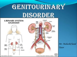
  • 5. anatomy and physiology of genitourinary system
  • 6.
  • 7. anatomy and physiology of genitourinary system
  • 9.
  • 10.
  • 11. anatomy and physiology of genitourinary system Nursing assessment: history, and physicalexamination 
  • 12. urological obstructions urethral strictures A urethral stricture is a scar in or around the urethra, which can block the flow of urine, and is a result of inflammation, injury or infection.
  • 13. Anatomy of the Male Reproductive System urological obstructions
  • 14. urological obstructions Risk factors- Urethral strictures are more common in men because their urethras are longer than those in women. Thus men's urethras are more susceptible to disease or injury.  A person is rarely born with urethral strictures and women rarely develop urethral strictures.
  • 15. urological obstructions Etiology - Stricture disease may occur anywhere from the bladder to the tip of the penis.  The common causes of stricture are trauma to the urethra and infections such as sexually transmitted diseases and damage from instrumentation. Trauma such as straddle injuries, direct trauma to the penis and catheterization can result in strictures of the anterior part of the urethra.
  • 16. urological obstructions Etiology - In adults, urethral strictures from instrumentation trauma may occur after prostate surgery and urinary catheterization.  In children, urethral strictures most often follow reconstructive surgery for congenital abnormalities of the penis and urethra, cystoscopy and occasionally may be congenital.
  • 17. urological obstructions Clinical features - painful urination. slow urine stream. decreased urine output. spraying of the urine stream. blood in the urine. abdominal pain. urethral discharge. urinary tract infections in men..
  • 18. urological obstructions Diagnostic evaluation- • History Collection • Blood test • Physicalexamination. • Urethral imaging (X-rays orultrasound). • Retrograde urethrogram
  • 19. Normal Urethrogram Obstructive Urethrogram urological obstructions
  • 20. urological obstructions Treatment- • Enlarging the stricture by gradual stretching (dilation). • Urethrotomy: Cutting the stricture with a endoscopic equipment • Urethroplasty surgical repair of the stricture with reconnection and reconstruction. • Urethral Stents where a biocompatible hollow tube is placed on the inside of the stricture to allow for free passage of urine.
  • 21. urological obstructions renal calculi Urolithiasis refers to stones (calculi) in the urinary tract. Stones are formed in the urinary tract when concentrations of substances such as calcium oxalate, calcium phosphate, and uric acid increase.
  • 22. urological obstructions Types of stone- Calcium stone Uric acid stone Cystiene stone Struvite stone
  • 23. urological obstructions Types of stone- Calcium stone: • Most stones (75%) are composed mainly of calcium oxalate crystals. • Increased calcium concentrations in blood and urine promote precipitation of calcium and formation of stones. • Cellulose sodium phosphate (Calcibind) may be effective in preventing calcium stones. • Therapy with thiazide diuretics may be beneficial.
  • 24. urological obstructions Uric acidstones: • 5% to 10% of all stones • Gout • Diet high in purines and abnormal purine metabolism. • Low-purine diet such as shellfish, asparagus, mushrooms, and organ meats are avoided. • Allopurinol may be prescribed to reduce serum uric acid levels and urinary uric acid excretion. • Proteins may be limited in diet
  • 25. urological obstructions Struvite stones: • 15% of urinary calculi • Struvite stones are made of magnesium, ammonium and phosphate • Form in persistently alkaline, ammonia-rich urine • Caused by the presence of urease splitting bacteria such as Proteus, Pseudomonas, Klebsiella, Staphy- lococcus, or Mycoplasma species.
  • 26. urological obstructions Cystine stones: 1% to 2% of all stones Occur in patients with a rare inherited defect in renal absorption of cystine (an amino acid). Penicillamine is administered to reduce the amount of cystine in the urine. Low-protein diet
  • 27. Urological obstrUctions Causes and predisposing factors: Chronic dehydration, poor fluid intake, andimmobility Living in mountainous and desert areas Infection, urinary stasis, and periods ofimmobility Medications- antacids, acetazolamide (Diamox), vitamin D, laxatives, and high doses of aspirin
  • 28. Urological obstrUctions Location of stones- Kidney Ureter Bladder Urethra
  • 30. Urological obstrUctions Clinical features- Pain that radiates to the lower abdomen and groin Heamturia Dysuria Oedema Pyuria Nausea, vomiting, diarrhea, abdominal discomfort Chills and fever (may)
  • 31. Urological obstrUctions renal calcUli Diagnostic evaluation- • History • Physical examination • Blood studies • Urinalysis • IVP • Cystoscopy • Radiographic studies
  • 32. Urological obstrUctions Management General Principles If small stone (< 4 mm) and able to treat as outpatient, 80% will pass stone spontaneously with hydration, pain control, and reassurance. Hospitalized for intractable pain, persistent vomiting, high-grade fever, obstruction with infection, and solitary kidney with obstruction. Medical management Surgical management Nursing management
  • 33. Urological obstrUctions Medical management: • Opioid analgesics or NSAIDs are administered. • NSAIDs provide specific pain relief because they inhibit the synthesis of prostaglandin E. • Hot baths or moist heat to the flank areas may also be useful. • Fluids are encouraged. This increases the hydrostatic pressure behind the stone, assisting it in its downward passage. • A high, around-the-clock fluid intake reduces the concentration of urinary crystalloids, dilutes the urine, and ensures a high urine output.
  • 34. Urological obstrUctions renal calcUli  N o n surgicalmanagement- Ureteroscopy ESWL (Extra Corporeal Shock wave lithotripsy) Endoscopic procedures Electrohydrolic lithotripsy Chemolysis
  • 35. Urological obstrUctions Ureteroscopy  Ureteroscopy involves visualizing the stone and then destroying it.  Access to the stone is accomplished by inserting a ureteroscope into the ureter and then inserting a laser, electrohydraulic lithotriptor, or ultrasound device through the ureteroscope to fragment and remove the stones.  A stent may be inserted and left in place for 48 hours or more after the procedure to keep the ureter patent
  • 36. Urological obstrUctions ESWL- ESWL is a noninvasive procedure used to break up stones in the calyx of the kidney In ESWL, a high-energy amplitude of pressure, or shock wave, is generated by the abrupt release of energy and transmitted through water and soft tissues. When the shock wave encounters a substance of different intensity (a renal stone), a compression wave causes the surface of the stone to fragment. Repeated shock waves focused on the stone eventually reduce it to many small pieces. These small pieces are excreted in the urine, usually without difficulty.
  • 38. Urological obstrUctions Endoscopic procedures-  A percutaneous nephrostomy or a percutaneous nephrolithotomy may be performed, and a nephroscope is introduced through the dilated percutaneous tract into the renal parenchyma. Depending on its size, the stone may be extracted with forceps or by a stone retrieval basket. Alternatively, an ultrasound probe may be introduced through the nephrostomy tube.
  • 39. Urological obstrUctions Electrohydraulic lithotripsy- an electrical discharge is used to create a hydraulic shock wave to break up the stone.  A probe is passed through the cystoscope, and the tip of the lithotriptor is placed near the stone  This procedure is performed under topical anesthesia.
  • 40. Urological obstrUctions Chemolysis- Chemolysis, stone dissolution using infusions of chemical solutions (eg, alkylating agents, acidifying agents) A percutaneous nephrostomy is performed, and the warm irrigating solution is allowed to flow continuously onto the stone.
  • 41. Urological obstrUctions Surgical management  Nephrolithotomy - Incision into the kidney with removal of the stone  Nephrectomy –  Pyelolithotomy - Ureterolithotomy - Cystostomy – removal of kidney removal of stone from renal pelvis removal of stone from ureter removal of stone from bladder Cystolitholapaxy - an instrument is inserted through the urethra into the bladder, and the stone is crushed in the jaws of this instrument
  • 42. DisorDers of kiDney glomerUlonephritis (acUte nephritic synDrome ) Definition – Acute glomerulonephritis refers to a group of kidney diseases in which there is an inflammatory reaction in theglomeruli.
  • 43. DisorDers of kiDney Risk factors and Causes– • Group A beta- hemolytic streptococcal infection in the throat • Impetigo (infection of the skin) • Acute viral infections-URTI, mumps, varicella zoster virus, Epstein-Barr virus, hepatitis B, and human immunodeficiency virus [HIV] infection). • Antigens outside the body (eg, medications, foreignserum) • High blood pressure • Diabetic kidney disease (diabetic nephropathy
  • 45. DisorDers of kiDney Clinical features- Hematuria - The urine may appear cola-colored be- cause ofred blood cells (RBCs) Edema andhypertension Oliguria Anemia from loss of RBCs into theurine
  • 46. DisorDers of kiDney Clinical features-  I n the more severe form of the disease, patients also complain of headache, malaise, and flank pain.  Elderly patients may experience circulatory overload with dyspnea, engorged neck veins, cardiomegaly, and pulmonary edema. Atypical symptoms include confusion, and seizures, which are often confused with the symptomsof a primary neurologicdisorder
  • 47. DisorDers of kiDney Diagnostic evaluation- History  O n examination- kidney is large, tender, edematous and congested Urinanalysis- protienuria, hematuria , oliguria Blood studies- Serum creatinine, BUN increased Hypoalbuminemia, hyperlipidemia  Antistreptolysin O titers are usually elevated in post streptococcal glomerulonephritis Electron microscopy analysis help identify the nature of the lesion. Kidney biopsy may be needed for definitive diagnosis.
  • 48. DisorDers of kiDney Complications- Hypertensive Encephalopathy Heart Failure Pulmonary Edema ESRD
  • 49. DisorDers of kiDney Management- Goal- To conserve renal function To treat complication adequately Types of management- Non pharmacological management Dietary management Pharmacological management Nursing management
  • 50. DisorDers of kiDney Non pharmacological management- Complete bed rest – as excessive activity may increase the proteinuria and hematuria. It should be encouraged until the urine clears and BUN, creatinine and BP return to normal. Strict intake out put charting. Fluid restrictions Plasmapheresis to decrease the serum anti body level. Dialysis if, uremic symptoms are severe.
  • 51. DisorDers of kiDney Dietary management- Protein restricted diet as the level of BUN and creatinine is high in blood  L o w fat diet due tohyperlipidemia Sodium restriction if hypertension, edema or congestive heart failure are present.  Increased carbohydrate diet to provide energy and to prevent the catabolism of protein.
  • 52. DisorDers of kiDney Pharmacological management- Residual streptococcal infection is suspected, penicillin is the agent of choice. Diuretics and antihypertensive agents may be given to control hypertension. Corticosteroids and cytotoxic agents are used to reduce the inflammation.  H 2 blockers (to prevent stress ulcers)  Phosphate binding agents (to reduce phosphate and elevate calcium).
  • 53. DisorDers of kiDney Nursing management- Monitor vital signs, intake and output, and maintain dietary restrictions during acute phase. Encourage rest during the acute phase as directed until the urine clears and BUN, creatinine, and blood pressure normalize. Administer medications as ordered, and evaluate patient's response to antihypertensives, diuretics, H2 blockers, phosphate-binding agents, and antibiotics (if indicated).
  • 54. DisorDers of kiDney Nursing management- Regular monitoring of blood pressure, urinary protein, and BUN concentrations Encourage patient to treat any infection promptly. Tell patient to report any signs of decreasing renal function and to obtain treatment immediately.
  • 55. DisorDers of kiDney nephrotic synDrome Definition- Nephrotic syndrome is a clinical disorder characterized by marked increase of protein in the urine (proteinuria), decrease in albumin in the blood (hypoalbuminemia), edema, and excess lipids in the blood (hyperlipidemia). These occur because of increased permeability of the glomerular capillary membrane.
  • 56. Etiology: Minimal change disease Diabetic kidney disease Systemic lupus erythematous Heart failure ( right side HF) Medications Certain infections ( hepatitis B and C, HIV,malaria)
  • 57.
  • 59. Diagnostic evaluation- Urinalysis- marked proteinuria, microscopic hematuria, 24-hour urine for protein (increased) Protein electrophoresis and immunoelectrophoresis of the urine to categorize the proteinuria Needle biopsy of kidney for histologic examination of renal tissue to confirm diagnosis. Serum chemistry- decreased total protein and albumin, normal or increased creatinine, increased triglycerides, and altered lipid profile
  • 60. DisorDers of kiDney nephrotic synDrome Management- Treatment of causative glomerulardisease Diuretics and angiotensinconverting enzyme inhibitors to control proteinuria Corticosteroids or immunosuppressant agents todecrease proteinuria General management ofedema Sodium and fluid restriction; liberalpotassium Dietary proteinsupplements
  • 61. DisorDers of kiDney nephrotic synDrome Nursing Management- Monitordaily weight, intake and output, and urine specific gravity. Monitor CVP (if indicated), vital signs, orthostaticblood pressure, and heart rate to detecthypovolemia. Monitor serum BUN and creatinine toassess renal function. Administer diuretics or immunosuppressants asprescribed, and evaluate patient's response. Infuse I.V. albumin asordered. Encourage bed rest for a few days to help mobilizeedema; however, some ambulation is necessary to reduce risk of thromboembolic complications.
  • 62. DisorDers of kiDney acute renal failure Definition- Acute renal failure is a sudden and almost complete loss of kidney function caused by failure of renal circulation or by glomerular or tubular dysfunction.
  • 63. DisorDers of kiDney acute renal failure Etiology-  P r e – renal (hypoperfusion of kidney) Intra – renal (actual damage to the kidneytissue) Post – renal (obstruction to urineflow)
  • 64. DisorDers of kiDney acute renal failure Etiology-  P r e –renal Hemorrhage Renal loses GI losses Impaired cardiac efficiency sepsis Anaphylaxis Antihypertensive medications
  • 65. DisorDers of kiDney acute renal failure Etiology- Intra –renal Prolonged renal ischemia Nephrotoxic agents Aminoglycosides agents Radiopaque contrast agents Heavy metals Solvents and chemicals
  • 66. DisorDers of kiDney acute renal failure Etiology- Post –renal Urinary tract obstructions Renal calculi Tumors BPH Blood clots Strictutres
  • 67. DisorDers of kiDney acute renal failure PHASES OFARF Initiating phase Oliguric phase Diuretic phase Recovery phase
  • 68. DisorDers of kiDney acute renal failure PHASES OFARF Initiating phase Begins with the initial insult and ends when oliguria develops Oliguric phase Urine output less than 400 ml/day Diuretic phase Urine out put become normal but nitrogenous waste products still remain elevated in blood Recovery phase It signifies the improvement of renal function It takes 3-12 months to return normal
  • 69. DisorDers of kiDney acute renal failure Clinical features- Vomiting and/or diarrhea, which may lead to dehydration. Nausea. Weight loss. pale urine. Less frequent urination, or in smaller amounts thanusual, with dark coloured urine Haematuria. Pressure, or difficulty urinating. Itching.
  • 70. DisorDers of kiDney acute renal failure Clinical features- Bone damage. Muscle cramps (caused by low levels of calcium whichcan cause hypocalcaemia) Muscle paralysis. Swelling of the legs, ankles, feet, face and/or hands. Shortness of breath due to extra fluid on the lungs Pain in the back orside Feeling tired and/or weak.
  • 71. DisorDers of kiDney acute renal failure Clinical features- Memory problems. Difficulty concentrating. Dizziness.  L o w bloodpressure. Anorexia Seizures (if blood urea nitrogen level is very high)
  • 72. DisorDers of kiDney acute renal failure Diagnostic evaluation- History regarding the etiological factors and risk factors. Physical symptoms Urine output – scanty, bloody, and low specific gravity Increased BUN and creatinine level in blood Hyperkalemia Metabolic acidosis Hyperphoshatemia Hypocalcemia Anemia
  • 73. DisorDers of kiDney acute renal failure Management- • Antihypertensive agents • Diuretics • Sodium polystyrene sulfonate (Kayexalate) • Fluid and electrolyte replacement • Dialysis • To correct dehydration. • To Keep other body systems working properly
  • 74. Chronic renal Failure CRF or ESRD is a progressive, irreversible deterioration in renal function in which the body’s ability to maintain metabolic and fluid and electrolyte balance fails resulting in uremia or azotemia.
  • 75. Etiology • Diabetes mellitus • Hypertension • SLE • Sickle cell disease • Glomerulonephritis • Pyelonephritis • ARF • Infections
  • 76. PATHPHYSIOLOGY DUE TO ETIOLOGICAL FACTORS DECREASED GFR HYPERTROPHY OF REMAINING NEPHRONS INABILITY TO CONCENTRATE URINE FURTHER LOSS OF NEPHRON FUNCTION LOSS OF NON-EXCRETORY AND EXCRETORY FUNCTION
  • 77. STAGES OF CRF 1) Reduced Renal reserve - BUN is high or normal - Client has no C/M - 40 to 75 % loss of nephron function 2) Renal Insufficiency -75 to 90 % loss of nephron function -Impaired urine concentration -Nocturia, mild anemia, increased creatinine and BUN
  • 78. 3) Renal failure - Severe azotemia - Impaired urine dilution - Severe anemia -Electrolyte Imbalances Hypernatremia Hyperkalemia Hyperphosphatemia 4) End Stage Renal Disease -10 percentage nephrons functioning -Multisystem dysfunction
  • 79. Clinical Manifestations of CRF Electrolyte and acid-base balance -Anemia -Bleeding Tendencies -Infection -Weight loss -Nausea and vomitting
  • 80. DIAGNOSTIC STUDIES  History and physical examination  Routine lab measurements - BUN - Serum Creatinine - Serum Electrolytes - Hematocrit and Hb levels - Urine Analysis - Urine Culture Radiological: - Renal Ultrasound - Renal Scan - C T Scan - Renal Biopsy